Signet-ring cell large B-cell lymphoma: A potential diagnostic pitfall with signet-ring cell carcinoma
Key Clinical Message
This study reveals the importance of recognizing uncommon histologic variants in diffuse large B-cell lymphoma, such as the signet-ring cell variant, which may result in an erroneous or delayed diagnosis with potential impact in patient treatment.
DEAR EDITOR
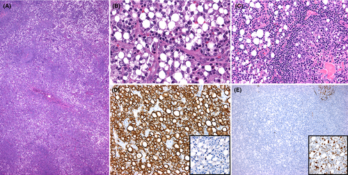
A 56-year-old man presented with a 4-cm left inguinal lymphadenopathy. Lymph node excision showed effacement of the architecture by an atypical infiltrate with few residual lymphoid follicles (Figure 1A). The infiltrate consisted of signet-ring cells and cells with multivacuolated cytoplasm (Figure 1B). The lymphoid follicles lacked polarization and contained signet-ring cells and >15 centroblasts per follicle (Figure 1C). By immunohistochemistry, the signet-ring cells were positive for CD20 (Figure 1D) and negative for pan-cytokeratin (Figure 1D, inset). CD21 highlighted follicular dendritic cell meshworks in lymphoid follicles but lack of them in diffuse areas (Figure 1E). The signet-ring cells were also positive for bcl-6 (Figure 1E, inset), bcl-2, CD10, and MUM1, and negative for T-cell markers. Ki-67 was 40%. Flow cytometry detected a CD10+ B-cell population with variable forward scatter and lack of surface light chains. Fluorescence in situ hybridization showed rearrangement of the BCL6 gene. A diagnosis of diffuse large B-cell lymphoma (DLBCL) with a minor component of follicular lymphoma was established. Signet-ring cell DLBCL is rare, with <100 reported cases to date.1, 2 Although this morphology has no prognostic impact, its recognition is crucial to avoid a misdiagnosis of metastatic signet-ring cell carcinoma to lymph node, leading to inappropriate management.1, 2

CONFLICT OF INTEREST
None declared.
AUTHOR CONTRIBUTIONS
VP and SPO: contributed equally to the preparation of this work.


