Exhaled breath diagnostics of lung and gastric cancers in China using nanosensors
Trial registration: ChiCTR1900026033, Chinese clinical trial registry (http://www.chictr.org.cn), retrospectively registered on September 18, 2019.
List of Abbreviations
-
- VOC
-
- volatile organic compounds
-
- LC
-
- lung cancer
-
- GC
-
- gastric cancer
-
- GC-MS
-
- gas chromatography connected to mass spectrometry
-
- GNP
-
- gold-nanoparticles
-
- SiO2
-
- silicon dioxide
-
- SU-8
-
- photosensitive polymer
-
- TD
-
- thermal desorption
-
- DFA
-
- discriminant factor analysis
-
- TP
-
- true positive
-
- TN
-
- true negative
-
- FP
-
- false positive
-
- FN
-
- false negative
-
- ROC
-
- receiving operating characteristics
-
- AUC
-
- area under the curve
Dear Editor,
Breath analysis is a promising diagnostic approach for various conditions [1, 2]. It is based on the identification of volatile organic compounds (VOCs) emitted in the breath, which creates a unique volatolomic signature [3]. Owing to their characteristics, VOCs can be measured non-intrusively from the breath or other body sources [3, 4]. Several studies have shown the diagnostic potential for a variety of conditions based on VOC analysis [5-9]. Malignant diseases, where early detection is crucial, are the main focus of VOC analysis, with lung cancer (LC) and gastric cancer (GC) being the most studied. LC and GC together were responsible for approximately 2.5 million deaths globally in 2018 [10]. The aim of VOC analysis of the breath using sensors is to identify a “VOC-print” comprising the total abundances and ratios of the compounds in the breath, giving an overall unique chemical pattern [11]. This technology can help to address specific challenges concerning LC screening and GC mortality [12, 13]. To facilitate real-world applications, different ethnic- and culture-based populations should be sampled. Here, we carried out a VOC-based clinical trial for GC and LC detection in China to classify these two major malignancies with different genetic and cultural backgrounds, using our developed sensors [1] with newly-developed sensor-printing and sampling methods.
A total of 545 breath samples (one/two samples per subject) were collected from 426 adult participants between January 2018 and July 2019 at the Jiangyin Hospital Affiliated to the Southeast University Medical College in China. The study population consisted of three groups: LC patients (n = 158), GC patients (n = 115), and healthy volunteers (n = 153), as detailed in Table 1. All participants gave their informed consent for inclusion before participating in the study.
| Lung cancer patients† | Gastric cancer patients† | ||||||||||||
|---|---|---|---|---|---|---|---|---|---|---|---|---|---|
| Characteristic | Whole population | Healthy volunteers | Total | Stage I | Stage II | Stage III | Stage IV | Unknown | Total | Stage I | Stage II | Stage III | Stage IV |
| Total (cases) | 426 | 153 | 158 | 96 | 12 | 7 | 36 | 7 | 115 | 24 | 8 | 17 | 66 |
| Age [years, median (range)] | 60 (23-81) | 47 (23-74) | 67 (36-81) | 62 (36-80) | 67 (49-81) | 65 (37-79) | 67 (45-80) | 67 (63-77) | 67 (30-80) | 65 (48-80) | 66 (48-72) | 69 (54-80) | 67 (30-80) |
| Gender [cases (%)] | |||||||||||||
| Male | 261 (61) | 107 (70) | 76 (48) | 32 (33) | 6 (50) | 6 (86) | 28 (78) | 4 (57) | 78 (68) | 18 (75) | 4 (50) | 11 (65) | 45 (68) |
| Female | 165 (39) | 46 (30) | 82 (52) | 64 (67) | 6 (50) | 1 (14) | 8 (22) | 3 (43) | 37 (32) | 6 (25) | 4 (50) | 6 (35) | 21 (32) |
| Smoking status [cases (%)] | |||||||||||||
| Yes | 91 (21) | 30 (20) | 37 (23) | 14 (15) | 3 (27) | 5 (71) | 13 (36) | 2 (29) | 24 (21) | 5 (21) | 3 (38) | 5 (29) | 11 (17) |
| No | 335 (79) | 123 (80) | 121 (77) | 82 (85) | 9 (75) | 2 (29) | 23 (64) | 5 (71) | 91 (79) | 19 (79) | 5 (63) | 12 (71) | 55 (83) |
| Alcohol use [cases (%)] | |||||||||||||
| Yes | 99 (23.2) | 37 (24) | 40 (25.3) | 13 (14) | 3 (25) | 5 (71) | 15 (42) | 4 (57) | 22 (19) | 4 (17) | 2 (25) | 2 (12) | 14 (21) |
| No | 326 (76.5) | 116 (76) | 117 (74.1) | 83 (86) | 9 (75) | 2 (29) | 20 (56) | 3 (43) | 93 (81) | 20 (83) | 6 (75) | 15 (88) | 52 (79) |
| Unknown | 1 (0.2) | 0 | 1 (0.6) | 0 | 0 | 0 | 1 (3) | 0 | 0 | 0 | 0 | 0 | 0 |
| Asthma [cases (%)] | |||||||||||||
| Yes | 3 (1) | 3 (2) | 0 (0) | 0 (0) | 0 (0) | 0 (0) | 0 (0) | 0 (0) | 0 (0) | 0 (0) | 0 (0) | 0 (0) | 0 (0) |
| No | 423 (99) | 150 (98) | 158 (100) | 96 (100) | 12 (100) | 7 (100) | 36 (100) | 7 (100) | 115 (100) | 24 (100) | 8 (100) | 17 (100) | 66 (100) |
| H. pylori infection* [cases (%)] | |||||||||||||
| Yes | 4 (1) | - | - | - | - | - | - | - | 4 (3) | 1 (4) | 1 (13) | 0 (0) | 2 (3) |
| No | 59 (14) | - | - | - | - | - | - | - | 59 (51) | 9 (38) | 3 (38) | 10 (59) | 37 (56) |
| Unknown | 52 (2) | - | - | - | - | - | - | - | 52 (45) | 14 (58) | 4 (50) | 7 (41) | 27 (41) |
| Number of breath samples | 545 | 153 | 222 | 136 | 16 | 7 | 54 | 9 | 170 | 31 | 13 | 24 | 102 |
- * The data of H. pylori infection were only available for patients with gastric cancer.
- † The tumor stage was determined according to the eighth edition of the Union for International Cancer Control (UICC) TNM classification of malignant diseases.
Exhaled alveolar breath was collected in a controlled manner. End-tidal expired air was directly trapped and pre-concentrated in Tenax® TA sorption tubes (Buchem BV, Apeldoorn, The Netherlands). These new tubes were specially constructed for direct sampling at the SunshineHaick Co. (China) (see Supplementary Methods).
The nanomaterial-based sensor system (nanosensor) was originally developed at Technion (Haifa, Israel) [5, 7] and was recently redesigned as a benchtop device for breath VOC analysis-based LC and GC detection in China (Figure S1). The collected samples were exposed to the nanosensor array. The sensors comprised layers of gold nanoparticles (GNPs) with 13 different organic ligands in two formats (manual and printed), resulting in 26 different sensors inserted in each nanosensor system. The printed method is a novel approach using an inkjet printer and a unique micro-barrier ring developed to overcome topological irregularities (Figures S2 and S3).
Fifteen sensors were chosen (Table S1) after checking that their responses were reproducible with no background noise [7, 9]. One or two sample tubes per volunteer were introduced to the sensor array chamber, which was specially assembled with a thermal desorption (TD) system, enabling the sample tube to be exposed directly to the sensor array.
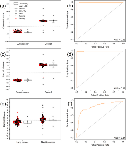
Exposure to the breath samples or the calibration compounds resulted in rapid and reversible changes of the sensors’ electrical resistance. Breath components were identified from the time-dependent resistance response of each sensor. Each sensor responded to all (or to a certain subset) of the VOCs found in the exhaled breath samples. Breath patterns were obtained from the collective response of the sensors by applying discriminant factor analysis (DFA). The DFA output variables constitute mutually orthogonal dimensions. We divided the dataset for each binary analysis (i.e., LC vs. control, GC vs. control, and GC vs. LC) into training (70% samples) and testing sets (30% samples). Leave-one-out cross-validation was used to calculate the classification success in terms of the numbers of true positive (TP), true negative (TN), false positive (FP), and false negative (FN) predictions. Given k measurements, the model was computed using k-1 training vectors. Receiver operating characteristic (ROC) analysis was used to test the performance of the training set data and to calculate the cut-off values (see details in Supplementary Methods supporting information).
Evaluation of the developed nanosensor system involved simulating the sample analysis by four similar nanosensor systems so that the reproducibility of different devices using known mixtures could be assessed. The clinical data were analyzed on the developed nanosensor system (Supplementary Methods and Figure S1).
The reproducibility of a diagnostic system is important. A reliable test requires high precision among different systems using the same sensing technology. Here, we ran 21 repeated breath samples collected over1 month from the same individual, as well as simulated breath mixtures from LC and GC patients, on four different systems running in parallel. The results showed that similar sensors from different systems gave comparable responses with a relative standard deviation of 0.1% for the response signals within the tested groups (Supplementary Methods and Figures S4, S5).
The new tube approach, i.e., direct sampling, was evaluated. Data on the capacity for breath collection were highly reproducible throughout the exposure on the device (Figure S5). Breath samples differed in humidity. Although the absorbent material Tenax could reduce the water content of the samples, it could not be removed completely. Therefore, to eliminate variation, the humidity was compensated using a linear regression of known humidity levels (Figure S6). The classification model based on a training set of LC versus control showed high accuracy, sensitivity, and specificity, with 0.99 area under the curve (AUC) in the ROC analysis (Table 2, Figure 1a, b). Likewise, all measures were 100% in the testing set. Similarly, GC versus control gave high levels of all performance measures for both the training and testing sets (Table 2, Figure 1c, d). The third classification model of LC versus GC gave high levels of measures, yet slightly lower, thought clinically acceptable (Table 2, Figure 1e, f). A number of outliers were found in the control group. The reason for such a response was unclear. We assumed that those people were unaware of the presence of cancer or they could be momentary electrical noises from specific sensors. Nevertheless, as can be seen, these outliers did not affect the performance of the model.
| Training set | Testing set | |||||
|---|---|---|---|---|---|---|
| Statistics | LC vs. C | GC vs. C | LC vs. GC | LC vs. C | GC vs. C | LC vs. GC |
| Accuracy (%) | 99 | 99 | 81 | 100 | 99 | 72 |
| Sensitivity (%) | 100 | 100 | 86 | 100 | 100 | 76 |
| Specificity (%) | 97 | 98 | 75 | 100 | 98 | 66 |
| PPV (%) | 98 | 98 | 82 | 100 | 98 | 75 |
| NPV (%) | 100 | 100 | 81 | 100 | 100 | 67 |
| TP (cases) | 156 | 117 | 133 | 66 | 53 | 52 |
| TN (cases) | 103 | 107 | 90 | 47 | 43 | 33 |
| FP (cases) | 3 | 2 | 30 | 0 | 1 | 17 |
| FN (cases) | 0 | 0 | 21 | 0 | 0 | 16 |
- PPV – positive predictive value; NPV – negative predictive value; TP – true positive; TN – true negative; FP – false positive; FN – false negative.

We further assessed the capacity of the system to distinguish early-stage (I, II) from late-stage (III, IV) cancers. For LC, the accuracy was 59% and 81% for the training and testing sets, respectively. For GC, the accuracy was 48% and 83% for the training and testing sets, respectively. The low accuracies could be attributed to the rather low numbers in each subgroup influencing the classifier. However, it is important to check whether stage I can be distinguished from stages II-IV, as stage I is considered localized and can be surgically removed in most cases. The latter classification for LC and GC gave 71% and 69% accuracy in the validation tests, suggesting that it was feasible to identify cases that remained localized and were suitable for surgical resection.
A number of confounding factors that could affect the analysis outcome were tested. The effects of gender, smoking and alcohol consumption were tested for LC and GC. Chronic conditions such as asthma are also important confounding factors [5, 6], but we could not test this in the present clinical study as only three subjects were recognized as asthmatic. The confounding factor analysis showed that most of the factors examined had a near-random influence on the classifier. For LC versus control, gender, smoking, and alcohol consumption gave AUCs of 0.62, 0.54, and 0.48 in the ROC analysis, respectively (Figure S7). The age factor gave AUC of 0.80, implying its influence on classification. Indeed, there was a rather big difference between the average age of LC patients (63.5 years) and healthy volunteers (47.5 years). However, the classifier for the comparison between LC and control gave a 99% accuracy; thus, even if age differences had some influence, most of the differences could be attributed to the health condition itself, i.e., sick versus healthy. For GC versus control, gender, smoking, and alcohol consumption gave AUCs of 0.45, 0.49, and 0.58, respectively, in the ROC analysis (Figure S8). Age gave an AUC of 0.88, again implying some influence on classification, but the sick versus healthy factor was much stronger (99% accuracy). Therefore, it is likely that age had a minor influence on the main classification, as for LC described above. No reliable statistical analysis was possible to determine whether the presence of Helicobacter pylori was a confounding factor, as only six GC patients were identified as H. pylori positive, though we previously showed that this factor had no influence on GC classification in a European population [9]. For LC versus GC, confounding factor analysis for gender, smoking, and alcohol consumption gave AUCs of 0.54, 0.55, and 0.53, respectively, in the ROC analysis. For the age factor, as in the two comparisons above for LC versus control and GC versus control, the AUC was 0.73 (Figure S9).
In conclusion, Patients with LC and GC have significantly different patterns of VOCs in breath opposed to healthy controls. Changes in breath VOCs can be easily measured using a nanosensor-array system. Fast and inexpensive sample collection can be done by direct breath sampling into dedicated Tenax tubes. The importance of confounding data was assessed and should continue to be tested in future studies. The data presented here are another step towards a real-world clinical diagnostic system for fast and affordable cancer detection and management.
DECLARATIONS
ETHICS APPROVAL AND CONSENT TO PARTICIPATE
The study was conducted in accordance with the Declaration of Helsinki, and the protocol was approved by the Ethics Committee of Jiangyin Hospital Affiliated to Southeast University Medical College, China (Project identification code No. 044). Chinese clinical trial registry is ChiCTR1900026033 (http://www.chictr.org.cn).
CONSENT FOR PUBLICATION
Not applicable
AVAILABILITY OF DATA AND MATERIALS
All data are available upon request.
COMPETING INTERESTS
The authors declare that they have no competing interests
FUNDING
This study received partial funding from the Horizon 2020 Information Communication Technology (ICT) Program under the breath Volatile Organic compound analysis for GAstric cancer Screening (VOGAS) project (grant agreement no. 824986). In addition, the project was partially funded by Jiangsu Sunshine Haick Co., which provided the breath sampling kits and systems.
AUTHORS' CONTRIBUTIONS
AG: data management, project design, data analysis, methodology, and manuscript revision; YYB: methodology, data interpretation, figure construction, and writing and revision of original draft; GY, WM, DS, LD, CW, QW, XS, JH, ZX, BH, and SW: clinical work and sample collection; YM and VKW: methodology and sensor preparation; HH: conception, coordination, funding acquisition, supervision of the project, and manuscript revision
ACKNOWLEDGMENTS
The authors acknowledge all the following for their support in discussion and technical advice concerning the study: Yidong Mi, Fei Shen, Xiaochen Wang, Jun Ge, Lin Gao, Feng Lin, Shaoping Liu, Qingfeng Lin, YeiLv, Wenjun Qian, Zhili Liu, Shuai Yan, Yuhong Zhang, ShaSha, Guoyi Shao, Weidong Zhong, Weiwei Tu, Xiangming Cao, Lei Xi, Simin Wang, Jie Zeng, Anqing Zhu, Yehua Mi.


