Ultrathin Covalently Bound Organic Layers on Mica: Formation of Atomically Flat Biofunctionalizable Surfaces
Abstract
Mica is the substrate of choice for microscopic visualization of a wide variety of intricate nanostructures. Unfortunately, the lack of a facile strategy for its modification has prevented the on-mica assembly of nanostructures. Herein, we disclose a convenient catechol-based linker that enables various surface-bound metal-free click reactions, and an easy modification of mica with DNA nanostructures and a horseradish peroxidase mimicking hemin/G-quadruplex DNAzyme.
The atomically flat nature of mica has made it the substrate of choice for microscopic visualization of dimensional parameters of various pre-fabricated nanomaterials such as DNA origami1 and protein conjugates,2, 3 among others. Mica is a hydrophilic aluminosilicate, which in saline solution is covered by a hydration layer with K+ ions that are tightly bound to the anionic silicate. While widely used for atomic force microscopy (AFM) studies,4 the surface modification and chemical functionalization of mica itself has received surprising little attention.5
Lately, catechol-based coatings including mussel-adhesive polydopamine proteins have found increasing attention for mica surface modification.6 Despite a general acceptance that the catechol moiety in mussel proteins is central to their adhesion ability,7 its exact adhesion mechanism is still unknown.8 Butler et al. recently showed that a pending amine functionality is central to binding.9 To demonstrate this, a symmetric trichrysobactin was synthesized that bears three 2,3-dihydroxybenzoyl moieties and a lysine tail. In this “two-punch” approach, the NH3+ moiety displaced the K+ ions, followed by surface attachment of the catechol moiety. Unfortunately, the six-step synthesis required for the scaffold hampers its widespread application. Even more, post-attachment functionalization of mica substrates with interesting biomolecules such as DNA and proteins is still an uncharted domain. Such an approach would require both facile and strong adhesion, combined with the presence of a moiety that can be routinely used for post-attachment functionalization. The current approaches7, 8 do not provide such a handle, and as such there is still need for a platform that allows various surface modification strategies and a stepwise observation of the assembly processes of biological nanostructures in a controlled fashion.10
Herein, we address this unresolved problem by a dual approach through the development of a simple molecule that allows both 1) covalent modification of mica, and 2) post-modification stepwise growth and study of molecular assemblies. To this end, we envisaged and synthesized a surface anchor (1) that possesses the structural characteristics required to combine optimal adhesion characteristics with a handle for modular functionalization (Figure 1). We extensively characterized the modified surfaces (M1) by static water contact angle (SCA), X-ray photoelectron spectroscopy (XPS), and AFM measurements. To illustrate the potential for surface functionalization, we explored several metal-free strain-promoted click reactions. Finally, to demonstrate biofunctionalization we pursued the stepwise formation of functional DNA constructs, such as G-quadruplex (GQ, G=guanine) structures on covalently modified surfaces by AFM.
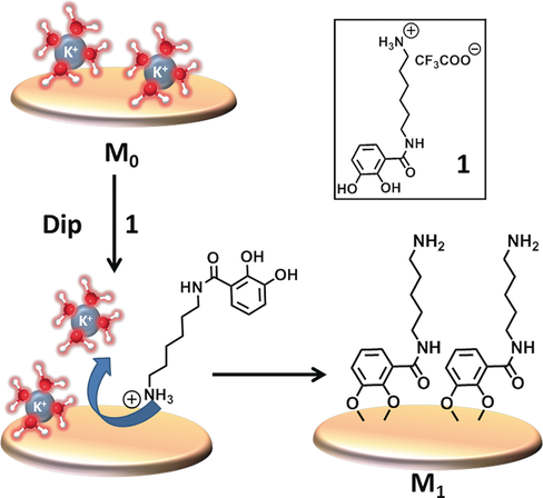
Tentative mechanism of mica modification by surface anchor 1.
As shown by Butler et al.9 and Hwang et al.,11 mica-adhesive molecules should likely contain both catechol and amino groups. However, in their case this resulted in a poorly defined polymeric layer possibly due to intramolecular Michael-type addition reactions between the catechol and free amino groups.12 Spencer et al. have tackled this issue of polymerization by inclusion of a strongly electron-withdrawing nitro group in the catechol motif to minimize auto-oxidation.13 Using this information, the design of our surface anchor 1 was based on the retention of the key components, that is a catechol moiety linked proximally by an electron-withdrawing amide group to a protonated amine (Figure 1).
A first step forward was the synthesis of surface anchor 1: 2,3-dihydroxybenzoic acid (DHBA) and mono-Boc-protected 1,6-hexanediamine were linked by a conventional amide coupling, followed by subsequent Boc-deprotection (Figure 2 a) to afford 1 in an overall yield of 66 % (0.78 g). A 1 mm solution of 1 in PBS buffer (pH 7.4) allowed for the simultaneous modification of large numbers of freshly cleaved mica surfaces in 24 h. A constant low water contact angle (55°; Figure 2 b), the emergence of N1s signal in XPS (Figure 2 c), and the XPS analysis of subsequent modifications on M1 confirmed modification of the mica surfaces with surface anchor 1 (see the Supporting Information). Importantly, molecules similar to surface anchor 1, but lacking one of the mentioned components, performed inferior than anchor molecule 1 (Table S1).
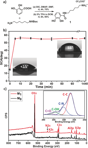
a) Synthesis of surface anchor 1. b) Increase of the water SCA during the modification process. c) XPS wide-range spectra of bare (M0) and modified (M1) mica (inset: narrow scan of the C1s signal).
XPS analysis of modified surfaces also showed the overall chemical composition of M1 to fit the molecular composition of 1 within experimental error (C/NXPS=6.8; C/NTheor=6.5; see the Supporting Information, Section S4.2). As shown by Hwang et al., the formation of polymeric layers on mica surfaces leads to the disappearance of the Al2p, K2s, and Si2p signals in the XPS spectrum.11 Interestingly in our case, the continued high intensity of these substrate-specific signals hinted at formation of ultrathin layers (Figure 2 c).
The deconvolution of the C1s narrow scan clearly revealed carbon atoms in distinct environments, namely, C−C, C−N, C−O, and C=O (Figure 2 c, inset). Experimental XPS binding energy results correlated nicely with simulated values obtained from DFT calculations (Section 6.1).14 AFM analysis of modified surfaces (M1) displayed a remarkably low surface roughness of 0.4 nm with a layer thickness of 2.5±0.5 nm (Figure S1–S3). This confirms exclusive formation of ultrathin layers (length of 1 in stretched-out fashion is ca. 1.3 nm) on mica surfaces. We note that the low (0.4 nm) roughness makes our modified surfaces amenable for reliable microscopic visualization of various covalently attached nanostructured biomolecules.
In view of the importance of metal-free click chemistry in bioconjugation chemistry, we performed various state-of-the-art “metal-free” strain-promoted click reactions (Figure 3). For this, amine-terminated layers M1 were converted to azide- functionalized coatings M2 (see Supporting Information) by a Cu-catalyzed azide transfer reaction.15 The resulting azide-terminated layers underwent a facile SPAAC reaction with a fluorinated labile BCN aromatic ester. This allowed easy characterization of the resulting surfaces M2 by XPS and direct analysis in real time high-resolution mass spectrometry (DART-HRMS),16 which showed the characteristic m/z peak of the surficial 4-perfluorinated butyl benzoate anion (339.0062) in the extracted ion chronogram (EIC; Figure S4). A clear XPS F1s signal was evident at 686.0 eV, indicating a successful reaction (50 %) (Figure 4 a). An inverse strategy for the surface-bound SPAAC reaction was also pursued with BCN immobilized on the surface (M4) and a reaction with an azide coupled to a fluorinated tail (Figure 3), to yield surface M5. The XPS F1s peak at 686.0 eV was used to demonstrate the extent of reaction (83 %; Figure 4 a and Section S4.10), while also the C1s and N1s spectra confirm the success of this reaction (Figure 4 b).
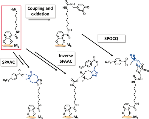
Functionalized mica as building block: General scheme showing the cycloaddition adducts of various metal-free click reactions on M1.
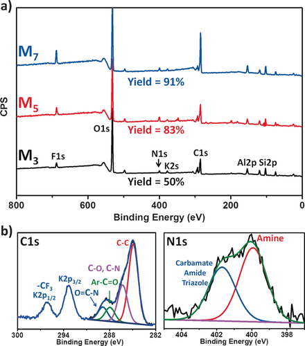
a) Stacked XPS wide scans of final cycloaddition adducts of SPAAC with azide on surface (M3; bottom), BCN on surface (M5; middle), and SPOCQ (M7; top). The F1s and N1s peaks were used for determining the yield of these reactions. b) XPS narrow scan C1s region (left) and N1s region (right) of SPAAC cycloaddition adducts (M5).
The strain-promoted click reaction between BCN and ortho-quinones (SPOCQ)17 was investigated as a second example on mica surfaces modified by our method. To this end, 2,3-dihydroxybenzyl amine was attached to surface M1 using a DIC coupling reaction, and subsequently oxidized to its corresponding 1,2-quinone (M6) using NaIO4. XPS analysis confirmed the presence of a modified anchor on mica (Section S4.13). The quinone underwent a facile SPOCQ reaction producing in excellent yield (91 %) surface M7 in 4 h, as evidenced by the F/N ratio in XPS (Figure 4 a and Section S4.14).
Having established prominent bioconjugation reactions on mica, we switched our attention to our other objective: the formation of AFM-observable covalently linked DNA nanostructures. Building on the insights from Famulok et al., a 168-bp DNA minicircle consisting of eight distinct DNA fragments18 was assembled in a step-wise fashion after one of the components was tethered covalently onto mica surface M1. Covalent attachment of strand D1 (Table S2) allowed its hybridization to a preformed assembly of oligomers D2–D8 (Table S2), forming circular structures. As shown in Figure 5 a, 3D height images of DNA-containing surfaces revealed circular structures that possess an internal cavity (Figures S8–S11). A large number of contiguous shapes with circular features were obtained, with an internal diameter of 20±2 nm (Figure S12), in accordance with literature (18.2 nm).18 When significantly lower concentrations of strands D2–D8 were applied (100-fold dilution, that is, 0.1 μm), we observed mostly open structures, and circles of which the larger internal diameter hinted to the formation of semi-closed dimeric and trimeric structures (Figures 5 b,c, S13 and S14). Control experiments with bare mica surfaces, that is, without the anchored DNA, did not show any circles or other (linear) structures, proving the need for a covalently attached anchor.
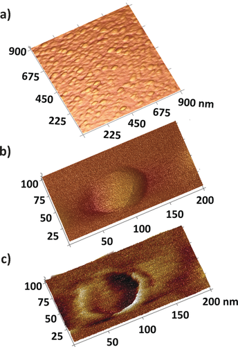
a) 3D height images of DNA circles assembled on mica. b) 3D height and c) quadrature image of a single DNA circle.
Lastly, we added another dimension to our method by the attachment of functional DNA, namely the horseradish peroxidase-mimicking hemin/G-quadruplex (hGQ) DNAzyme.19 Such a hGQ DNAzyme can oxidize a variety of organic substrates in the presence of H2O2.20 To demonstrate the feasibility of hGQ catalytic activity of DNAzymes on mica surfaces, we attached the guanine-rich sequence EAD2 (Table S2) covalently to M1 mica surfaces. Covalent attachment of DNA was confirmed with XPS analysis by the emergence of P2s and P2p signals in the XPS wide spectra at 189.0 eV and 133.0 eV, respectively (Figure S4.17). Furthermore, complexation of hemin [FeIII-protoporphyrin IX chloride] to the formed GQ under K+-rich conditions was confirmed by the emergence of characteristic XPS Fe2p peaks20b (710.0 and 721.0 eV) (Figure S4.17, inset).
After validation of the covalent attachment of DNA, we assessed its HRP-mimicking ability using 2,2′-azino-bis(3-ethylbenzothiazoline-6-sulfonate) (ABTS2−), which is converted into ABTS.− in the presence of the hGQ DNAzyme and H2O2.20a Indeed, upon addition of H2O2 to an ABTS-solution that contained just a single hGQ-functionalized mica slide (12 mm diameter), within 5 min a green-colored solution with λmax=414 nm (Figure 6) was formed. Control experiments (without ABTS, without modified mica, or without H2O2) did not show formation of this oxidation product. Therefore, the oxidation of ABTS2− was solely attributable to the presence of catalytically active hGQ DNAzyme EAD2 on the surface.
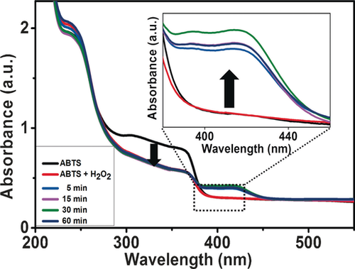
UV/Vis spectrum of ABTS oxidation catalyzed by EAD2-hemin DNAzyme, inset: changes in the spectrum upon ABTS oxidation.
To summarize, we report the facile and covalent modification of mica using a catechol-based surface anchor with a flexibly linked amino group. This yielded robustly bound, low-roughness, and ultrathin layers that are amine-terminated. We demonstrate that this approach allows for highly flexible surface modification, using a range of metal-free click reactions as case in points. In addition, we display the potential of our strategy for microscopic imaging of functional DNA constructs, and the potential for following the formation of such constructs in a stepwise fashion. We thus believe our work opens up new avenues for the immobilization and successive visualization of a wide variety of biomolecule conjugates on mica and related surfaces.
Acknowledgements
The authors thank The Netherlands Organization for Scientific Research (NWO) for funding via ECHO project number 712.012.006. We also thank Medea Kosian, Dr. Sidharam Pujari, and Dr. Jorge Escorihuela for stimulating discussions.
Conflict of interest
The authors declare no conflict of interest.


