Glial localization of antiquitin: Implications for pyridoxine-dependent epilepsy
Abstract
Objective
A high incidence of structural brain abnormalities has been reported in individuals with pyridoxine-dependent epilepsy (PDE). PDE is caused by mutations in ALDH7A1, also known as antiquitin. How antiquitin dysfunction leads to cerebral dysgenesis is unknown. In this study, we analyzed tissue from a child with PDE as well as control human and murine brain to determine the normal distribution of antiquitin, its distribution in PDE, and associated brain malformations.
Methods
Formalin-fixed human brain sections were subjected to histopathology and fluorescence immunohistochemistry studies. Frozen brain tissue was utilized for measurement of PDE-associated metabolites and Western blot analysis. Comparative studies of antiquitin distribution were performed in developing mouse brain sections.
Results
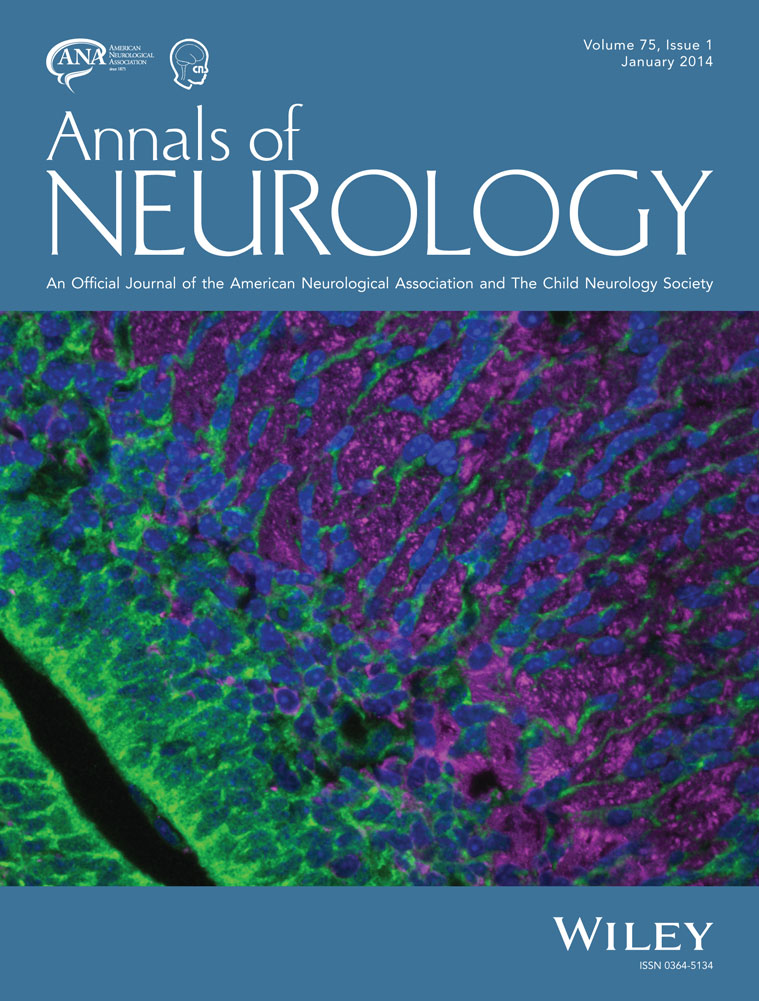
Histologic analysis of PDE cortex revealed areas of abnormal radial neuronal organization consistent with type Ia focal cortical dysplasia. Heterotopic neurons were identified in subcortical white matter, as was cortical astrogliosis, hippocampal sclerosis, and status marmoratus of the basal ganglia. Highly elevated levels of lysine metabolites were present in postmortem PDE cortex. In control human and developing mouse brain, antiquitin immunofluorescence was identified in radial glia, mature astrocytes, ependyma, and choroid plexus epithelium, but not in neurons. In PDE cortex, antiquitin immunofluorescence was greatly attenuated with evidence of perinuclear accumulation in astrocytes.
Interpretation
Antiquitin is expressed within glial cells in the brain, and its dysfunction in PDE is associated with neuronal migration abnormalities and other structural brain defects. These malformations persist despite postnatal pyridoxine supplementation and likely contribute to neurodevelopmental impairments. ANN NEUROL 2014;75:22–32