Journal list menu

 Issue
IssueJournal of Morphology: Volume 282, Issue 1
1-168January 2021
Export Citations
Download PDFs
COVER IMAGE
Cover Image
- First Published: 23 December 2020
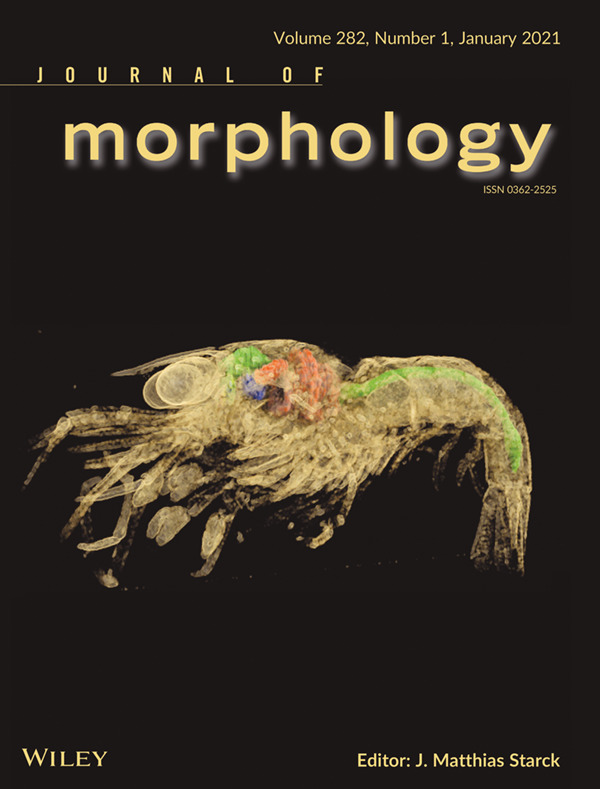
Cover illustration. Neocaridina davidi is a freshwater shrimp that is commonly bred all over the word. It has pelagic larvae that differ considerably in their morphology and habits from adults. In this issue of the Journal of Morphology, Sonakowska-Czajka and collaborators (pp. 48-65) document the 3D-morphology of the midgut of larval stages using X-ray microtomography, and, using light and transmission electron microscopy, provide a detailed description of structural and ultrastructural changes of the midgut epithelium (intestine and hepatopancreas) that occur during postembryonic development. The cover images shows a zoea III larva of Neocaridina davidi with the long intestine (green) and hepatopancreas (red).
ISSUE INFORMATION
RESEARCH ARTICLES
Skull osteology of Vipera walser (Squamata, Viperidae): Description, variability, ontogeny, and diagnostic characters in comparison to other Italian vipers
- Pages: 5-47
- First Published: 15 October 2020

The description of the skull of Vipera walser provides new information on the ontogeny and intraspecific variability of this species, along with some skull features which allow to distinguish isolated skull bones of V. walser from those of the other Italian vipers.
Postembryonic development and differentiation of the midgut in the freshwater shrimp Neocaridina davidi (Crustacea, Malacostraca, Decapoda) larvae
- Pages: 48-65
- First Published: 19 October 2020
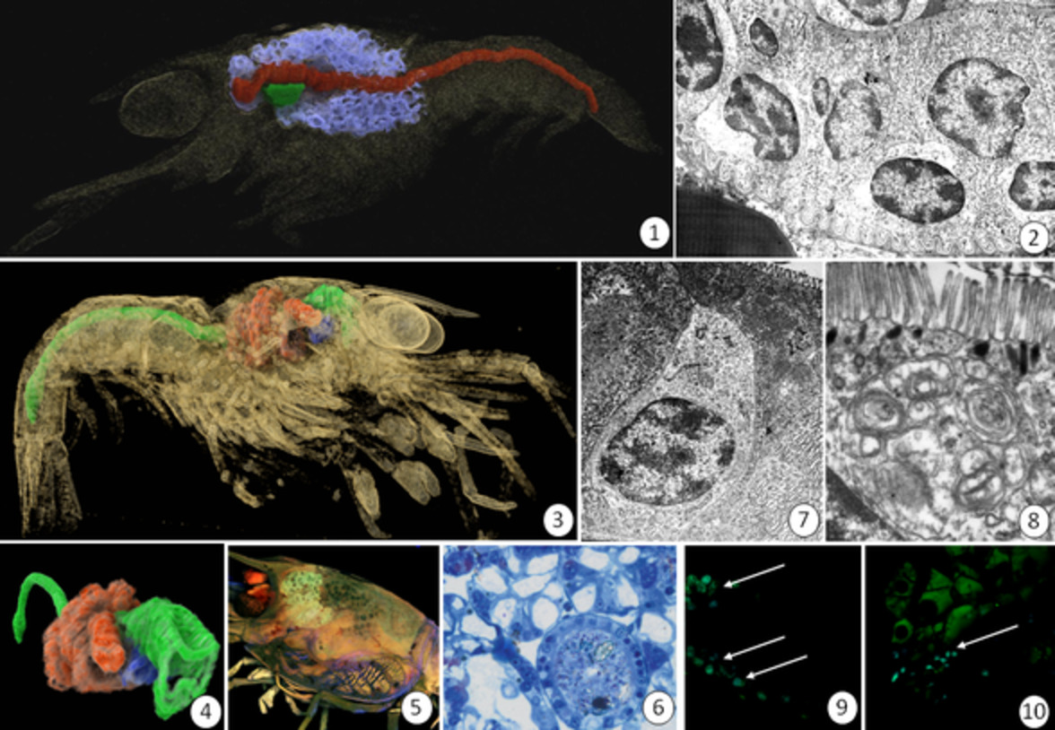
(1) Zoea I of Neocaridina heteropoda. Midgut composed of the long intestine (red) and hepatopancreas (blue). A fragment of the foregut (green). XMT. (2) Zoea I. The anterior region of intestine with regenerative and digestive cells. TEM. (3) 3D view of zoea III body with the midgut composed of the long intestine (green) and hepatopancreas (red). A fragment of the foregut (blue). XMT. (4) Zoea III. 3D view of the isolated midgut composed of the long intestine (green) and hepatopancreas (red). A fragment of the foregut (blue). XMT. (5) Hepatopancreas in I zoea body. XMT. (6) Zoea III of N. heteropoda. Cross section through the intestine and hepatopancreatic tubules. LM. (7) Zoea III. The anterior region of intestine with regenerative and digestive cells. TEM. (8) Zoea III. The apical cytoplasm of the digestive cells in intestine with large autophagosome. TEM. (9–10) Zoea III. Fluorescence micrographs showing anti-phosphohistone H3 (green) regenerative cells (arrows) during mitotic division. Longitudinal section. Hoechst 33342 staining (blue). FM.
Do exaggerated chelicerae function as weapons or genitalia in a long-jawed spider? Functional allometric analysis yields an answer
- Pages: 66-79
- First Published: 19 October 2020
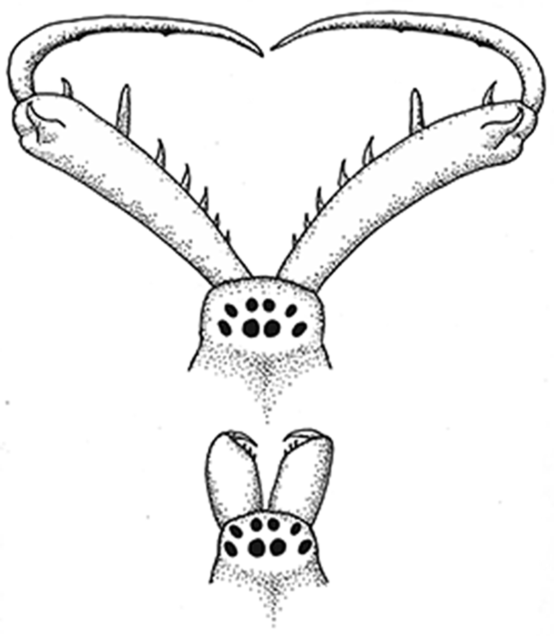
In the long-jawed orbweaver Tetragnatha elongata, chelicerae grow from a typical orbweaver length in penultimate spiders (bottom) to the extreme form only seen in the adult (top) spider. Sexual selection has shaped the chelicerae in both sexes for an optimal “mechanical mesh” because these spiders interlock chelicerae during mating.
3D heart morphological changes in response to developmental temperature in zebrafish: More than ventricle roundness
- Pages: 80-87
- First Published: 14 October 2020
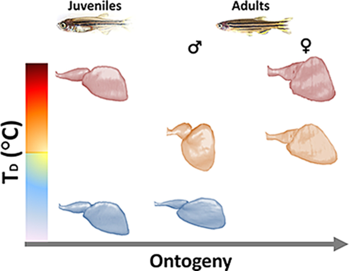
We examined the effect of developmental temperature on the three-dimensional heart shape of zebrafish. We found that temperature experienced during the embryonic and larval stages permanently alters ventricle sphericity and other significant heart morphometric indices in juvenile, male and female fish that have not been described so far.
The skin of birds' feet: Morphological adaptations of the plantar surface
- Pages: 88-97
- First Published: 24 October 2020
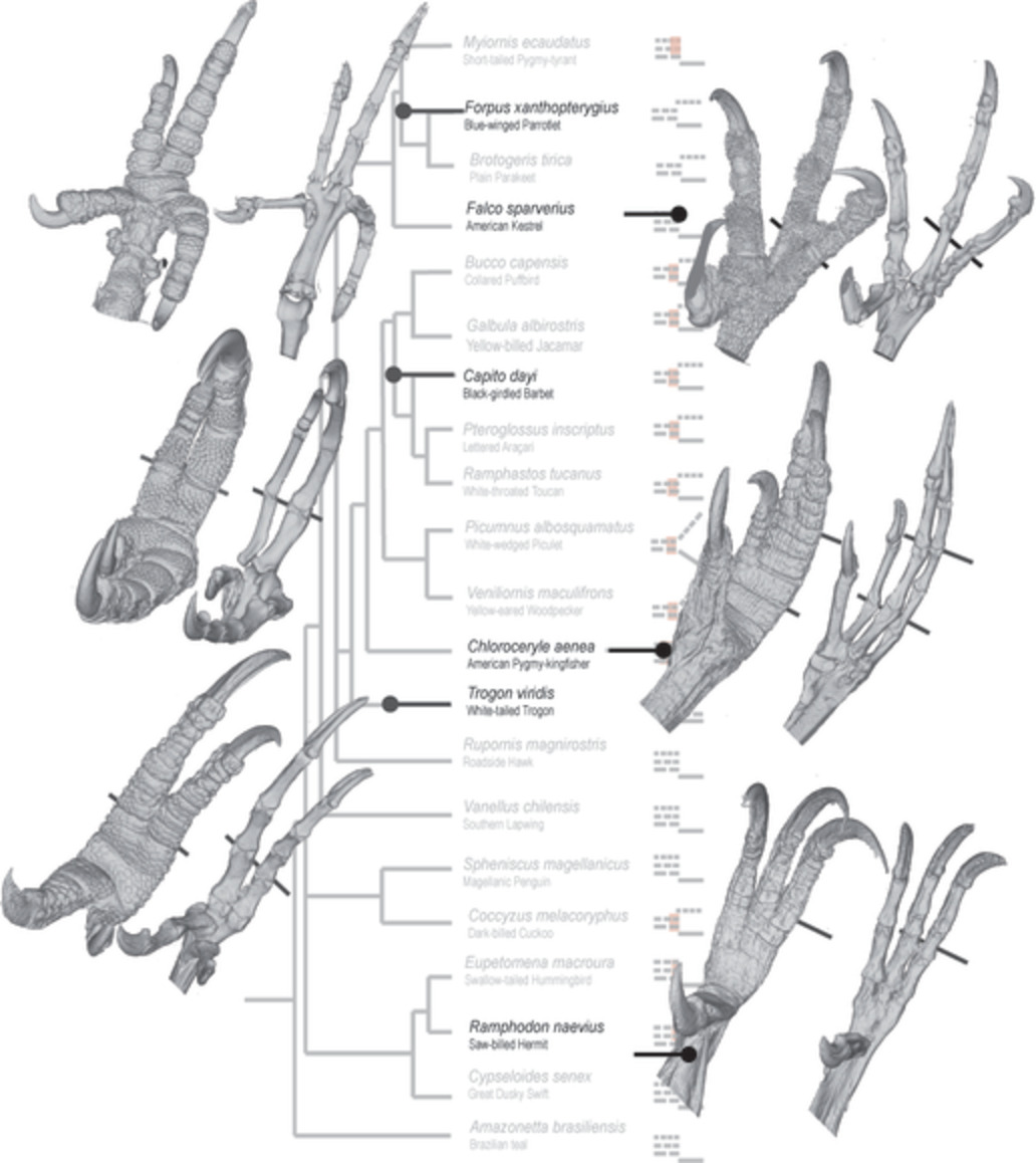
In the feet of birds, the morphological adaptations involve two systems: the osteomuscular and the tegumentary. In perching species, the foot may be syndactyl with the skin forming a sheath that closely associates the phalanges of two or three toes. In Psittacidae, which manipulate with the feet, the toes are free. In raptors and walking birds, loose skin may involve the base of two toes, or in swimmers the entire length of the toes forming a web.
Microorganization of ovaries and oogenesis of Haplotaxis sp. (Clitellata: Haplotaxidae)
- Pages: 98-114
- First Published: 19 October 2020
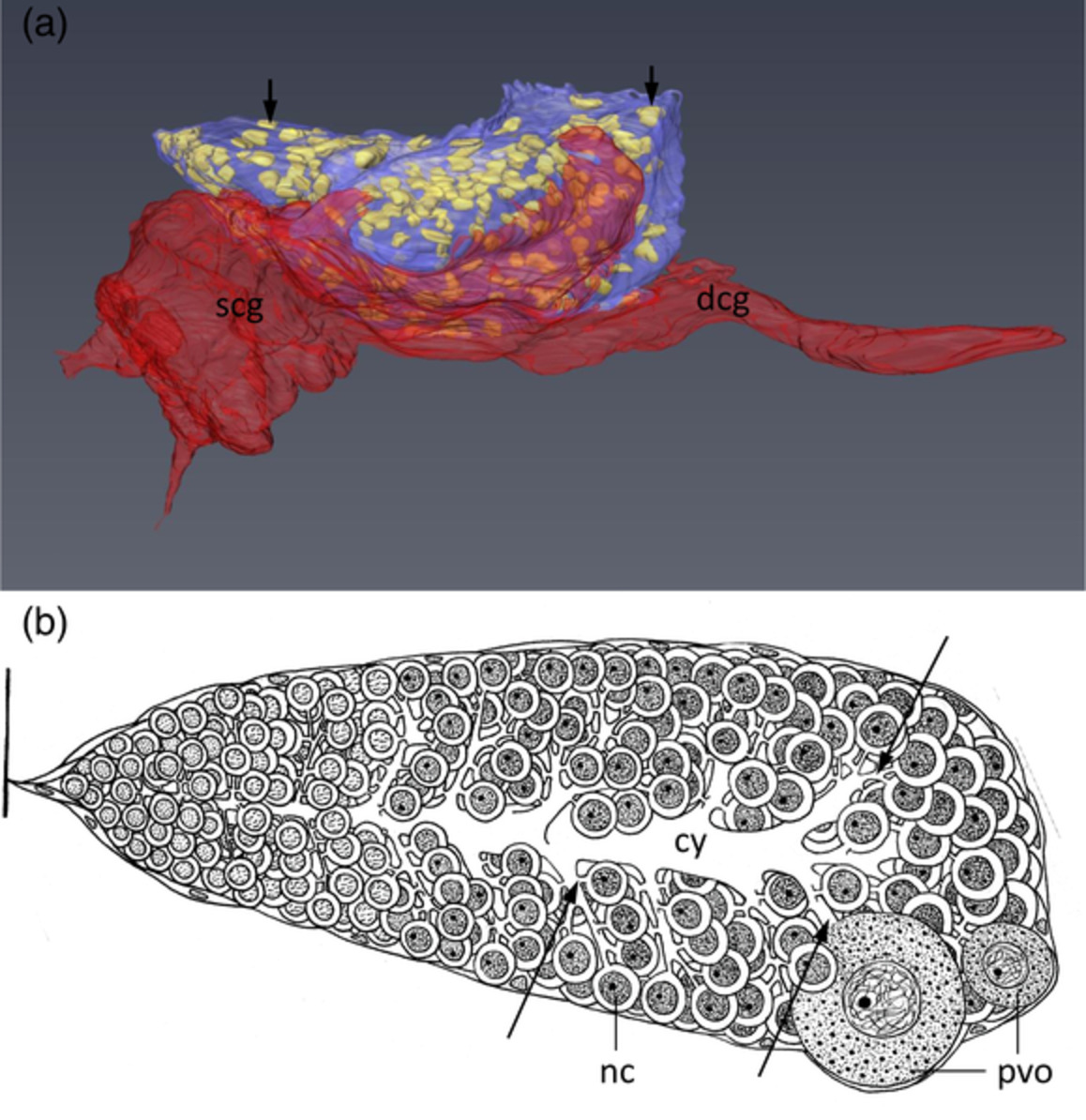
In Haplotaxis sp., the ovary (purple) was in close contact with copulatory gland (red). On the ovary surface, a thin envelope made up of somatic cells is present; short arrows indicate somatic cell nuclei. Copulatory gland is composed of a secretory part (scg) and a duct (dcg) that opens at the ventral side of the body wall. In the active ovary, a multicellular germ-line cyst with the developmental gradient of germ cells is present (anterior end on the left, posterior end on the right). Clustering germ cells differentiate into nurse cells (nc) and oocytes. Young previtellogenic oocytes (pvo) protrude at the ovary surface. Each germ cell has one intercellular bridge (long arrows) connecting it to the common elongated and branched mass of cytoplasm termed the cytophore (cy). Such ovary micromorphology is broadly similar to the “Tubifex” ovary type that is known from several other microdrile taxa. It seems that the “Tubifex” ovary type is the basal morphological type of ovaries in microdriles and allied taxa. Our study also supports the close relation of haplotaxids sensu stricto to the lumbriculids + branchiobdellids + hirudinids clade.
Tadpole of the Amazonia frog Edalorhina perezi (Anura: Leptodactylidae) with description of oral internal and chondrocranial morphology
- Pages: 115-126
- First Published: 20 October 2020
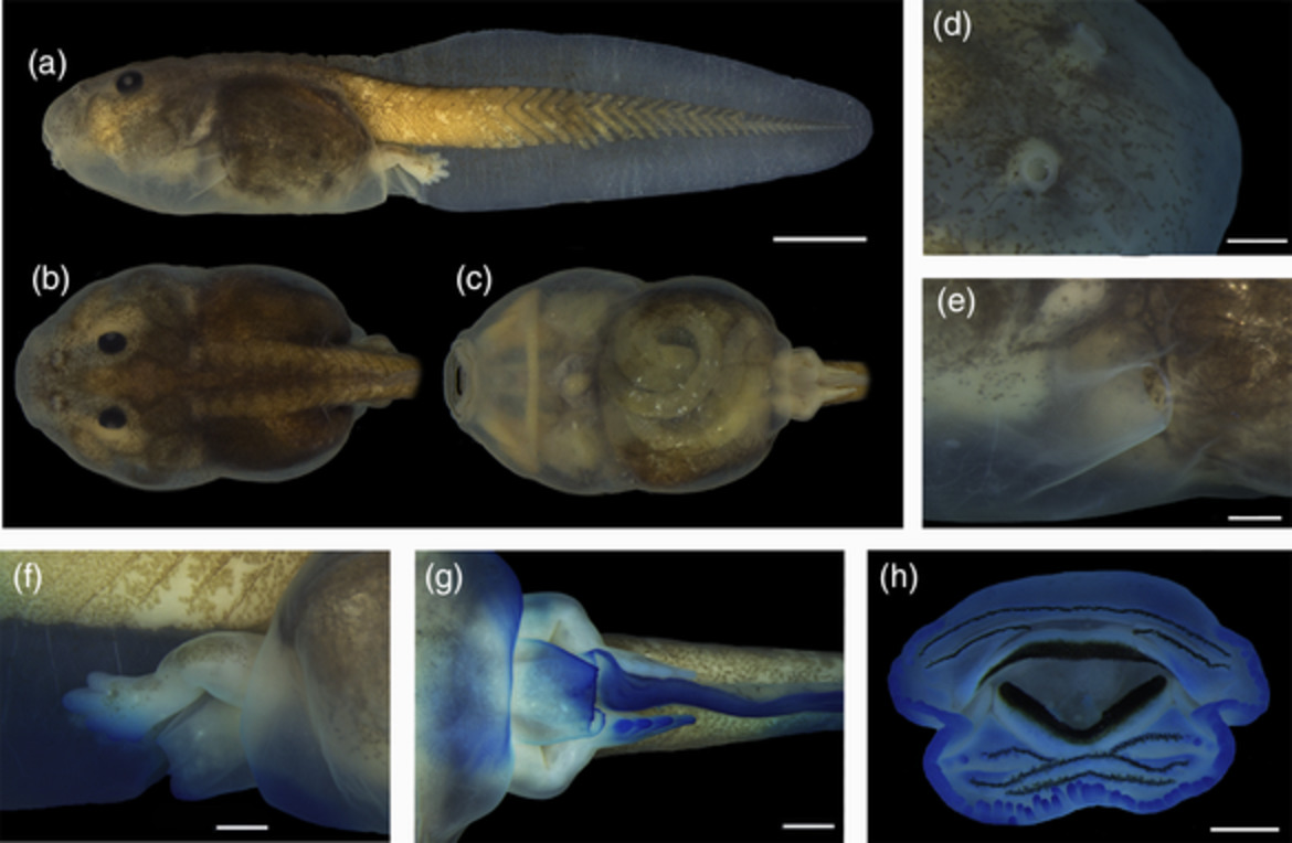
The tadpole of the frog Edalorhina perezi is described. The presence of a dextral vent tube is a synapomorphy for the clade consisting of Edalorhina, Engystomops, and Physalaemus. The combination of two lingual papillae, a filiform median ridge, and the lack of buccal roof papillae are diagnostic of E. perezi and putative synapomorphies of Edalorhina. The skeleton also presents some unique characteristics.
The functional morphology of lingual prey capture in a scincid lizard, Tiliqua scincoides (Reptilia: Squamata)
- Pages: 127-145
- First Published: 22 October 2020

Most scincid lizards use the jaws to capture prey, but the blue-tongued skink (Tiliqua scincoides) uses its tongue. It employs extensive hydrostatic deformation of its unusually broad tongue to maximize the area of tongue-prey contact and the strength of wet adhesion.
Ultrastructural aspects of spermatogenesis in Calyptogena pacifica Dall 1891 (Vesicomyidae; Bivalvia)
- Pages: 146-159
- First Published: 26 October 2020

The spermatogenesis in Calyptogena pacifica is similar to that in shallow-water bivalves. Proacrosomal vesicles are associated with outer electron-dense material. The axoneme is located near the nucleus until late spermatid stage.
Morphological and histological examination of short-wing formation in the winter moth Protalcis concinnata (Insecta: Lepidoptera, Geometridae)
- Pages: 160-168
- First Published: 24 October 2020

We studied morphological and histological characteristics of short-wing formation in the females of the winter moth Protalcis concinnata (Wileman, 1911) and compared with sex differences in the shape and size of wings during pupal-adult development.