Vibration Analysis of Multilayered Human Anatomy in Seated Posture: Implications for Health and Comfort
Abstract
The influence of machinery and transportation systems plays a crucial role in the development of human life. Humans encounter various kinds of vibration from the different environmental conditions such as air turbulence and bumpy roads; operating an excavator, machinery equipment, and so on causes a discomfort which affects the human health intensely. An attempt was made to reduce the impact of vibration on the human body by developing a 5-layered seated human anatomical structure that consists of the skeleton (bone), viscera (organs), nerves, muscles, and skin with the consideration of the Indian anthropometric dimensions belonging to the 95th percentile of the human male community and associated properties of the human tissue cells of different layers of the human body. The different modes of vibration that correlated with the natural frequencies of a human subject in a sitting posture were determined by performing the natural frequency analysis (modal analysis) using FEM approach. The resonant frequency of the multilayered human body was discovered in the range of 2–5 Hz with severe deformation observed in the hand, forelimb, and head due to low-frequency vibration that shall cause motion sickness and discomfort in the human subject. The frequency of resonance values of the present study was validated with the numerical study results available in the existing literature.
1. Introduction
The incorporation of machinery and transportation systems into technological development has become an intrinsic part of the human life; whether commuting by automobiles, airplanes, trains, ships, or other modes of transportation, operating heavy machinery, navigating aircraft, humans are continuously exposed to various vibrations. The human body experiences vibrations from different sources in transportation systems that include engine operation, atmospheric turbulence, state and roughness of road, and others. Along with that, some crucial vibrational impact of machinery components, motion, terrain conditions, dynamic loads, structural design, and others raises a significant concern about the discomfort and human welfare. The discomfort caused by the vibration affects the human health–related problems, that is, headache, motion sickness, back pain due to prolonged journey, spinal cord injury (SCI), numbness, vibration white finger, cardiovascular diseases, and musculoskeletal disorders [1]. A research study was carried out to investigate the human body responses exposed to whole-body vibration (WBV). Dong and Guo [2] performed static, modal, and transient dynamic analyses on a computational model of the seated whole human body to predict the effect of different factors affecting the biodynamic characteristics of the spine with three different postures of intervertebral discs (IVDs) in the lumbar spine. Pankoke et al. [3] developed a 2D dynamic FE model with adjustable posture for a seated man and calculated the internal forces and undamped eigenvalues to know the degeneration effect of height and mass of the body on the lower lumbar spine. Guo et al. [4] investigated the three-dimensional (3D) T12 spine–pelvis FE model to analyse the vibration effect by performing modal and harmonic response analyses. The different modal shapes of the human spine are extracted with the resonant frequencies, and the rotation of the lumbar spine vertebral column was seen during the WBV. Dewangan et al. [5] analysed the apparent mass response for the 58 participants exposed to vertical WBV in different frequency ranges with different parameters of body mass, fat, and stature for the masculine and feminine candidates seated without any backrest. Singh et al. [6] evaluated the eigenvalues of the seated 54-kg lumped parameter model acquired from the Indian anthropometric data for 50th percentile male people without a backrest. Kumar and Saran [7] studied the seat-to-head (STHT) transmissibility for the 10 male human subjects exposed to different magnitudes excited in the frequency range of Gaussian random vibration under three independent directions. Nawayseh and Griffin [8] experimented the response of 12 human participants and developed a 3DOF model to predict the influence of apparent mass on the human body dynamics caused by WBV on seat transmissibility. Lu et al. [9] conducted an investigation of the entire human spine that has been affected by the soft tissue of muscles and lower limbs and analysed the vertical random vibration along with the different resonant frequencies of the human model to stimulate the modal displacement and stress induced in the spinal IVDs. Wang et al. [10] suggested the transmissibility of STHT transmission of the 12 seated human male people with the influence of three different back support conditions when exposed to WBV in the frequency range of different vibration excitations. Nawayseh [11] attempted to study the effect of the different seating conditions with and without a backrest and headrest along with the posture of the body and leg in sitting conditions on the vertical vibration transmitted through a car seat. Harsha et al. [12] assessed the transmissibility of STHT and back support-to-head (BTHT) with the influence of different postures, vibration magnitudes, and frequency ranges of the seated human body under WBV in the vertical direction to improve the vehicle seat design and suspension system and optimize the comfort analysis. Bhatia et al. [13] examined the vibration analysis of the human body joints to calculate the natural frequency of male people of Indian community in standing posture and then evaluated the transmissibility effect and induced stresses on knee and neck joints. Thuong and Griffin [14] estimated the discomfort of 12 standing human people exposed to 4-Hz sinusoidal vibration in the three different translational directions with 10 different magnitudes of r.m.s. value. Sharma et al. [15] proposed a human CAD model weighing 76 kg mass with different anatomical layers corresponding to the 95th percentile of the Indian anthropometric data and analysed the different natural frequencies with the deformation of each anatomical layer in the standing human body. Morioka and Griffin [16] determined the frequency dependence response of the human body in sitting posture caused by foot vibration with different ranges of magnitudes and frequencies in the fore-aft, lateral, and vertical directions. The primary resonance of a human subject in sitting posture lies in the range of 2–6 Hz in the study conducted by Dong et al. [17], Thuong and Griffin [14], and Singh et al. [18]. Also, as per ISO 2631-1:1997 [19], the fundamental resonance for WBVs occurs in the range of 4–5 Hz for seated posture. The low-frequency vibrations with a range of 2–6 Hz for prolonged time show the effects like motion sickness, discomfort, and fatigue to the human subject [14]. The current research work presents the natural frequencies of the five-layered human anatomical structure, that is, skeleton, viscera, nerves, muscles, and skin, in a sitting posture with the deformation observed in the different vibration modes using finite element analysis (FEA).
2. Materials and Methodology
A computer-aided design of a seated human multilayered model has been created with the use of 3D modelling software, that is, SolidWorks 2021, representing a 76-kg human subject by considering Indian anthropometric data belonging to the 95th percentile male community [20]. The model includes different anatomical structures such as the skeleton, viscera, nerves, muscles, and skin [21–27]. Figure 1 shows the 3D human CAD model of five different anatomical layers in sitting posture. The layers are as follows: 1st layer—skeletal system; 2nd layer—skeletal and visceral systems; 3rd layer—skeletal, visceral, and nervous systems; 4th layer—skeletal, visceral, nervous, and muscular systems; and 5th layer—skeletal, visceral, nervous, muscular, and integumentary systems.
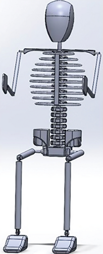
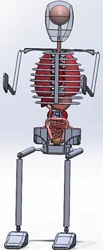
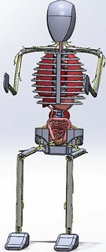
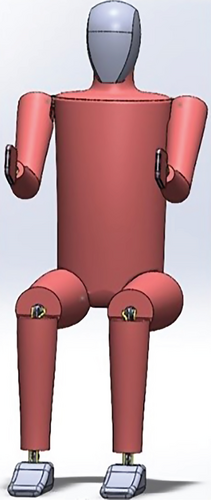
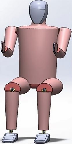
The skeletal structure of the human body (i.e., bones) constructed of the axial skeleton and appendicular skeleton, namely, head, vertebral column (cervical, thoracic, lumbar, sacral, and coccygeal), trunk and thorax, shoulder girdle, upper limb (humerus, ulna, and radial), lower limbs (femur, tibia, and fibula), and pelvis (pubis, ischium, and ilium) [23–25], represented as the 1st layer of a human skeletal system providing support and movement to the body as shown in Figure 1a. The upper limb and lower limb bones were assumed to consist of the humerus and femur with uniform bone thickness throughout the entire body having an average diameter of 14–26 [28] and 29.4–37 mm [29]. The 2nd layer, that is, viscera (internal organs) assembled with the skeletal system shown in Figure 1b, comprises the thoracic cavity organs (heart, bronchi, and lungs) and abdominal cavity organs (liver, stomach, small intestine, kidneys, ureter, large intestine, and urinary bladder) undergoing the exposure of vibration. The heart is situated in the midplane of the mediastinum with a conical hollow form of the muscular organ circumscribed with the pericardium. The measurement of the heart is about 120∗90 mm, and it weighs around 300 g in males [24]. Most of the thoracic cavity segment is covered by the lungs, leaving compact space for the heart which forms a cavity of the left lungs. The texture of the lungs is spongy with brown or grey in colour, and they are a pair of respiratory organs oriented in the thoracic cavity. The upper end (apex) is blunt, and the base is concave and crescent-shaped weighing around 700 g of the right lung which is about 50–100 g heavier than the left lung [24]. The liver is a wedge-shaped, large, and solid gland placed in the right upper quadrant of the abdominal cavity, occupies the right hypochondrium portion with a reddish brown in colour, and weighs about 1600 g in the male body [25]. The stomach is a very distensible organ with a muscular sack of the digestive tube that lies diagonally in the upper and left parts of the abdomen engaged with the epigastric, umbilical, and left hypochondrium portion; the majority of the stomach is covered with the left coastal margins and the thoracic cage. The stomach shape depends upon the degree of the muscular tone of the persons usually in J-shaped with about 250 mm long and having the 1.5–2 L or more mean capacity in adults [25]. The structure of the small intestine is adjustable for the absorption and digestion which is extended from the pylorus to the ileocaecal junction with a great length of around 6 m and the large intestine is expanded from the ileocaecal junction to the anus with 1.5-m-long cecum [25]. The kidneys are pair of excretory organs with closely packed bean-shaped structure located on the posterior abdominal wall, augmented vertically from the twelfth vertebra border to the third lumbar vertebrae centre of the body. The size of each kidney is about 110 mm length, 60 mm wide and 30 mm thick with a reddish brown in colour. The average weight of the kidney around 150 g in male with little bit longer and narrower in left kidney than the right one. The function of the ureter is to convey the urine from the kidneys to the urinary bladder through a pair of narrow and thick-walled muscular tubes. Urinary bladder lies in the anterior part of the pelvic cavity act as a muscular reservoir of urine [25]. Figure 1c represents the 3rd layer, that is, the nervous system which is the chief controlling and coordinating system of the body. The human nervous system is the most intricate part of evolution. It is divided into central nervous system (CNS) and peripheral nervous system (PNS). CNS is responsible for combining, and synchronizing the sensory information and commanding the suitable motor actions which are functioned through brain and spinal cord, the average weight of adult brain in air is 1500–2000 g whereas the floating of brain in cerebrospinal fluid weighs only 50 g. An adult brain contains about 180–200 billion neurons (very rich) [22]. While the PNS involves the cranial nerves and spinal nerves, the peripheral autonomic nervous system (ANS) is dissected into sympathetic and parasympathetic nerves which consists of neurons, ganglia, plexuses, nerve fibres and nerve cell bodies, and others [22, 27]. The brain receives information from and assess the activities of the head and neck, expand with thoracic and abdominal cavity organs which are implemented by cranial nerves. There are 31 pairs of spinal nerves (8 cervical, 12 thoracic, 5 lumbar, 5 sacral and 1 coccygeal) through the CNS sense the information and dominated the activities of the trunk and limbs [27]. Median, ulnar, and radial nerves which are regulated through the musculocutaneous nerve, sciatic nerve, and femoral nerve are the thickest and chief nerves of the human body through the lower limb functioned via cutaneous nerves, superficial nerve, peroneal nerve, and tibia nerve [22, 26, 27]. The muscular system is assumed as the 4th layer depicted in Figure 1d, supporting the movement of the body with flexibility in joints. The muscle thickness varies for each and every person due to the genes, age, weight, workout, and so on. The variation of thickness of muscles was found to be in the average range of 13.85 to 67.7 mm [18], the anterior human abdomen region is modelled with curvature surface to avoid the overlapping of the thigh and lower torso in the present study. As the outermost sensory organ of the body, that is, the human skin displayed in Figure 1e, the integumentary system deals with the epidermis, hypodermis, and so on to prevent the human body from bacteria, injury, sensory information of pain, touch or feel, temperature of the body, and so on; the surface area of the adult skin is 1.5–2 m2 [21]. The thickness of the skin surface was considered to be 1-mm thickness across the body [17].
The movement of the body is associated with the different types of joints linked with various segments of the bone such as pivot joint, hinge joint, ball and socket joint, cartilaginous joints, and so on. Table 1 represents the joints allied with the skeletal CAD model of the human body with the allowable movement corresponding to the number of degree of freedom (DoF). The pairing of the humerus and ulna with humeroulnar joint, radial and palm with wrist or radiocarpal joint, femur and tibia with tibiofemoral joint, tibia and foot with ankle or talocrural joint which are related to the hinge joint. The connectivity of clavicle (shoulder girdle) and humerus with glenohumerul joint, pelvic and femur with hip or acetabulofemoral joint incorporated with ball and socket joint and the pivot joint is attached between the cranial and cervical with atlanto-axial joint.
| Articulation of ligament | Related joint | Planes of movement | Number of degree of freedom (DoF) | |
|---|---|---|---|---|
| Cranial (head) bone | Cervical (neck) bone | Pivot joint | Vertical axis (rotation movement) | 1 |
| Clavicle (shoulder) bone | Humerus (upper arm) bone | Ball and socket joint | All three axes (circumduction movement) | 3 |
| Humerus (upper arm) bone | Ulna (forearm) bone | Hinge joint | Transverse axis (flexion and extension movement) | 1 |
| Radial (forearm) bone | Palm (hand) bone | Hinge joint | Transverse axis (flexion and extension movement) | 1 |
| Pelvic (hip) bone | Femur (thigh) bone | Ball and socket joint | All three axes (circumduction movement) | 3 |
| Femur (thigh) bone | Tibia (lower leg) bone | Hinge joint | Transverse axis (flexion and extension movement) | 1 |
| Tibia (lower leg) bone | Tarsal (foot) bone | Hinge joint | Transverse axis (flexion and extension movement) | 1 |
| Rib bone | Sternum bone | Cartilaginous joint | Slight movement | 0 |
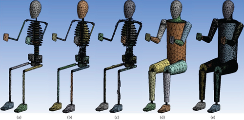
The human subjects consisting of multiple system having different cells of bone, viscera, nerves, muscles and skin studied in the field of biomechanics. The properties of the human tissue in terms of biomechanical values are allocated to the different segment of the human body and considered to be homogenous and isotropic in nature [38] shown in Table 2. Meshing is the conversion of the infinite geometrical shape into the finite number of elements that elements are coordinated with a junction or point called as a node [39]. The automatic tetrahedral mesh was generated with default mesh element size, tetrahedral meshing is used for complicated structure, stiffer and flexible when compared to other mesh element types [40]. Figure 2 express the finite element model of the 5-layered seated human subjects in vertical erect posture without any backrest support. The mesh was produced with 82,710 no. of elements and 2,23,074 no. of nodes for skeletal system shown in Figure 2a, 1,08,242 no. of elements and 2,71,419 no. of nodes for skeletal and visceral system shown in Figure 2b, 2,22,244 no. of elements and 5,10,141 no. of nodes for the skeletal, visceral and nervous system shown in Figure 2c, 2,37,601 no. of elements and 5,40,584 no. of nodes for the skeletal, visceral, nervous and muscular system shown in Figure 2d and 3,14,614 no. of elements and 7,00,287 no. of nodes for the skeletal, visceral, nervous, muscular and integumentary system shown in Figure 2e. The boundary condition of the human in sitting posture has been considered that the buttocks were assumed to be fixed with the seat and feet linked with the floor was supposed to be rigid without any external load [18, 41]. It is assumed that a human subject is sitting with a free-hanging posture, that s, not holding anything in the hands or putting their hands on the arm seat which means the vibrations are transmitted through the seat and feet for WBVs. The assumption of free-hanging posture ensures that the upper limbs are exposed fully to the effects of vibrations, preventing any additional constraints that can alter the resonance of the whole body. Also, this type of posture can find more deformations in the hands due to a lack of structural support that represents the discomfort in real conditions where unrestricted movement of limbs provides discomfort due to vibrations.
3. Results and Discussion
The research work was carried out to study the structural dynamics behaviour of the multilayered human anatomical structure by performing the modal analysis of the 5-layered human CAD model system include the skeletal (bone), viscera (organs), nerves, muscles and skin to determine the natural or resonant frequency associated with the vibration patterns of the 76 kg weigh Indian human anthropometric data belonging to the 95th percentile of male people [20] in sitting posture under un-damped free vibration by utilizing the tetrahedral meshing parameters and boundary conditions of hip and feet as a fixed constraint in FEM analysis, that is, ANSYS 22.1 software.
3.1. Modal Analysis of the Skeletal System (1st Layer)
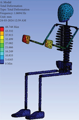
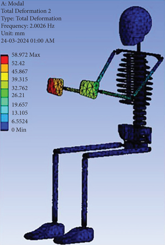
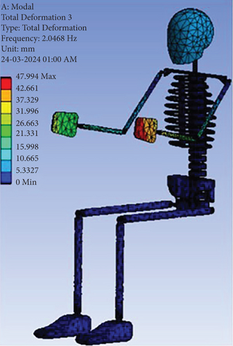
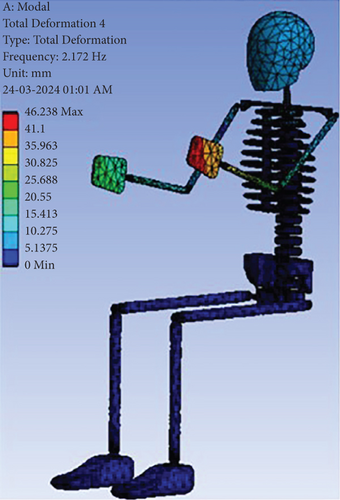
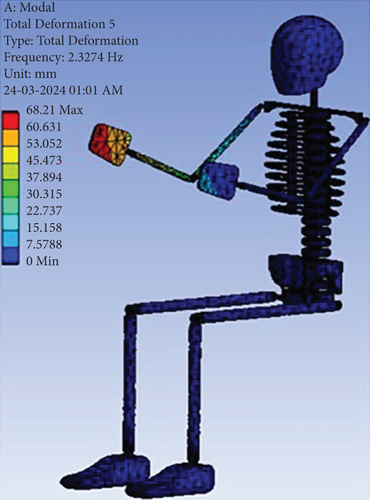
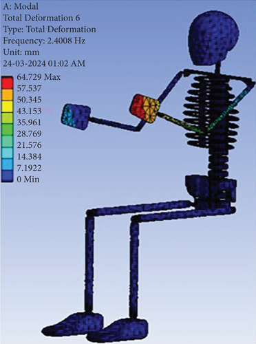
The fundamental frequency of 1.87 Hz, the oscillatory motion of the upper limb with the maximum deformation observed at the phalanges, metacarpal, wrist joint of the right hand and left hand, then the little vibration effect was seen in the head transmitted via ulna, radial and humerus bone, shoulder girdle shown in Figure 3a. The second fundamental frequency was observed at 2.03 Hz shown in Figure 3b, the flexion motion injury of the upper limb in the transverse axis with superior deformation in the right palm, ulna and radial bone and fewer deformation in the left palm, ulna and radial skeleton. Contrast to the second mode, the injury of the left upper limb due to extension movement with maximum wear in the hand, forearm bone at the natural frequency of 2.05 Hz in the third mode shape shown in Figure 3c. In fourth vibration mode shown in Figure 3d, the excursion movement with extreme deformation of the left hand, forearm and upper arm in the lateral and vertical direction was observed at 2.17 Hz frequency. The natural frequency of 2.33 Hz, the acquired deformity was spotted in the right palm, forelimb and brachium of the body due to vertical vibration injury in the fifth natural mode shown in Figure 3e. Similar to the fifth mode of vibration, the acquired deformity was obtained with the injury of left hand, lower arm and upper arm of the body in the vertical direction at a natural frequency of 2.40 Hz shown in Figure 3f.
3.2. Modal Analysis of the Skeletal and Visceral Systems (2nd Layer)
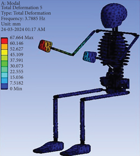
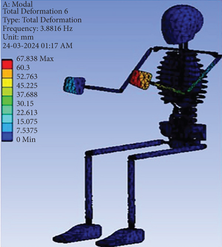
From Figure 4a, the resonance frequency was obtained at 2.22 Hz, the oscillation movement was occurred from the right-to-left in the transverse axis with the deformation of thoracic and abdominal cavity organs (i.e., lungs, heart, liver, and stomach) and extreme at the right hand followed by brain, head (skull), forearm and upper arm. Figure 4b shows the equivalent mode of first vibration, while the second resonance frequency was procured at 2.34 Hz, the oscillation movement from left-to-right was appeared in the lateral direction with severe deformation of the left palm, cranium, brain, lower arm and upper arm of the skeletal system along with less deformation in the respiratory system and digestive system. The first and second mode of vibration causes some diseases like chest pain or breathing problems, digestion problems, stomach pain, fatigue, and so on due to the impact of vibration. Figure 4c exposes the vibratory motion of the hand and forelimb about an elbow joint and the maximum deformation of the right palm at the frequency of 3.23 Hz. Figure 4d shows the extensor movement of the hand in the lateral direction and the left-hand deformed max at the natural frequency of 3.40 Hz in the fourth mode shape. Following to the fourth modal shape, the deformation of right hand was observed to be maximum with the up and down motion in the vertical axis with the frequency of 3.80 Hz shown in Figure 4e. From Figure 4f, the vertical vibration was seen in the left hand with the superior deformity at 3.88 Hz frequency.


3.3. Modal Analysis of the Skeletal, Visceral, and Nervous Systems (3rd Layer)
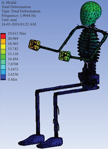
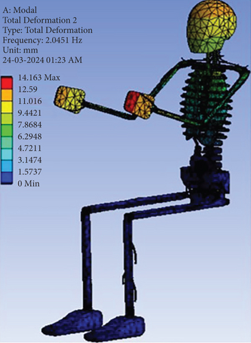
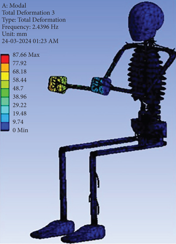
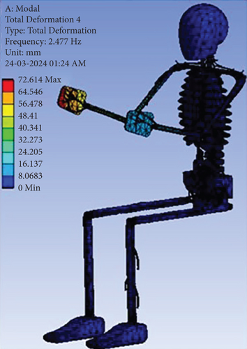
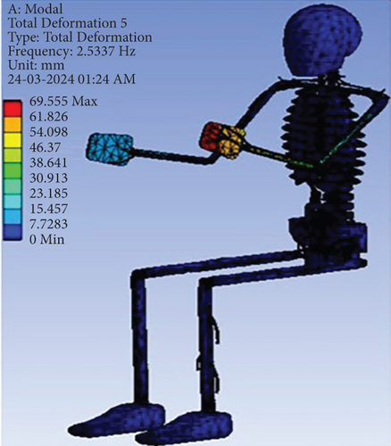
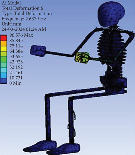
The first natural mode was found at 1.90 Hz fundamental frequency revealed in Figure 5a, with the deformation of the ulnar, radial, and median nerves being seen maximum in the right hand. As shown in Figure 5b, the second natural mode shape was noticed with deformation of the ulnar nerve, radial nerve and median nerve situated in the left hand and head with brain deform due to the regulation of musculocutaneous sensory nerve in the second fundamental frequency of 2.04 Hz. Figure 5c represents the third mode of vibration at the natural frequency of 2.44 Hz with the deformation of the nerves in the right-hand causes shakiness due to hand-arm vibration. The right- and left-hand nerves deform due to the vertical vibration in the frequency of 2.48 Hz and 2.53 Hz shown in Figure 5d,e. At the natural frequency of 2.64 Hz, the left-hand nerve deformed with an oscillatory motion in the lateral direction in the final mode shown in Figure 5f. From the analysis, it was found that the impact of vibration on the nervous system can cause a loss of nerve senses, numbness effect, paralysis of the body parts, spinal injuries, and so on. Most of the nerve damage happened in the hand and neck due to the hand–arm vibration; it can lead to hand–arm vibration syndrome (HAVS).
3.4. Modal Analysis of the Skeletal, Visceral, Nervous, and Muscular Systems (4th Layer)
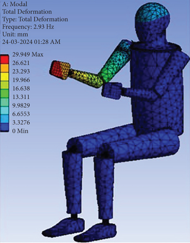
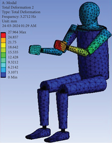
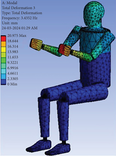
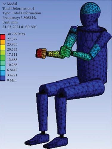
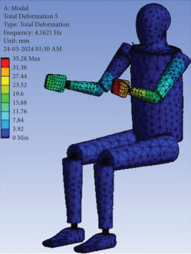
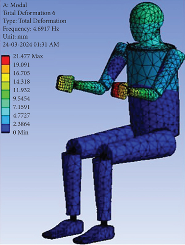
In first mode shape, the resonating frequency was analysed at 2.93 Hz with the maximum right hand muscles deformation due to the oscillation movement in the transverse axis shown in Figure 6a. The frequency of 3.27 Hz was occurred in the second pattern of vibration, distorted maximum in the left hand due to vertical vibration shown in Figure 6b. The slight bending movement of the upper trunk of the body shown in Figure 6c, deformed the both hand in the fore-aft direction and lesser amount of deformity seen in the forearm, biceps and face muscles at 3.43 Hz. From Figure 6d, the deflection of the right-hand muscles causes extensor tendon injuries due to vibration with the natural frequency of 3.81 Hz. As shown in Figure 6e, vibratory movement of upper limb in the lateral direction at 4.16 Hz frequency with left hand muscles contorted in the fifth vibration mode. The natural frequency of 4.69 Hz, the turning movement of the upper body with the deformation seen superior at left hand, inferior at right hand, lower arm, upper arm, chest, and head of the human body shown in Figure 6f. The outcomes of the muscular system analysis show that the reduction of the strength in hand muscles tends to have a motor reflex, muscle contraction and relaxation, and so on due to the prolonged vibration exposures. The effect of vibration seen on the hand muscles, because in hand mass of the muscle fibre is less as compared to other body parts that causes a sarcopenia disease (i.e., weakness of muscles and loss of muscle function).
3.5. Modal Analysis of the Skeletal, Visceral, Nervous, Muscular, and Integumentary Systems (5th Layer)
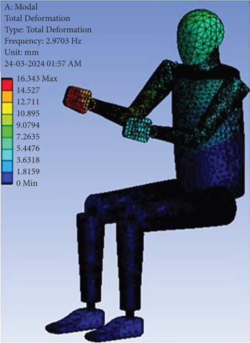
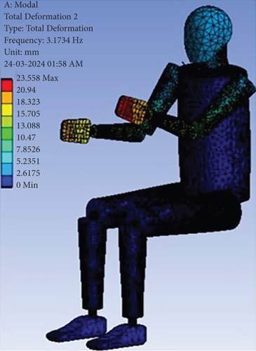
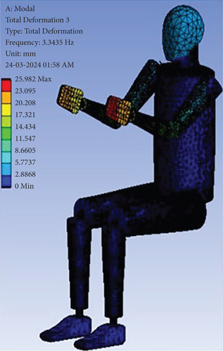
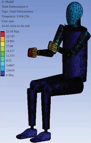
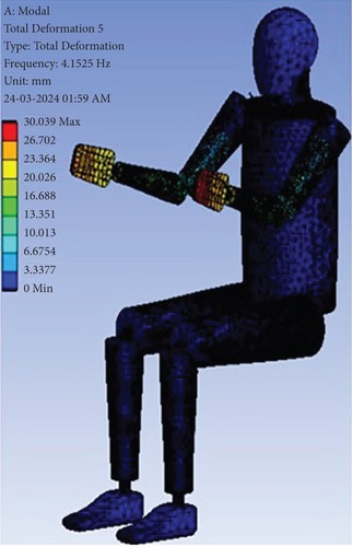
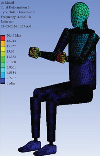
The primary harmonic resonance was achieved with the maximum deformation of the right hand in the vertical axis shown in Figure 7a at 2.97 Hz with somewhat deformation of the forelimb, upper arm, head and upper segment of the body. At 3.17 Hz as shown in Figure 7b, the secondary harmonic resonance was attained in the second natural mode shape, the left upper limb has extreme distortion with less severe deflection of the right hand and forearm. The flexural movement of the both upper limb towards the median plane at the frequency of 3.34 Hz presented in Figure 7c, the deformity of the left hand was superior to the right hand, lower arm and upper arm in the third mode of vibration. From Figure 7d, the right hand and forearm bend with utmost deformation seen at the natural frequency of 3.92 Hz in the fourth pattern of vibration. Abduction movement was occurred at the frequency of 4.15 Hz demonstrated in Figure 7e, where both the upper extremity from the median axis of the body in the fifth vibration mode. The bending of the upper trunk was observed about a pivot point of the thoracic vertebra with a maximum contortion at left hand then reduced at right hand, lower and upper arm of both hand and head or skull part of the body with the natural frequency of 4.56 Hz in the final natural vibration mode shown in Figure 7f. The analysis of the integumentary system (i.e., skin) of the human body exposed to vibration causes some skin problems such as itching, redness or red patches, vision impairment, loss of consciousness or dizziness, pale or white finger, and so on.
The natural frequency analysis of the present research work has been validated with the Dong et al. [17] and Singh et al. [18] presented in Figure 8. Dong et al. [17] conducted a study on 3-D FE model of 50th percentile of the Chinese human male body in sitting posture (vertical seated human body) composes of skeleton, muscle soft tissue, viscera, ligament, IVD, and skin has been developed in ANSA v15.2.0 software and analyse the response of vertical seated body (VSD) in the different vibration modes with the Abaqus 6.14 simulation software. Singh et al. [18] developed the 3-D CAD model of 95th percentile of the Indian male human subjects consisting of three-layers of skin, muscles and bone in SolidWorks 2016 and evaluated the natural frequency corresponding mode shapes in sitting posture by performing modal analysis in ANSYS 16.0.
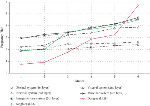
The resonant frequency was obtained at 0.74 Hz in the flexion-extension direction and 1.9 Hz with fore-aft motion in the first natural mode shape by Dong et al. [17] and Singh et al. [18]. The second pattern of vibration was occurred in the lateral bending direction at 0.91 Hz frequency by Dong et al. [17] whereas Singh et al. [18] found at 2.0 Hz frequency with to and fro motion. Dong et al. [17] achieved the third mode shape at the frequency of 1.76 Hz in the vertical axis and Singh et al. [18] observed the natural frequency at 3.5 Hz with the right-hand deformation. The fourth mode of vibration was found with the frequency of 2.87 Hz and 3.7 Hz by Dong et al. [17] and Singh et al. [18]. The pre-final vibration mode of Dong et al. [17] and Singh et al. [18] was seen at 3.24 Hz frequency in transverse axis and at 4.0 Hz frequency in vertical axis. The natural frequency of Dong et al. [17] appeared at 5.72 Hz and Singh et al. [18] spotted at 4.5 Hz. There might be having some variation in the results of the present study with the Dong et al. [17] study and Singh et al. [18] study due to the consideration of the human model geometry, anthropometric dimension, meshing parameter, different anatomical layer, different FEA and modelling software, and so on. The present research study focused on the modal analysis of the 5-layered human CAD model system including skeletal system (bone), visceral system (organs), nervous system (nerves), muscular system (muscles) and integumentary system (skin) which reflects the actual human body. The natural frequency of all five-layer human body has been found within the range of 2 to 5 Hz with the maximum deformation observed at the hand, forearm and the head along with that there was no vibrational effect seen below the lumbar body segment (i.e., thigh, hip, lower leg, and leg) due to the fixed constraints with the floor and seat, lower body beneath the lumbar vertebral column acted as a rigid body. The overall analysis of the study results was validated and found to be in correlation with the available literature study of Dong et al. [17] and Singh et al. [18].
4. Conclusion
In the research work, a seated human model weighing 76 kg with five anatomical layers incorporates the skeleton, viscera or organs, nerves, muscles, and skin has been constructed with the Indian male anthropometric dimensions and the properties of human tissue cells are allocated to the different layers of the human body which are available from the existing literature and then modal analysis was performed for the all 5-layered human system to determine the impact of the vibration on the human body using FEM analysis. The natural frequency was discovered in the range of 2 to 5 Hz with the severe deformation of the hand, forearm, and head contrast to different parts of the body and there was no vibration effect seen in the lower body segments. The range of this frequency results in human discomfort, motion sickness, and headache when it is exposed to vibrations for long intervals of time. The skeletal system (1st layer), visceral system (2nd layer) and nervous system (3rd layer) show low resonance frequency compared to the muscular and integumentary system (4th and 5th layer) due to the presence of mass in the body. All the results are compared and validated with the existing literature. The obtained resonance frequency values will be helpful in the development of the product design, suspension system, seat ergonomics, machinery equipment, and so on to reduce the impact of vibration on the human body.
Conflicts of Interest
The authors declare no conflicts of interest.
Funding
The authors received no specific funding for this work.
Open Research
Data Availability Statement
The data will be available upon request from the corresponding author.


