Effects of Bacteriophage on Antibacterial Properties, Nonspecific Immune Responses, and Gut Microbiota in Litopenaeus vannamei Post Vibrio parahaemolyticus Infection
Abstract
Effective immune regulation and balanced gut microbiota play important roles in preventing pathogen infections in Litopenaeus vannamei farming. Bacteriophages are a promising candidate in pathogen control for their specific antibacterial properties. While previous studies focused on the direct antibacterial effects of phages, their effects on nonspecific immune responses and gut microbiota after infection remains to be less explored. In this study, a lytic Vibrio parahaemolyticus phage was isolated from wastewater with a broad host range (66.7% lytic efficiency), low multiplicity of infection (MOI; 0.1), and high environmental tolerance (pH: 3–11; temperature: 4–60°C). Whole genome analysis revealed a 93,814 bp double-stranded linear DNA molecule with 45.1% GC. Both the in vitro cocultivation (24 h) and in vivo shrimp cultivation trails (7 days) demonstrated that phage could effectively reduce the quantities of Vibrio (>99%). The in vivo phage fed shrimp exhibited elevated levels of nonspecific immune-related enzymes like alkaline phosphatase (AKP), catalase (CAT), superoxide dismutase (SOD), phenoloxidase (PO), and lysozyme (LZM) and upregulated immune-related gene expression including those of antimicrobial peptides, pattern recognition receptors (PRRs), and pattern recognition proteins. Additionally, phage treatment improved the diversity of the gut microbiota (Shannon-10 index) after Vibrio infection, indicating restored microbial balance in shrimp. These results suggest that phage therapy promotes nonspecific immune responses and repair intestinal dysbacteriosis in shrimp after Vibrio infection, elucidating a promising strategy to treat pathogenic Vibrio in shrimp aquaculture.
1. Introduction
Due to increasing demands, the Litopenaeus vannamei aquaculture industry has experienced fast expansion over the last 30 years. However, intensive and high-density shrimp farming often occurs simultaneously with bacterial diseases [1]. Several researchers have reported that the annual losses in shrimp production caused by pathogenic Vibrio bacteria, such as Vibrio parahaemolyticus, Vibrio alginolyticus, and Vibrio harveyi, are as high as 20%, resulting in approximately USD 1 billion in global economic losses [2, 3]. Among these bacteria, V. parahaemolyticus is considered the most virulent (24 h LC50 to shrimp: 103–104 cfu/mL) and has become the primary focus of prevention and control in shrimp farming [2].
Like other invertebrates, shrimps lack an adaptive immune system, they strictly rely on a fixed nonspecific immunity mechanism to prevent pathogen entry and spread [4]. The defense mechanism of shrimp includes pattern recognition receptors (PRRs), antioxidant enzymes, and reactive oxygen species mediated microbicidal action [5, 6]. Once foreign pathogens are detected, the immune response is activated and tries to eliminate the pathogens [7]. In addition to the nonspecific immunity response, the gut microbiota could also reduce the risk of pathogen infection to shrimp by forming biological barrier in intestine, competitively eliminating the colonization of pathogens [8]. Therefore, both the normal regulation of immune response and balance of intestinal microbiota play important roles in preventing pathogen infection in shrimp and are easily influenced by exogenous substances (pathogens or immunoenhancers) [9, 10].
The pathogenicity of V. parahaemolyticus to shrimp depends on the toxics it produces, it has been confirmed that some virulence genes are present on plasmids of V. parahaemolyticus, such as the photorhabdus insect-related toxin A/B (pir A/pir B) and Vibrio toxic component genes (vtcc), the toxins encoded by these virulence genes are one of the key pathogenic factors in causing shrimp disease [11]. Once V. parahaemolyticus outbreaks in shrimp farming, these toxin genes can encode massive exotoxins, adhesion factors, and endotoxins [12], negatively affect immune function and intestinal bacterial composition [13], thus increase the morbidity and mortality rate. Usually, to stimulate immune system of shrimp and reduce Vibrio infection, antibiotics and disinfectants are extensively used in aquaculture, but these chemicals have become more and more ineffective because of the occurrence of antibiotic-resistant genes and have negative impacts on the environment, such as antibiotic residues and chemical pollution [14–16]. Therefore, products that claim to be environmentally friendly, such as probiotics, vaccines, and bacteriophages, aimed at inhibiting Vibrio endlessly emerge [17–19].
In comparison, bacteriophages have been favored for their high specificity, lack of drug residue, and ease of application [20]. Nowadays, phages are explored in the medical [21], agricultural [22], food-related [23], and environmental fields [24]. There are more than 600 papers on the application of phages in agriculture and over 90% of these studies show that phages are highly effective in reducing specific pathogen concentration [25–27]. However, most of the researches only focused on the direct antibacterial function of phages, less is known about the effects of phage application on the nonspecific immunity response and intestinal microbiota in shrimp after Vibrio infection. Only Alagappan et al. [28] and Solís-Lucero et al. [29] demonstrated that phage application could mediate antioxidant enzymes activity. To date, there is no in-depth research on the effect of phage on the immune response of shrimp, such as immune-related gene expression (PRRs or antimicrobial peptide system), much less about the influence of phage on the intestinal microbiota in shrimp under Vibrio infection.
Given the importance of normal immune regulation and intestinal microbiota balance in shrimp to prevent pathogen infection, it is crucial to investigate the effects of phage application on the nonspecific immunity response and intestinal microbiota in shrimp after Vibrio infection. In practical farming, once shrimp are severely infected with Vibrio, the immune system regulation will be inhibited [9] and intestinal dysbacteriosis will occur simultaneously [30]. So, whether phage application can enhance the immune system response of shrimp infected by Vibrio? Can phage alleviate the imbalance of gut microbiota in shrimp infected with Vibrio?
To answer the above questions, the present study investigated the effects of phage on antibacterial properties, nonspecific immune responses, and intestinal microbiota in L. vannamei after V. parahaemolyticus infection. First, a lytic phage was isolated and genomically sequenced; the host range, multiplicity of infection (MOI), one-step growth curve, optional pH, and temperature of the phage were also confirmed. In vitro and in vivo experiments were then conducted to assess the phage’s antibacterial ability. Its effect on the nonspecific immune response in shrimp was also evaluated by detecting the antioxidant and immune related enzymes activity and gene expression. Finally, this study analyzed the potential influences of phage application on the composition and diversity of shrimp gut microbiota by genomic sequencing. In addition to the direct antibacterial ability, these results gave a more detailed description on the immune enhancement and gut microbiota rebalance capacity of phages in preventing pathogenic Vibrio in shrimp and provided new insights for the rational application of phages in aquaculture.
2. Materials and Methods
2.1. Shrimp and Culture Conditions
Healthy 1-month-old shrimp (L. vannamei; weight = 0.1 ± 0.02 g) were purchased from Guangdong Hisenor Group Co., Ltd., China. The shrimp were fed with commercial feeds from Qingyuan Haibei Bio-Tech Co., Ltd., China. During the experiments, the water temperature, pH, and salinity were maintained about 26–28°C, 7.8–8.0, and 20–22‰, respectively.
2.2. Vibrio Strains and Culture Conditions
V. parahaemolyticus 43 (Vp 43) and the other strains used, including V. parahaemolyticus, V. owensii, and V. harveyi (Table 1), were taken from stocks in our laboratory. The hepatopancreases of diseased shrimp and water used for Vibrio isolation were obtained from several aquacultural regions of China using the methods reported in prior research [31]. The virulence genes pir A/B and vtcc (Table 1) in these strains were detected with a PCR assay using the commercial kit from Guangdong Haid Animal Husbandry and Veterinary Research Institute Co., Ltd. Typically, the Vibrio strains were reserved at −80°C in tryptose soya broth (TSB) with 30% glycerol. When used, the Vibrio strains were incubated with the following standard laboratory conditions: TSB, 37°C, and 220 rpm aeration.
| Bacteria | Strain | Region | Source | pirA/B | vtcc | Purpose of this research |
|---|---|---|---|---|---|---|
| Vibrio parahaemolyticus | 43 | Jiangsu | Water | + | + | Phage isolation |
| 97 | Jiangsu | Hepatopancreas | − | + | Phage specificity test experimental strains | |
| 95 | Jiangsu | Hepatopancreas | − | + | ||
| 84 | Jiangsu | Hepatopancreas | − | − | ||
| 76 | Jiangsu | Hepatopancreas | − | + | ||
| 69 | Fujian | Hepatopancreas | − | + | ||
| 32 | Fujian | Hepatopancreas | − | + | ||
| Vibrio owensii | 27 | Shandong | Hepatopancreas | + | − | |
| 09 | Shandong | Water | + | − | ||
| Vibrio harveyi | 07 | Fujian | Water | + | − |
2.3. Phage Isolation, Purification, and Host Range Determination
A conventional double-layer agar procedure was used to isolate the phage [32]. The strain Vp 43 mentioned above was used as the host to isolate the phage. The waste water samples were obtained from Huangsha aquaculture market (Guangzhou, China). The water sample (25 mL) was filtered through sterile cotton and mixed with 25 mL sterilized 2 × TSB medium, and then 2 mL of Vp 43 culture (~108 CFU/mL) was added. The medium was cultured with 220 rpm for 18 h to obtain the phages, then, it was filtered through a 0.22 μm membrane to remove the bacteria after centrifuged at 8000 × g for 10 min. A total of 100 μL of the Vibrio culture combined with 100 μL of filtrate was added into 5 mL of melted TSA top agar (0.75% agar added in TSB), and then, the mixture was subsequently spread evenly over the surface of a preprepared TSA bottom plate (1.5% agar added in TSB). The formation of phage plaques was viewed following 16–18 h culture at 37°C.
A single spot was added to 1 mL of sterilized phosphate-buffered saline (PBS) buffer and stored at 4°C for 6–8 h to harvest the phages. Subsequently, tenfold dilutions were performed with a PBS buffer to obtain distinct single plaques on a double-layer agar plate. The process was duplicated five times to obtain a pure phage. The phage proliferation solution was obtained using cocultivation with a single phage and host strain in TSB under 37°C with 220 rpm, and then, the solution was centrifuged and filtered to remove the cell debris. The enrichment solution was then reserved at 4°C for the next researches. The double-layer agar method mention above was used to detect the titer of the enriched phage (plaque-forming units (PFUs) per mL filtrate (PFU/mL)).
A standard spot tests assay was used to determine the phage’s host range [33]. All the strains mentioned above (Table 1) were tested. In brief, every bacterium (~108 CFU/mL, 100 μL) was mixed with 5 mL of melted TSA top agar and was spread evenly onto the TSA bottom plate, and then, the phage stock (~109 PFU/mL, 10 μL) was dripped onto the plate and cultured for 18 h. Once a clear phage lytic spot was observed on the plate, the host range of a tested phage was thus confirmed.
2.4. Conventional Phage Study
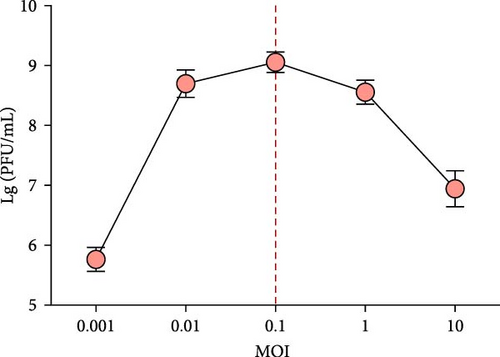
The optimal MOI for the phage was confirmed using previous methods [34]. In brief, phage (~109 PFU/mL) were added to 5 mL of Vp 43 (~108 CFU/mL) to get MOIs of 0.001, 0.01, 0.1, 1, and 10, respectively, the mixture was cultured at 220 rpm and 37°C for 16 h. The supernatant was further centrifuged and filtered with a 0.22 μm membrane and the double-layer agar method mentioned above was used to confirm the titer of the phage. This experiment was repeated three times. The MOI of the highest phage titer was defined to be optimal.
A one-step growth curve test was implemented as previously depicted in prior study [35] with some changes. First, the phage (107 PFU/mL) was blended with the Vp 43 culture (~108 CFU/mL, MOI = 0.1) and cultured for 30 min at 37°C to enable the phage to absorb the host bacteria. The mixture was then centrifuged at 12,000 × g for 60 s to wipe of the unabsorbed phages after incubation, and then, the precipitates were purified with TSB twice. Next, the suspension was transferred to 20 mL of TSB, followed by immediate incubation at 37°C at 220 rpm. This time was defined as t = 0 s, 1 mL sample was obtained every 10 min for up to 120 min. The double-layer agar method was conducted to confirm the phage titer. All the experiments were conducted three times.
To assess temperature stability, phage (0.5 mL) was cultured for 1 h at the following temperatures: 30, 40, 50, 60, 70, and 80°C. Similarly, to assess the pH stability, phage (0.1 mL) was mixed with PBS buffer (0.9 mL) to get a pH gradient (2, 3, 4, 6, 7, 8, 9, and 11, which was adjusted by HCl or NaOH) and was cultured for 1 h at 37°C. At the same time, the effect of acid–base on the Vp 43 was also assessed, Vp 43 (~109 CFU/mL, 0.1 mL) was mixed with TSB buffer (0.9 mL) to get a pH gradient (2, 3, 4, 6, 7, 8, 9, and 11, which was adjusted by HCl or NaOH) and was cultured for 1 h at 37°C. The double-layer agar and serial plating method mentioned above were used to confirm the phage titer and Vibrio concentration, respectively. All the experiments were repeated three times.
2.5. Phage DNA Extraction, Genome Sequencing, Assembly, and Phylogenetic Analysis
To improve our cognition of this phage at the genomic level, phage DNA was extracted using the Virus Genomic DNA Rapid Extraction Kit (Guangdong Haid Animal Husbandry and Veterinary Research Institute Co., Ltd). The purified phage was sent to Suzhou GENEWIZ Biotechnology Co., Ltd. (Suzhou, China) for genome sequencing, the Illumina Hiseq sequencing platform was used to library construction and sequencing. The velvet was used to assemble the filtered reads, and SSPACE and Gapfiller were used to fill the gap [36, 37]. Then, the whole genome sequence of the phage was uploaded to GenBank and the sequence read archive (SRA) database with these respective login numbers PP027964 and SRR27308897. The BLAST procedure in National Center for Biotechnology Information (NCBI) nonredundant (NR) database was used to annotate the coding genes. The phage genomes for phylogenic analysis were downloaded from NCBI and aligned using MEGA 11 software, the software was also used to construct a phylogenetic tree.
2.6. Evaluation of Phage Antibacterial Effect In Vitro and In Vivo
The in vitro cocultivation was used to assess the lytic ability of the isolated phage. Vp 43 culture (~106 CFU/mL) was mixed with the phage stock to result in an MOI of 0.1. Then, the mixture was cultured for 24 h at 37°C. The host culture without the phage was conducted as the control group. The samples were obtained at 0, 1, 1.5, 3, 4, 5, 7, 10, and 24 h, and the Vibrio content was measured using serial dilution plating. The inhibitory effect of the phage on Vibrio was assessed via the growth of Vibrio in the medium.
To validate the phage’s ability to control pathogenic bacterial infections in practical applications, 480 shrimp were cultured in a 50 L tank with 24 L seawater. The temperature was maintained at 28°C, the strain Vp 43 was added into the tank to obtain a final concentration of ~105 CFU/mL for a 6 h challenge. After the exposure, the shrimp were split into six tanks (80 shrimp per tank) with 8 L seawater. The experimental samples contained blank control (240 normal shrimp in three tanks), phage control (240 normal shrimp in three tanks), Vibrio challenge (240 challenged shrimp in three tanks), and phage treatment groups (240 challenged shrimp in three tanks). The phage control and phage treatment groups were fed an artificial diet made of ~109 PFU/g phage four times a day, while the blank control and Vibrio challenge groups were fed a normal artificial diet.
To test the removal of Vibrio from shrimp via phage feeding, on days 0, 3, and 7, the shrimp (n = 3) in each group were randomly sampled and then immediately transported into sterile centrifuge tubes with sterile steel balls and 0.01 mol/L PBS inside; the mixture was then homogenized and gradient diluted with PBS (10−1–10−4), and then, 10 μL of these dilutions was dropped on the medium. After this, the medias were cultured for 18 h at 37°C. Vibrio inhibition by phage was evaluated based on the estimated bacterial numbers.
2.7. Evaluation of Phage Application on the Regulation of Nonspecific Immune Response in Shrimp
During phage feeding in vivo test, on day 0, 3, and 7, the shrimp (n = 6) from different tanks were collected (three samples in total at each time), and the hepatopancreases collected using a pipette tip were quickly transported into a sterilized tube. Briefly, to ensure the homogeneity of the sample, a tissue homogenizer was used to homogenize the hepatopancreases. Low temperature was requested during homogenate to prevent enzyme activity loss. The compounds were then centrifuged at 3000 rpm for 10 min at 4°C and the supernatants were collected and stored at 4°C for further detection. The levels of relevant immune response and antioxidant enzymes, malondialdehyde (MDA), alkaline phosphatase (AKP), catalase (CAT), superoxide dismutase (SOD), lysozyme (LZM), and phenoloxidase (PO) in these samples were detected by commercial test kits (Nanjing Jiancheng Bioengineering Institute, Nanjing, China) according to the manufacturer’s instructions. Each enzyme was detected in triplicate.
The nitroblue tetrazolium (NBT) reduction was detected to determine the production [39]. Briefly, the hemolymph (100 μL) was centrifuged (800 rpm, 20 min, 4°C) and the supernatant was removed, the zymosan (100 μL) was added to induce production. Then NBT was added and set for staining (30 min). The reaction was terminated with methanol (100%) and the hemolymph was washed three times with 70% methanol. Finally, the formazan was dissolved in KOH (120 μL, 2 M) and DMSO (140 μL). Absorbance was measured at 630 nm.
On day 7, the hepatopancreases of shrimp from different groups were collected to detect the relative expression of the immune-related genes. The primers were listed in Table S1. For qRT–PCR analyses, total RNA was extracted with Trizol Reagent and treated with DNase I. The purity and quantity of the RNA were examined by gel electrophoresis and NanoDrop. Then, RNA was reverse-transcribed by ReverTra Ace qPCR RT Master Mix with gDNA Remover (TOYOBO, Japan). Transcript levels were investigated with a CFX96 Connect Real-Time PCR Detection System (Bio–Rad). The PCR program was 95°C for 1 min, followed by 40 cycles of 95°C for 15 s and 60°C for 1 min. The abundance of the EF1α gene was used as an internal standard for standardization of results. The quantitation-comparative CT (2−∆∆CT) method was used to calculated the relative expression levels of immune-related genes [40].
2.8. Evaluation of Phage Application on the Composition of Gut Microbiota in Shrimp
During phage feeding in vivo test, on day 7, the shrimp (n = 12) from different groups were collected and the gut tissues were transported into the sterile centrifuged tube. To investigate the effect of phage application on the composition of the shrimp gut microbiota, the 16S rDNA gene sequence analysis was conducted. In brief, approximately 30 mg of gut tissue was collected from each sample to get genomic DNA by a TIANamp Stool DNA extract kit according to the manufacturer’s instructions. The the Nanodrop was used to measure the quality of the extracted genomic DNA samples. The samples were then sent to Guangdong Magigene Biotechnology Co., Ltd, for gene sequencing and species identification. The V3–V4 variable regions primer pairs F: (5′-ACTCCTACGGGAGGCAGCA-3′) and R: (5′-GGACTACHVGGGTWTCTAAT-3′) were used for amplifying the 16S rDNA gene of gut bacterial. The Illumina MiSeq platform was used to sequence the purified amplicons.
2.9. Statistical Analyses
Statistical analyses and figure preparation were conducted using Graphpad Prism 9 software. Student’s t-test was used to detect the significance (p < 0.05 and p < 0.01) during different groups (blank control vs. phage control, blank control vs. Vibrio challenge, and Vibrio challenge vs. phage treatment).
3. Results
3.1. Origin and Virulence Genes of the Different Vibrio Strains
In this work, 10 Vibrio strains from different aquacultural regions of China were used. The virulence-associated genes pir A/B and vtcc were studied using a PCR assay to investigate the virulence of these strains. The results revealed that 40% of the strains carried the pir A/B gene, while 60% contained the vtcc gene (Table 1). Among them, only Vp 43 carried the pir A/B and vtcc genes; thus, it was selected for lytic phage isolation for its high risk of pathogenicity.
3.2. Isolation of Phage and Host Range Determination
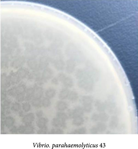
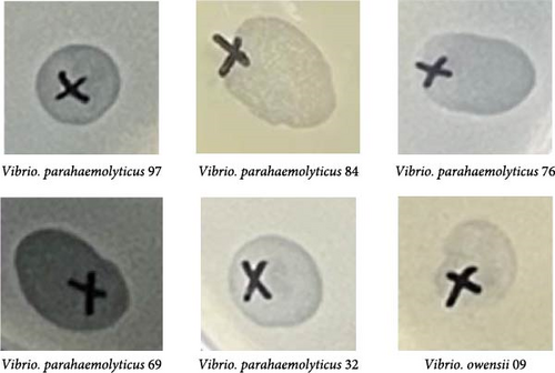
One lytic phage was successfully obtained from the waste water sample using the Vp 43 strain as the host and the phage was further purified by the double-layer agar method. After overnight culture at 37°C, the phage formed clear plaques on the plate (Figure 1A). Among the nine Vibrio strains used in this study, five V. parahaemolyticus strains and one V. owensii strain formed plaques on the plate (Figure 1B), indicating that the Vp 43 phage owned 66.7% lytic efficiency and lysed multiple capsular serotypes.
3.3. Host Phage Optimal MOI, One-Step Growth Curve, Temperature, and pH Stability
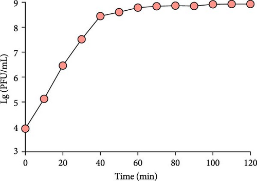
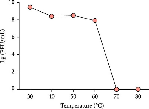
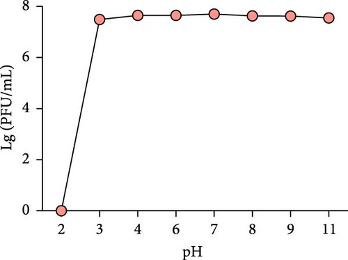
With the Vp 43 strain as the host, the phage titer was the highest (1.1 × 10⁹ PFU/mL) at an MOI of 0.1 (Figure 2A). Thus, the optimal MOI of the phage was 0.1, indicating that the phage can achieve a high yield and excessively proliferate. The one-step growth curve is exhibited in Figure 2B; the latent period of the phage lasted less than 10 min, in contrast, on time 0–50 min, the phage concentration increased sharply, indicating that the lysis period of the phage continued for 50 min. This result confirmed that the phage has a very strong lysis ability. The thermal stability assessment showed that the phage contents had no obvious change at 30–60°C; however, there was no observed phage at 70–80°C, this result revealed that the phage was steady at 30–60°C, but it was entirely inactivated at 70°C (Figure 2C). The pH stability assay showed that the phage contents was comparable at pH 3–11, demonstrating that the phage remained active over a broad pH range, 3.0–11.0, for at least 1 h (Figure 2D), similarly, Vibrio maintains alive at pH 6–11 (Figure S1), but once in harsh pH conditions, the phage (below pH 3.0) and Vibrio (below pH 4.0) were completely inactivated.
3.4. Genome Characterization and Phylogenetic Analysis of Phage
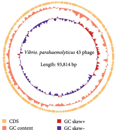
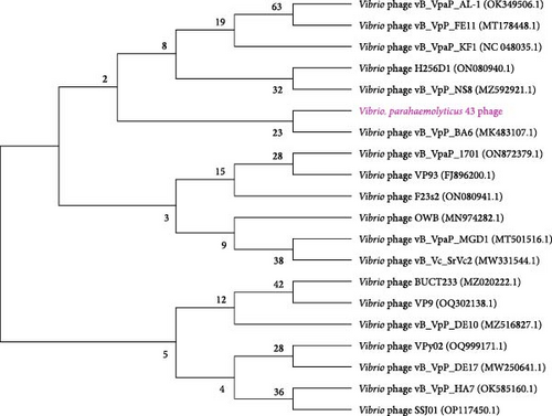
The complete phage genome was obtained through the assembly and annotation of sequencing data. A phage genome map is shown in Figure 3. The phage genome consists of 93,814 bp linear double-stranded DNA, the G + C content of the phage is 45.1%. A total of 115 open reading frames (ORFs) were systematically identified and 48 ORFs (41.7%) were annotated with specific functions, which encode the proteins associated with phage structure and packaging proteins, DNA replication recombination and regulation, metabolism, and host cell lysis (Table S2). Another 60 ORFs (52.2%) were all assigned as hypothetical proteins. A phylogenetic tree is shown in Figure 4. The Vp 43 and Vibrio vB_VpP_BA6 (MK483107.1) phages were grouped together on a single branch, indicating that the former is a Vibrio phage. The Vp 43 and vB_VpP_BA6 phages were only 50% homologous. Thus, the Vp 43 phage might be a novel Vibrio phage strain.
3.5. In Vitro and In Vivo Antibacterial Effect of Phage on Vibrio
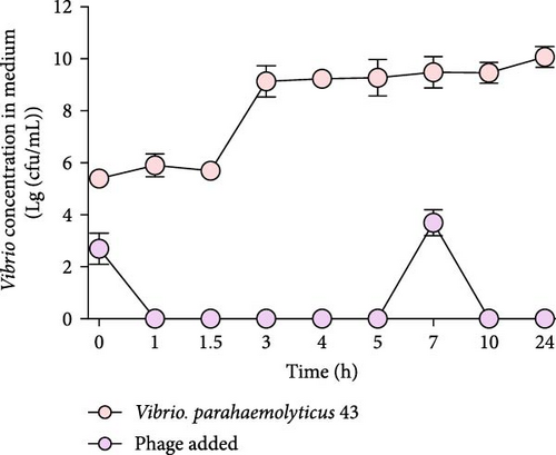
The result of the in vitro tests is shown in Figure 5A. The strain concentration kept increasing in the control group and it amounted from 2.5 × 10⁵ to 1010 cfu/mL at 24 h. In contrast, apart from the temporary increase at 7 h, the strain concentration of the phage treatment reduced significantly in the first few hours, indicating the fast inhibition efficiency of the Vibrio by the phage.
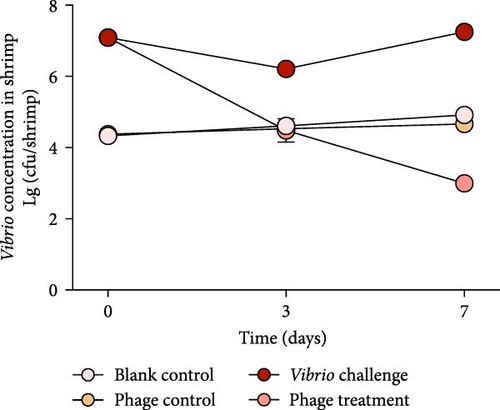
In the trials of the in vivo laboratory shrimp cultivation, on days 0–7, the Vibrio content of the blank control group was 2.14 × 10⁴−8.50 × 10⁴ cfu/shrimp; during phage feeding in the control group, the Vibrio concentration (2.4 × 10⁴−4.6 × 10⁴ cfu/shrimp) was comparable with that of the blank control group (Figure 5B). After the shrimp were challenged and infected with Vp 43, the Vibrio content was 1.22 × 10⁷−1.79 × 10⁷ cfu/shrimp. When the phage was administered, the Vibrio content in the shrimp reduced to 3.58 × 10⁴ cfu/shrimp on day 3 and <2000 cfu/shrimp on day 7, suggesting that the phage could effectively inhibit Vibrio content growth in vivo.
3.6. Influence of Phage Application on the Nonspecific Immune Levels in Shrimp
3.6.1. Immune Parameters
During the in vivo Vibrio challenge and phage feed test, on day 0, 3, and 7, the antioxidant and immune-related enzyme responses in the hepatopancreases or hemolymph were monitored in four groups. As shown in Figure S2, in the blank control group, the MDA, AKP, CAT, SOD, LZM, PO contents, phagocytic activity, and were all steady during the experiment. However, after Vibrio infection, the production of MDA and was significantly increased on day 0–7. In contrast, the AKP, CAT, SOD, LZM, PO content, and phagocytic activity in shrimp exhibited obvious decrease. Compared with the blank control group (Figure 6), the MDA levels and production significantly increased (p < 0.01; t-test) after the Vibrio challenge; in contrast, the AKP, CAT, LZM, PO, and phagocytic activity showed a drastic decrease (p < 0.05; t-test), which indicated that pathogenic Vibrio increase the level of immune stress in shrimp.
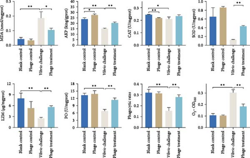
After phage feeding, on day 7, the antioxidant and immune-related enzyme responses in the shrimp hepatopancreases or hemolymph were shown in Figure 6. Phage feeding significantly improved the AKP expression level (p < 0.05; t-test) and reduced the CAT level (p < 0.01; t-test) comparing with those of the blank control group; however, there were no obvious differences in the MDA, SOD, LZM, PO, phagocytic activity, and between the two groups. When the phage was administered after Vibrio challenge, the MDA activity level and production reduced significantly (p < 0.05; t-test); in contrast, the AKP, SOD, LZM, PO, and phagocytic activity levels improved significantly (p < 0.01; t-test), demonstrating that the phage could effectively relieve the immune stress caused by Vibrio infection.
3.6.2. Immune-Related Gene Expression
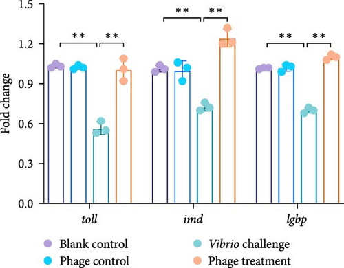
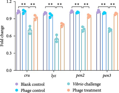
After the in vivo Vibrio challenge and phage feeding trail, the relative expression levels of toll, imd, lgbp, cru, lyz, pen2, and pen3 genes in the shrimp hepatopancreases among different treatment groups are shown in Figure 7. There was no significant difference in relative gene expression between the blank control and phage control group. Compared with the blank control group, the expression of PRR system and pattern recognition protein, including that of toll, imd, and lgbp were all significantly downregulated (p < 0.01; t-test) after Vibrio infection on day 7. Similarly, four antimicrobial peptides—cru, lyz, pen2, and pen3, were also significantly downregulated (p < 0.01; t-test) in the Vibrio challenge group comparing with those of the blank group. However, after phage feeding, the expression of PRR system, pattern recognition protein, and antimicrobial peptides genes were all significantly upregulated (p < 0.01; t-test) on day 7 comparing with those of the Vibrio challenge group.
3.7. Influence of Phage Application on the Composition of Gut Microbiota in Shrimp
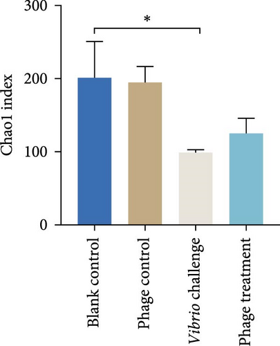
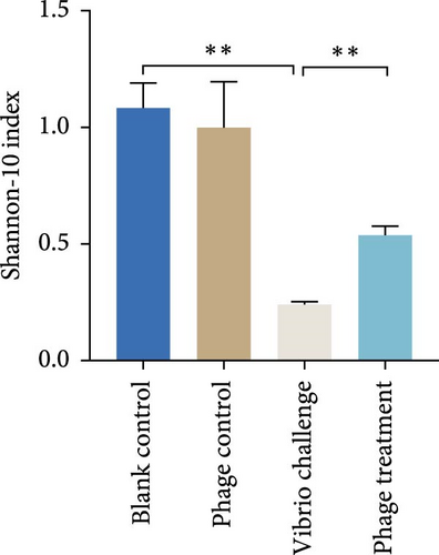
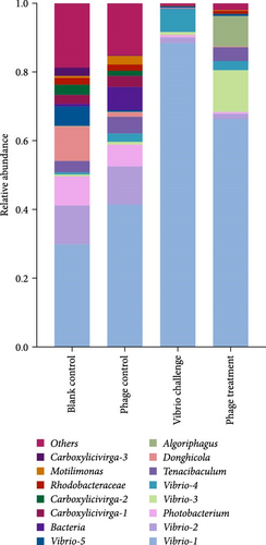
After 7 days of phage feeding, the Chao1 and Shannon-10 indexes of the gut microbiota were similar with those of the blank control group (Figure 8A,B), indicating that phage application had no significant impact on the diversity and abundance of gut microbiota in normal shrimp. In the control group (Figure 8C), Vibrio 1 (47.9%) was the leading genus, followed by Donghicola (10%), Photobacterium (8.5%), Carboxylicivirga (8%), and then others. After the Vibrio challenge, the relative abundance of the species Vibrio 1 (88.4%) increased significantly and the Chao1 and Shannon-10 indexes reduced significantly (p < 0.05 and p < 0.01, respectively; t-test; Figure 8A,B). Results from these revealed that pathogenic Vibrio infection significantly reduced the abundance and diversity of the shrimp gut microbiota. In contrast, after phage feeding, the Shannon-10 index obviously recovered (p < 0.01; t-test; Figure 8B). which indicated that phage application could improve the diversity of the gut microbiota in shrimp.
4. Discussion
Vibrio parahaemolyticus is a major pathogenic bacteria that can infect various animals, like shrimp, shellfish, and fish, resulting in mass mortality and vast economic losses [41]. In invertebrates, the resistance of pathogens mainly relies on nonspecific immunity and regulation of gut microbiota. As natural antibacterial agents, phages exist extensively in natural environments and have developed an impressive array of systems in controlling pathogens. But to date, several studies only focused on the direct antibacterial effects of phages [25–27], less is known about its indirect effects, such as the influences on the nonspecific immunity and composition of gut microbiota in shrimp after Vibrio infection. Given the importance of the normal regulation of the immune system and the balance of gut microbiota in controlling pathogens and maintaining the normal growth process of shrimp, the crucial factors need to be investigated, such as phages’ direct antibacterial effect, influences on nonspecific immunity, and composition of gut microbiota in shrimp after Vibrio infection.
By screening against Vp 43, a lytic phage was obtained. As an important criteria for biological control, the wider the range of a phage is, the more actual application prospects it has [42]. In this study, 66.7% of the Vibrio of different genera were lysed by the phage, this host range was much larger than those of other phages specific for Vibrio [43, 44], indicating that it has high potential for application. In addition to the lytic range, a smaller MOI and a shorter incubation period are also preferred to reduce the cost of phage application [45]. In this work, the phage was confirmed to have an optimal MOI of 0.1 and an incubation period of 10 min, which are considerably better than those of other Vibrio [46, 47]. Finally, the tolerance of phages to extreme environments also influences the broad applicability of their use. In this study, the phage kept stable at 4–60°C, indicating that it can tolerate higher or lower temperatures. Similarly, the phage was steady at pH 3–11, though the host Vibrio was inactive at pH 3–4. Extremely high temperatures (>60°C) and low pH (<3) can inactivate the phage by rapidly denaturing its nucleic acids and proteins. Compared with previously reported Vibrio phages [48, 49], this phage was more tolerant to harsh and extreme environments, indicating that it own better activity as a biocontrol agent for application in aquaculture; the extremely strong acid resistance also indicated that the phage may have a high inhibitory effect on acidophilic microorganisms and can be potentially used in the food industry [50].
In general, the inhibition efficiency of pathogenic Vibrio is one of the key indicators to evaluate the application of phages. In this study, after 24 h cocultivation in vitro, there was no visible Vibrio in the mixture, indicating that pathogenic Vibrio can be inhibited in a short time, this is consistent with Chen et al.’s [32] result that the Vibrio phage could inhibit pathogenic Vibrio within the first 2 h. We noticed that the content of Vibrio increased momentarily at 7 h, leading us to speculate that some Vibrio may have undergone adaptive evolution in short time; however, under limited nutrient environment, they are unable to proliferate. In addition to the in vitro test, in order to simulate the application of a phage that is more suitable for aquaculture, we also conducted in vivo shrimp phage experiment. The in vivo test showed that after the application of the phage for 7 days, the content of Vibrio decreased by 4–5 log cfu/shrimp. The efficiency of lytic phages in preventing different types of pathogens in shrimp has been demonstrated by several studies. Alagappan et al. [28] reported that a phage could significantly reduce the levels of Vibrio infection in shrimp, thus, reducing the mortality rates. Jun et al. [51] also highlighted the lytic activity of phage against V. parahaemolyticus and their efficacy in protecting shrimp. All of these results indicate that the phages can effectively reduce the Vibrio concentration by directly inhibiting pathogenic Vibrio in shrimp.
As invertebrates, the shrimps’ nonspecific immune system is crucial in preventing Vibrio and maintaining healthy farming processes, it is easily disturbed by pathogenic Vibrio and activated by immunoenhancers [9, 10]. Among the several pathways involved in the regulation of the nonspecific immune response, PRRs, antioxidant enzymes activity, and reactive oxygen species-mediated microbicidal activity play important roles in defending against exogenous pathogens [52]. In this work, besides the phage inhibiting Vibrio, our results also showed that pathogenic Vibrio or the phage significantly changed the shrimp’s antioxidant and nonspecific immune levels. In the presence of a Vibrio challenge, the MDA level and production increased sharply; however, the AKP, CAT, SOD, LZM, PO content, and phagocytic activity in shrimp exhibited obvious decrease. As the most important product of lipid peroxidation and oxidative damage, the MDA and contents reflect the lipid peroxidation and superoxide anions level in the organism and an increase in their contents represents a reduction in the body’s antioxidant capacity [53, 54]. The results of this work indicated that pathogenic infection interfered the shrimps’ nonspecific immune systems regulation. Compared with the Vibrio challenge group, after phage application, the AKP, SOD, LZM, PO, and phagocytic activity in the shrimp increased significantly, in contrast, the MDA and contents significantly reduced, this is consistent with Alagappan et al.’s [28] results that a phage could enhance the immune response, working as immunomodulators in shrimp. AKP plays important roles in the dephosphorylation of proteins (enzymes), it not only participates in the digestion, absorption, and transport of some nutrients, but constitutes an important detoxification system within the shrimp [55]; in addition to AKP, SOD and LZM also play crucial roles in the oxidation and antioxidation balance of shrimp [56]. Furthermore, in the proPO system, the PO activity is regarded as a critical enzyme, which participates in invertebrate humoral defense mechanism to eliminate pathogens [57], and phagocytic activity is also mentioned as common indicator to measure immune responses in shrimp [58]. It can be inferred that infection with Vibrio reduced the expression level of the nonspecific immune system in shrimp, while the bacteriophages may act as an immunomodulatory and reduce the amount of Vibrio by activating the nonspecific immune response in shrimp.
During the Vibrio challenge and phage feeding, the related gene expression of PRR system, pattern recognition protein, and antimicrobial peptides were also monitored. The toll, imd, and lgbp gene were responsible for the regulation of PRR system [59, 60], while the cru, lyz, pen2, and pen3 gene were related with the synthesized of antimicrobial peptides, they play crucial roles in the innate immune system by exerting an immunomodulatory effect [61, 62]. In this work, all of these genes in shrimp were downregulated after Vibrio challenge, indicating that pathogen infection interrupted the normal regulation of PRR system and synthesis of antimicrobial peptides. Tanji et al. [63] showed that the absence of toll and imd signaling pathways causes a deficiency in antimicrobial peptides production, leading to more susceptibility to pathogenic bacteria. In contrast with Vibrio infection, after phage feeding, these genes were all significantly upregulated, suggesting that phage application could activate the PRR system and antimicrobial peptides synthesis pathway. To date, there has been no relevant research on the effects of phages on the expression of PRRs and antimicrobial peptide synthesis related genes in shrimp. We speculate that phage activated the production of nonspecific immune related enzymes in shrimp by upregulating the expression of PRR and antimicrobial peptides synthesis system related genes, thereby enhanced the resistance of shrimp to pathogenic bacteria, but the indirect effect mediated by phage needs to be investigated further.
Similar with the regulation of the nonspecific immune system in shrimp, the ecological balance of the gut microbiota also plays crucial roles in shrimp farming, and this balance is easily interfered by exogenous factors (bacterial or viral). Our results proved that the phage had no significant effect on the abundance and diversity of gut microbiota in healthy shrimp. However, pathogenic Vibrio significant reduced the abundance and diversity of shrimp gut microbiota, thus, disrupting the dynamic equilibrium; this is consistent with Aguilar-Rendón et al. [64] and González-Gómez et al. [13] results that state that V. parahaemolyticus is the most predominant species in the gut microbiota after inoculation. After the phage treatment, the relative abundance of V. parahaemolyticus in the shrimp reduced sharply, while the Shannon-10 index increased significantly; these results clearly demonstrated that the phage effectively alleviated the dysbiosis of the shrimp gut microbiota caused by pathogenic Vibrio. Similar results obtained by Deng et al. [65] also demonstrated that the microbial community composition and function in shrimp can be directly or indirectly modulated by the phage–prokaryote interactions. We speculate that the phage may increase the diversity of other bacterial communities and rebalance the composition of shrimp gut microbiota by directly reducing the number of pathogenic Vibrio in the intestines of shrimp and more detailed repair mechanisms mediated by phages need to be researched in the future.
5. Conclusions
In this work, a potential Vibrio lytic phage was identified and functionally investigated. The phage presented a series of practically applied features and was proved to be effective in preventing Vibrio via in vitro and in vivo test. This phage can enhance the antioxidant and nonspecific immune ability of shrimp by upregulating the expression of PRRS and antimicrobial peptides synthesis system related genes. This phage could also enhance gut microbiota diversity of shrimp by inhibiting pathogenic Vibrio. This work verifies the practical efficacy of phages and elucidates their effects of normal regulation of nonspecific immunity and gut microbiota in shrimp, thus, providing theoretical recommendations for the rational application of phages in aquaculture.
Conflicts of Interest
The authors declare no conflicts of interest.
Author Contributions
Dongdong Song: methodology, investigation, software, data curation, writing–original draft, funding acquisition. Baojun Shi: conceptualization. Jinlu Huang: writing–review and editing, funding acquisition. Jian Wang: conceptualization, supervision, writing–review and editing, funding acquisition.
Funding
This research was financially supported by the China Postdoctoral Science Foundation (Grant 2023M740757) and the 2023 Qingyuan Qingxin District Science and Technology Plan Project (Grant 2023QJ01001).
Acknowledgments
This research was financially supported by the China Postdoctoral Science Foundation (Grant 2023M740757) and the 2023 Qingyuan Qingxin District Science and Technology Plan Project (Grant 2023QJ01001).
Supporting Information
Additional supporting information can be found online in the Supporting Information section.
Open Research
Data Availability Statement
The data obtain in the present study are available from the corresponding author on reasonable request.


