A Case of Syrian Child with Cerebral Infarction as an Extraintestinal Manifestation of Ulcerative Colitis
Abstract
Thromboembolic complications are rare but well-recognized manifestation of ulcerative colitis, especially because of their associated high mortality. We report a case of a Syrian child admitted to Damascus Hospital with a one-day complaint of sudden onset of numbness followed by weakness in the left lower and upper limbs, right mouth angle deviation, and loss of sphincters’ control. Earlier, she was diagnosed with ulcerative colitis and treated with immunosuppressants. CT and MRI scans revealed focal infarction around the M2-M3 segments of the right middle cerebral artery; she was treated with Aspirin. On discharge, she had significant improved neurological examination and was able to walk. Subsequent proctocolectomy was performed. We highlight the importance of thromboembolism in ulcerative colitis as there is paucity in the literature regarding its management and its symptoms may be overlooked especially in high-load central hospitals. We conducted a brief literature search and summarized findings of similar reported cases.
1. Introduction
Extraintestinal manifestations of idiopathic inflammatory bowel disease (IBD) have been reported in 25% to 36% of patients [1]. Common manifestations include sacroiliitis (14%) and peripheral arthritis (10.7%), while rare manifestations include ocular (8%), mucocutaneous (2.7%), and vascular (2%) [2].
Neurologic manifestations in IBD appear to be more common than previously estimated with a reported incidence of cerebrovascular complications in 0.12% to 4% of all patients with IBD [3, 4]. Generally, it occurs as a postoperative complication and found more in Crohn′s disease than ulcerative colitis (UC) [5]. Thromboembolic complications of UC are reported at an incidence of only 1.2%-7.5%, but are well recognized because of their associated high mortality [6–9] which occurs in 60% of cases [4].
We report a rare case of a Syrian child who was suffered a cerebrovascular accident (CVA) as a complication of ulcerative colitis. To the best of our knowledge, this is the first documented case in Syria.
2. Case Presentation
A 15-year-old Syrian female was admitted to the hospital on November 2016 with a one-day complaint of sudden onset of numbness in the left lower and upper limbs, followed by weakness in the same areas, right mouth angle deviation, and loss of sphincters’ control. She did not experience headache, nausea, vomiting, convulsions, or coma.
Eight months earlier, she developed massive rectal bleeding, colonoscopy was performed, and the patient was diagnosed with ulcerative colitis (UC). She was treated with mesalazine 1 gram three times daily, azathioprine 50 milligram daily, prednisolone 40 milligram daily, and cefuroxime 500 milligram tab twice daily for a week.
She has no history of smoking, alcohol abuse, or illicit drug use. She did not report any suspected allergies and she has no other history of hypertension, diabetes mellitus, cardiac, rheumatological, or hematological disease.
On examination, her vital signs are blood pressure 100/60 mmhg, Pulse 110/minute, respiratory rate 36/minute, and temperature 37.5°C. General examination revealed conjunctival pallor and pitting edema in the left lower limb and purple stretch marks extends on the whole lower limbs till the sacrum.
On neurological examination, there was no impaired consciousness and the patient was awake and alert. Cranial nerves exam was only significant for left facial nerve palsy. Motor examination showed 5/5 strength in the right upper and lower limbs, 3/5 left upper limb, and 0/5 left lower limb; there was also hypotonia on the left limbs and normal tone on the right limbs without any atrophy. Reflexes examinations scored 2/4 for the right limbs (normal) and 1/4 for the left limbs (hyporeflexia). Right toes showed planter flexion and absence of the flexion for the left toes. No cerebellar abnormalities were noted in the right side; cerebellar exam was not performed on the left side due to limbs weakness. She scored 10 on National Institutes of Health Stroke Scale (NIHSS). Sensory examination revealed loss superficial and deep sensations on the left side and normal sensations on the right side. Other systematic examinations, including cardiac, respiratory, and gastrointestinal systems, were all normal.
Investigations including blood tests showed evidence of pancytopenia (hemoglobin 4.4 g/dL, platelets: 66 x1000/mm3 dropped to 3 x1000/mm3 after in two days of admission, WBCs: 1.4 x1000/mm3 with 35% neutrocytes, 61% lymphocytes, 3% monocytes, and 1% eosinophils); urinalysis values were within normal ranges. Thrombophilic and immunological screening including homocysteine, factor V Leiden, protein C, protein S, antithrombin, lupus anticoagulant factor, and antiphospholipid antibodies were all insignificant.
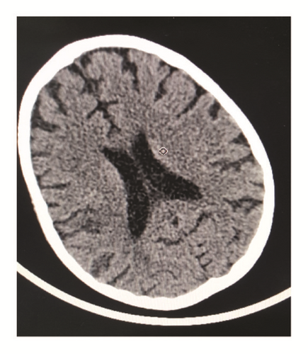
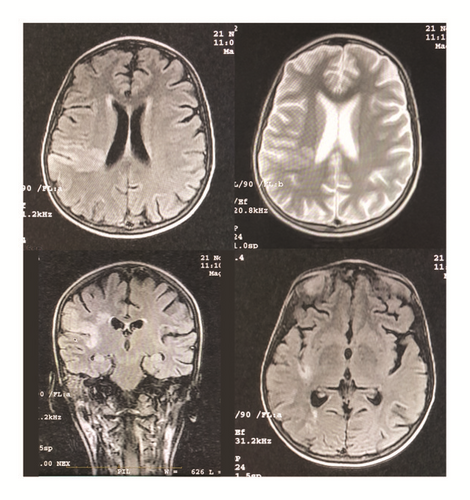
An emergency computerized tomography (CT) scan (Figure 1) showed small hypodensity foci situated in the cortical and subcortical area in right partial lobe. Magnetic resonance imaging (MRI) (Figure 2) showed cortical and subcortical areas in the right temporoparietal fossa with high signal on T2 and FALIR studies. T1 study showed isointense foci in the cortical and subcortical area in the posterior part of the parietal lobe extending deeply through the posterior horn of the right lateral ventricle. Based on these findings, the accident is complicated with focal infarction around the M2-M3 segments of the right middle cerebral artery. Aspirin 162mg was given upon these findings and the prednisolone treatment was continued.


Cardiac echocardiogram and carotid arteries Doppler ultrasound study were both normal. The patient did not complain of any symptoms related to her UC when she had the CVA, which indicates that the UC was not in active stage.
On December 2016, the patient was able to walk and her neurological examination dramatically improved (NIHSS: 0); she was then discharged and referred to physical therapy. On January 2017, the patient suffered from overt rectal bleeding, she was admitted again to the hospital, and proctocolectomy was performed.
A written informed consent was obtained from the patient before writing this report, Syrian Private University and Damascus Hospital Ethical Committee approved the report, and both are available upon request.
3. Discussion
Increased coagulability and thrombosis due to IBD were first described in 1936 [29]. Intestinal inflammation may lead to increased risk for thrombosis through several pathways: activation of coagulation cascade, decreasing anticoagulant activity, inducing hypofibrinolysis, malabsorption, and hypercatabolism with vitamin deficiencies [4]. Most patients with IBD do not have demonstrable specific coagulation defects [30]. Dehydration, immobility, sepsis, surgery, and corticosteroid therapy are also risk factor for thrombosis in IBD patients [31]. The precise mechanism of these factors remains unclear. Arterial thrombosis particularly strokes may be considered a rare condition [17] but with high morbidity and mortality [32–34].
Males and females may be equally affected which correlates with previously reported cases. The cerebral vascular involvement seems more frequent among younger IBD patients [4]. Conventional CT scan or MRI is used to define the cerebral affected areas. At this moment, no guidelines are available for the treatment of cerebral thrombosis and strokes in IBD [17].
By reviewing previous literature, Schneiderman et al. were the first to report similar case with thrombosis of the left internal carotid artery (ICA) and occlusion of the left distal basilar artery in two separate patients with UC respectively [26]. The youngest patient reported was a 1-year-old girl with UC, who was complicated with bacterial endocarditis and subsequent infarctions of both middle cerebral arteries (MCA) [27].
We have conducted a brief literature search and summarized cases reported on Cerebral Arterial Thrombosis associated with UC using modified version of Katsanos et al. [35] table (Table 1).
| Author | Date | Sex | Age | UC Activity | IBD Treatment | Neurological symptoms | Site of cerebral injury/neurological findings | Risk Factor | Thrombophilia screening | Anticoagulation/antiplatelet treatment | Follow-up/outcome |
|---|---|---|---|---|---|---|---|---|---|---|---|
| Calabro et al. [10] | 2011 | F | 49 | No | Right hemiparesis, aphasia, altered conscious | Left frontotemporal infarct, lesion in the left mesencephalic area, left ICA stenosis | Hypercholesterolemia, elevated Lpa | LMWH (nadroparine 0.6 ml/d), aspirin (300 mg/d) / clopidogrel (75 mg/d) | Improved | ||
| Casella et al. [11] | 2013 | M | 25 | Yes | Left hemiparesis, confusion, sensitive impairment, bladder incontinence | Right MCA infarct | Anemia | Local thrombolysis/ LMWH and aspirin (100 mg/d) | Complete recovery | ||
| Chetri et al. [12] | 2002 | M | 42 | Yes | Steroids, 5-ASA | Left hemiplegia | Right MCA infarct and right CCA occlusion | Anemia | Improved | ||
| Fukuhara et al. [13] | 1993 | M | 18 | No | Steroids | Left hemiparesis | Right paramedian branch of the basilar artery infarct, Ventromedial pons | Anemia | Complete recovery | ||
| Hilton-Jones et al. [14] | 1985 | M | 20 | No | Basal ganglia infarct | Anemia, abnormal platelet aggregation | Recurrent thrombotic episodes, death | ||||
| Houissa et al. [4] | 2011 | M | 24 | Yes | Steroids | Left hemiplegia | Left MCA infarct | Neg | LMWH | Complete recovery | |
| Houissa et al. [4] | 2011 | F | 25 | Yes | 5-ASA | Left hemiparesis | Tenticular & right thalamic infarcts | Thrombocytosis | Neg | LMWH | Partial recovery (Residual right hemiparesis) |
| Jorens et al. [15] | 1990 | M | 31 | Yes | 5-ASA | Right hemiparesis, nonconfluent aphasia | Recent ischemic lesion in left internal capsule and old ischemic lesion in left basal ganglia | Anemia, thrombocytosis, pro-C, pro-S and prothrombin deficiency, history of TIA | Partial recovery | ||
| Jorens et al. [16] | 1991 | M | 32 | Yes | Left hemiparesis, stupor, hemiataxia | Temporoparietal cerebrovascular ischemic lesion | Complete recovery | ||||
| Joshi et al. [17] | 2008 | M | 55 | Yes | Steroids, 5-ASA | Left hemiparesis | Right parietal lobe infarction | Thrombocytosis | Neg | LMWH | Partial recovery |
| Joshi et al. [17] | 2008 | M | 24 | Yes | Steroids, 5-ASA | Headache, altered conscious, global aphasia | Left MCA infarct | Thrombocytosis | Neg | Complete recovery, subsequent epilepsy | |
| Keene et al. [18] | 2001 | F | 13 | Yes | Steroids, subtotal colectomy | Seizure | Multiple bilateral cerebellar and corona radiata infarcts | Anemia | Complete recovery | ||
| Kelly et al. [19] | 2014 | M | 38 | No | Steroids, AZA, infliximab | bilateral lower limb claudication, acute confusion with associated ataxia and diplopia | right medial temporal lobe extending posterior to the occipital lobe | Neg | Warfarin | Controlled | |
| Lloyd-Still and To masi [20] | 1989 | M | 5 | Yes | Steroids, 5-ASA | Right hemiparesis, seizure | Left MCA arteritis | Anemia | Partial recovery, epilepsy developed 10 years later | ||
| Mayeux and Fahn [21] | 1978 | M | 17 | Yes | Steroids | Right posterior frontal area infarction | Slowly improved | ||||
| Nogami et al. [22] | 2007 | F | 26 | Yes | Steroids | Left hemiparesis | Right MCA Infarct and right CCA occlusion | Severe anemia | Heparin (15.000 U/d) | Massive GI bleeding, no improvement | |
| Paradis et al. [23] | 1985 | F | 12 | Yes | Right hemiparesis, seizure | Left major cerebral vessels occlusion | Anemia, thrombocytosis | Complete recovery | |||
| Patterson et al. [24] | 1971 | M | 11 | Yes | Cerebral emboli | Colectomy | |||||
| Richard et al. [25] | 2014 | F | 42 | Yes | Steroids, AZA | Sudden right hemiplegia | Right MCA | Slight hyperhomocysteinemia | LMWH, Enoxaparin sodium, Aspirin | ||
| Salloum et al. (our case) | 2016 | F | 15 | No | Steroids, 5-ASA, AZA | Left hemiparesis, 7th CN palsy | M2-M3 segments of the Right MCA | Pancytopenia | Neg | Aspirin 162mg | Colectomy, complete recovery |
| Schneiderman et al. [26] | 1979 | M | 34 | Yes | Right hemiplegia, nonfluent aphasia | Left ICA thrombosis | Thrombocytosis | Thrombectomy, heparin/warfarin | |||
| Schneiderman et al. [26] | 1979 | F | 12 | Yes | Steroids, 5-ASA | Hemianopia, headache, seizures | Distal basilar artery defect extending to the left PCA | Elevated fVIII | Death | ||
| Tomomasa et al. [27] | 1993 | F | 1 | Yes | Steroids, 5-ASA | Right hemiplegia, altered conscious, seizures | Left anterior & MCA infarct/ right MCA infarct | Thrombocytosis, bacterial endocarditis | No improvement | ||
| Yassinger et al. [28] | 1976 | F | 15 | Yes | Steroids | Right frontal lobe infarction | Recovered |
- M = male; F = female; 5-ASA = 5-aminosalicylic acid; AZA = azathioprine; CN = cranial nerve; ICA = internal carotid artery; MCA = middle cerebral artery; CCA = common carotid artery; PCA = posterior cerebral artery; LPa = lipoprotein; Pro-C = protein C; Pro-S = protein S; TIA = transient ischemic attack; fVIII = factor VIII; LMWH = low molecular weight heparin; GI = gastrointestinal.
About half of the patients were on corticosteroid treatment and more than one-third of them were being treated with 5-aminosalicylic acid (5-ASA) at the time of the cerebrovascular event [35]. The predominant neurological symptom on admission in most of the case reports was left or right sided hemiparesis; our patient was also admitted with left hemiparesis as the main complication. In addition, the right or left MCA was the most frequent sites of cerebral arterial thromboembolism depicted on imaging studies similar to our patient that manifested with cerebral arterial thromboembolism around the M2-M3 segments of the right MCA as confirmed by imaging studies [35]. As previously reported, the risk of arterial thromboembolic events may be increased in patients with active disease [4], although our patient did not have an active disease by the time of CVA.
Thrombocytosis and anemia were the most commonly observed potential risk factors for cerebral arterial infarction in the laboratory analysis [35], although there is no sufficient evidence supporting the theory of solitary thrombocytosis causing thromboembolic phenomena [26]. Our patient contrary presented with pancytopenia which may be due to immunosuppressants given [36].
Our patient did not have a demonstrable specific coagulation defects, in comparison; hyperhomocysteinemia [4] and other acquired deficiencies of antithrombin III and protein S have previously been reported in similar cases [30, 35]. Finally, smoking and severe dehydration have been mentioned in only two cases contrary to our patient.
Our patient received Aspirin as a treatment after she was diagnosed with the cerebrovascular accident. She had improved neurological examination on discharge and she was able to walk; she was then discharged and referred to physical therapy, although as reported in previous cases also most of the patients also recovered either without any or with minor neurological deficits; either they received anticoagulation or antiplatelet treatment or not [35] (Table 1).
Clinical experience treatment of arterial ischemic cerebral lesions in patients with IBD is very limited due to lack of enough trials [37]. More studies are required to clarify the correlation between IBD and the thrombophilias and to evaluate the role of anticoagulant therapy and proctocolectomy in the management of these patients. Similarly, there is also lack of steady evidence and official guidelines for stroke management in both children and adults with IBD comorbidity. Both American Heart Association and European Stroke Organization guidelines for stroke management and prevention in the general population are currently presented as a reference point for the treatment of IBD patients who are complicated by an ischemic cerebral event [38–40].
Conflicts of Interest
The authors declare that they have no conflicts of interest.
Authors’ Contributions
Mohamad Anas Almidani and Riham Salloum approached the patient, Riham Salloum and Nawras Alhalabi drafted the initial manuscript, and Riham Salloum, Nawras Alhalabi, and Mohamad Anas Almidani revised and wrote the final version of the manuscript and approved publication.
Acknowledgments
We appreciate the collaboration of the patient and the patient’s family; we express gratitude to Dr Anas Jawhar and Dr Tareq Al Saadi for their kind collaboration. In addition, we would like to express our sincere thanks and appreciation to the dean of faculty, Professor Nizar AlDhaher, and the university president, Professor Nazir Ibrahim, the Faculty of Medicine of Syrian Private University Research Group, and We Research Team for their endless encouragement and scientific motivation.


