Volume 2014, Issue 1 505968
Meeting Abstract
Open Access

Whole Slide Image Analysis System for Quantification of Liver Fibrosis

Tokiya Abe

Tokiya Abe

Department of Pathology, Keio University School of Medicine, 35 Shinanomachi, Shinjuku-ku, Tokyo 160-8582, Japan keio.ac.jp

Search for more papers by this author
Yuri Murakami

Yuri Murakami

Global Scientific Information and Computing Center, Tokyo Institute of Technology, 2-12-1 Ookayama, Meguro-ku, Tokyo 152-8550, Japan titech.ac.jp

Search for more papers by this author
Masahiro Yamguchi

Masahiro Yamguchi

Global Scientific Information and Computing Center, Tokyo Institute of Technology, 2-12-1 Ookayama, Meguro-ku, Tokyo 152-8550, Japan titech.ac.jp

Search for more papers by this author
Yoshiko Yamashita

Yoshiko Yamashita

Medical Solutions Division, NEC Corporation, 7-1, Shiba 5-chome, Minato-ku, Tokyo 108-001, Japan nec.com

Search for more papers by this author
Tomoharu Kiyuna

Tomoharu Kiyuna

Medical Solutions Division, NEC Corporation, 7-1, Shiba 5-chome, Minato-ku, Tokyo 108-001, Japan nec.com

Search for more papers by this author
Ken Yamazaki

Ken Yamazaki

Department of Pathology, Keio University School of Medicine, 35 Shinanomachi, Shinjuku-ku, Tokyo 160-8582, Japan keio.ac.jp

Search for more papers by this author
Akinori Hashiguchi

Akinori Hashiguchi

Department of Pathology, Keio University School of Medicine, 35 Shinanomachi, Shinjuku-ku, Tokyo 160-8582, Japan keio.ac.jp

Search for more papers by this author
Yutaka Yasui

Yutaka Yasui

Department of Gastroenterology and Hepatology, Musashino Red Cross Hospital, 1-26-1 Kyonan-cho, Musashino, Tokyo 180-8610, Japan jrc.or.jp

Search for more papers by this author
Masayuki Kurosaki

Masayuki Kurosaki

Department of Gastroenterology and Hepatology, Musashino Red Cross Hospital, 1-26-1 Kyonan-cho, Musashino, Tokyo 180-8610, Japan jrc.or.jp

Search for more papers by this author
Namiki Izumi

Namiki Izumi

Department of Gastroenterology and Hepatology, Musashino Red Cross Hospital, 1-26-1 Kyonan-cho, Musashino, Tokyo 180-8610, Japan jrc.or.jp

Search for more papers by this author
Michiie Sakamoto

Corresponding Author

Michiie Sakamoto

Department of Pathology, Keio University School of Medicine, 35 Shinanomachi, Shinjuku-ku, Tokyo 160-8582, Japan keio.ac.jp

Search for more papers by this author
First published: 15 December 2014

Background and Aims

Histological evaluation of fibrosis after a liver biopsy is crucial for evaluating the pathology of patients with chronic liver disease. We have reported image analysis allowing quantification of liver fibrosis using Elastica van Gieson (EVG) stained whole slide images (WSIs) of liver biopsy specimens [1]. In this paper, a whole slide image analysis system for quantification of liver fibrosis was developed to apply a large number of cases in routine practice.

Method

Our system was composed of 2 steps: color correction and tissue classification. Firstly, the color correction was performed by transforming the color distribution from a target WSI into a reference WSI, where the distribution was estimated by two triangle pyramids. Next, the tissue classification was performed by using quadratic discriminant function generated from RGB signal data sets in reference WSIs. After 2 steps, the WSI pixels were classified into five classes corresponding to four tissue areas: collagen fibers, elastic fibers, nucleus, and cytoplasm and one nontissue area (i.e., glass slide). Finally, the area ratios of collagen and elastic fibers were automatically quantified.

Results

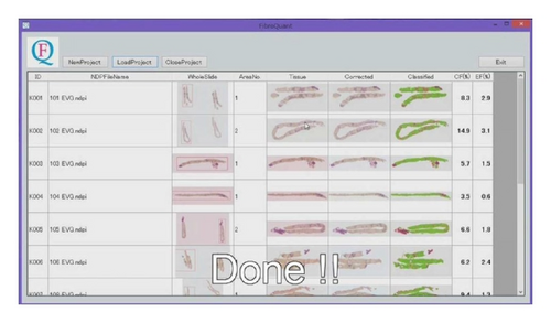
WSIs of liver biopsy specimens collected from 102 patients with hepatitis C were analyzed by our system [2]. The system successfully corrected the color of any WSI to that of the reference WSI and precisely extracted fine collagen and elastic fibers from portal and periportal areas (Figure 1). The averaged calculation time for WSIs with hundred-millions of pixels solution was about four minutes.
Details are in the caption following the image
Whole slide image analysis system for quantification of liver fibrosis. The figure shows the color corrected and classified WSI and the area ratios of collagen and elastic fibers obtained by the system.

Conclusions

The whole slide image analysis system could provide quantification of liver fibrosis in biopsy specimens with different color distributions, which can display continuous value, and represent a progression of liver disease.

Acknowledgment

This study was supported by grants from the New Energy and Industrial Technology Development Organization (Project no. P10003).

      The full text of this article hosted at iucr.org is unavailable due to technical difficulties.