Design and Evaluation of Photo-Induced Biofeedback Fabric for the Enhancement in Sleeping Sense
Abstract
Based on overcoming the sleeping obstacle for people, the purpose of this study is to design a photo-induced biofeedback fabric which is a kind of optical fiber fabric with the function of enhancing sleeping sense and to evaluate its effect. The fabrics with two layers including background layer and pattern layer were designed firstly. The pattern layers with 3 kinds of wavelengths of sine waves and the light controller with 3 kinds of flashing frequencies were then prepared. Guiding the light into the optical fiber, it will emit out of the optical fiber and stimulate our visual system to change the form of brain wave. Finally, EEG and sleeping scale were applied to evaluate the effect of enhancing sleeping sense. The results were shown that human’s brain wave can be changed from sober status to shallow-sleeping status and the effect of enhancing sleeping sense can be achieved for all pattern layers in frequencies of 0, 5 and 10 Hz. Furthermore, female is more significant than male and participants age from 30 to 49 are the most significant. Besides, the stronger the participant’s stress is, the more significant the sleeping sense is.
1. Introduction
According to the investigation of Taiwan Society of Sleep Medicine (TSSM), more than 4.5 million people have sleeping obstacle and 2.5 million people have suffered from chronic insomnia in Taiwan in 2006. Only a few people would like to consult doctors for this general issue. Therefore, how to enhance sleeping sense by using an effective and natural method has become an important issue and needs to be studied. Because the sleeping quality is strongly relative to stress [1, 2], stress relaxation and providing sound sleeping quality are the main aims of this paper.
In general, methods of stress relaxation are listed as follow: medical control treatment, exercise and homework treatment, breathing control treatment, musical treatment, Biofeedback treatment, and so forth [3, 4]. The principle of Biofeedback was adopted in this paper and was applied to train the people with sleeping obstacle to realize that it is their own ability to exercise self-control and self-adjustment, as well as it is also to make them understand that it is one’s own responsibility to overcome this stress problem [5–7].
Referring to the aforesaid method of biofeedback treatment, a unique home decoration fabric with the functions of stress relaxation and sleeping sense enhancement was created by the combination of textile, photoelectric system, and medical technology. This is in a way with the hope to stimulate the activity of the brain wave by the light emitting out of the optical fibers which were weaved into fabric and sewn on it, other than by medication. It is expected that such innovation can release the stress exerted in our busy life and provide a new natural way choice of stress relaxation and enhancing sleeping sense for the people.
2. Materials and Methods
2.1. Biofeedback Fabrics Design
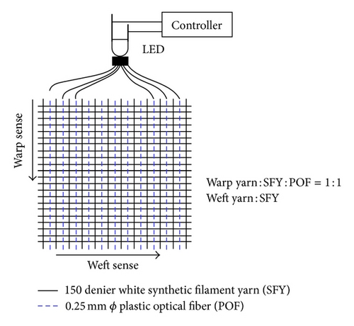
There are two layers in this biofeedback fabric including pattern layer and background layer. The background layer is woven with the warp and the weft densities of 36 ends per inch by 150 denier white synthetic filament yarn (SFY) and plastic optical fiber (POF) of 0.25 mm diameter. By sewing the Plastic Optical Fibers (POFs) of 1.5 mm diameter on the surface of the background layer in a form of sine wave with various wavelengths as the pattern layer, the light can be guided into one end of the POFs and then emit out of the lateral of the POFs. The emitting light can stimulate a change of the brain wave by the reaction of man’s visual system. Besides, a light controller was designed for obtaining various flashing frequencies of light. The details of the design for our study were described as follows.
(1) Background Layer. The background layer draft was shown in Figure 1. A plain weave fabric was applied to this background layer. The warp yarn of the background layer is composed of synthetic filament yarn and plastic optical fiber (POF) in ratio of 1 : 1 for color appearance in background layer. The weft yarn is applied with the same SFY as the warp yarn. All of the POFs of the warp yarn were gathered into a strand and connected to a 0.3 watt natural LED light with a flashing frequency adjusted by a light controller.

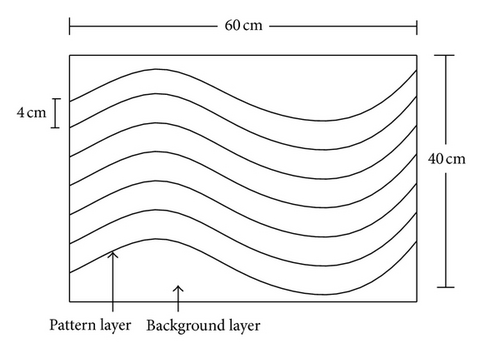
(2) Pattern Layer. The pattern layer draft was shown in Figure 2. For the curvature of pattern layer, plural sine waves with wavelengths (λ) of 60 cm in sample 1 (S1), 30 cm in sample 2 (S2), and 10 cm in sample 3 (S3) in a ratio of amplitude to wavelength 1 : 2, were arranged horizontally with a distance of 4 cm between each line. The plastic optical fibers of 1.5 mm diameter with significant side emitting light characteristic were sewn on the background layer with transparent Polyamide 6 filament yarns of 0.2 mm diameter. All of the POFs of pattern layer were gathered into a strand and connected to a 0.3 watt blue LED light with a flashing frequency adjusted by a light controller.

(3) Light Controller. There are four types of light intensities and were provided including dark, glimpse, moderate, and strong. Besides, three types of the flashing frequencies were provided including 0 HZ (f1), 5 HZ (f2), and 10 HZ (f3). The LED can be connected with the strand of POFs by using a black thermoplastic tube.
2.2. Evaluating Procedure and Method
(1) Biofeedback Apparatus for Measuring Brain Wave. This experiment used PED international Limited Electroencephalogram (EEG) measurement instrument model no: 1-330C-2, J + J engineering as the main equipment to measure and collect data. This instrument is equipped with Physiolab USE3’S Software for data storing and data analyzing.
- (1)
B1 is Alpha wave ranged from 8 to 14 HZ;
- (2)
B2 is Beta wave ranged from 14 to 40 HZ;
- (3)
B3 is Theta wave ranged from 4 to 8 HZ;
- (4)
B4 is Delta wave ranged from 0.4 to 4 HZ.
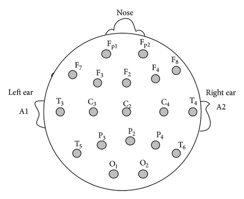
The brain wave electrodes are located in F8 as positive, A2 as negative behind the right ear, and A1 as ground behind the left ear as shown in Figure 3. The biofeedback reaction will go into the encoder unit via the above said EEG measurement instrument. The brain wave reaction of the participant will simultaneously indicate on the computer screen.

(2) Sleeping Scale. We widely collected 130 adjectives related to sleeping sense and relaxation from the literature. The 21 preferable adjectives were selected by using KJ (Kawakita Jiro) method which is a grouping method performed by 6 members. Finally, 7 adjectives were chosen including Relaxed (Q1), Soft (Q2), Comfortable (Q3), Stretchable (Q4), Light (Q5), Free (Q6), and Sleepy (Q7); those 21 adjectives by using SDT (Semantic Differential Technique) of 5 scales performed by 30 subjects. After transferring these 7 adjectives into 7 questions and a sleeping scale of SDT with 11 scales (0 to 10), they were utilized to evaluate the sleeping sense in this study. For further data analysis, the 6 questions of Q1 to Q6 can be grouped together standing for Relaxed as Q1–Q6 expressed in Table 9 to Table 12.
- (1)
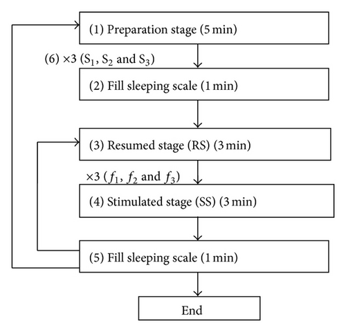
5 minutes before the evaluation is conducted, the participants have to be informed of the purpose and the procedure in preparation stage and the samples should be prepared as showed in Figure 5.
- (2)
For the comparison of sleeping sense after the stimulated stage, filling sleeping scale is conducted in 1 minute before starting the resumed stage.
- (3)
Closing the eyes and taking a rest for 3 minutes is necessary in resumed stage (RS), it can assure that the participant’s condition has recovered from the former evaluated stage (SS).
- (4)
In the stimulated stage (SS), the participants open their eyes and look at the biofeedback fabric for 3 minutes. During this period of time, the participants’ visual system will be stimulated and their brain waves was changed and then recorded at every 10-seconds interval.
- (5)
Filling sleeping scale in 1 minute after each stimulated stage for the comparison of sleeping sense and repeating (3) to (5) steps for 3 types of the flashing frequencies (f1, f2, and f3) of the biofeedback fabric are done, and then the experiment is completed for every single sample.
- (6)
Repeating (2) to (5) steps for all the 3 samples of the different curvatures (S1, S2, and S3) of biofeedback fabric are done, and then the experiment is completed for single subject.


(4) Evaluating Condition. Before the starting of this experiment, the relevant conditions and factors which may affect the result of the experiment should be kept constantly and remained throughout the whole experiment. The biofeedback fabric is placed at 100 cm above the ground level and the distance between the biofeedback fabric and participant is 200 cm. The participant sits straight at eyes level with the objective board in horizontal level as shown in Figure 5. The light strength of the evaluating room is kept under 0.001 lx. The participant should be motionless, keeping silence and quiet in the evaluating room in order not to be disturbed. A total of 30 participants are divided into 3 groups by age in this experiment. The age of the first group (Age 1) is 18 to 29, the second group (Age 2) is 30 to 49, and the third group (Age 3) is 50 to 65. Each group consists of 10 participants; male and female are in the same quantity.
3. Results and Discussions
The EEG instrument was applied to measure the brain wave’s strength at different flashing frequencies and to evaluate the biological state of the participants at resumed stage and stimulated stage. The measuring data obtained will be further analyzed and compared based on different stimulation condition, participant’s age, and gender. Therefore, all the participants’ brain waves need to be stabilized at resumed stage of phases of f1, f2, and f3 for the comparison of stimulated stage. The participants’ brain waves at resumed stage of phases of f1, f2, and f3 for sample 1, sample 2, and sample 3 were shown in Table 1. The P values of the differences among all the phases of f1, f2, and f3 for all samples and all brain waves (B1, B2, B3, and B4) are all larger than 0.05; that is, there are no significant differences of their brain wave activities in all phases according to ANOVA test. We can confirm that the brain wave after 3 minutes rest of closing eyes can be stabilized and be able to resume the original status before starting the measurement of stimulated stage.
| Sample | Brain wave | f1 | f2 | f3 | F | P | |
|---|---|---|---|---|---|---|---|
| Sample 1 | B1 | M | 4.99 | 5.19 | 4.76 | .201 | .818 |
| Alpha | SD | 2.20 | 2.73 | 2.94 | |||
| B2 | M | 3.90 | 4.14 | 3.89 | .310 | .734 | |
| Beta | SD | 1.36 | 1.31 | 1.46 | |||
| B3 | M | 3.52 | 3.69 | 3.29 | .498 | .610 | |
| Theta | SD | 1.42 | 1.64 | 1.64 | |||
| B4 | M | 6.36 | 7.37 | 6.21 | .824 | .442 | |
| Delta | SD | 3.50 | 4.23 | 3.69 | |||
| Sample 2 | B1 | M | 6.00 | 6.09 | 5.93 | .035 | .966 |
| Alpha | SD | 2.32 | 2.28 | 2.33 | |||
| B2 | M | 4.13 | 4.23 | 4.38 | .458 | .634 | |
| Beta | SD | 0.86 | 1.01 | 1.15 | |||
| B3 | M | 4.08 | 4.30 | 4.16 | .303 | .739 | |
| Theta | SD | 1.04 | 1.16 | 1.15 | |||
| B4 | M | 4.27 | 8.61 | 7.88 | .820 | .444 | |
| Delta | SD | 3.19 | 4.98 | 4.27 | |||
| Sample 3 | B1 | M | 5.93 | 5.89 | 5.65 | .128 | .880 |
| Alpha | SD | 2.26 | 2.31 | 2.28 | |||
| B2 | M | 4.52 | 4.39 | 4.27 | .395 | .675 | |
| Beta | SD | 1.02 | 1.10 | 1.08 | |||
| B3 | M | 4.29 | 4.30 | 4.49 | .165 | .848 | |
| Theta | SD | 1.01 | 1.14 | 2.06 | |||
| B4 | M | 6.89 | 7.76 | 7.79 | .931 | .398 | |
| Delta | SD | 2.62 | 3.50 | 2.51 | |||
(1) Analysis of Brain Wave at Different Stimulated Stage. The brain wave differences between the resumed stage (RS) and the stimulated stage (SS) in phases of f1, f2, and f3 were analyzed according to the t-test. The results were listed in Table 2 and described as follows. For all the phases of f1, f2, and f3, Alpha waves (B1) are decreased significantly and transferred into Theta wave (B3) and Delta wave (B4) standing for the slights sleeping status. The less the brain wave’s frequency (e.g., B4 < B3), the more significant increasing is the brain wave’s strength (e.g., B4 > B3); that is, the reaction of the brain wave can be changed when the participants was stimulated by the light out of the optical fiber in biofeedback fabric, as well as the brain wave strength is more obviously and easily enhanced for the lower frequency of brain wave. The results demonstrated that the brain wave can be changed and reached to a status of slight sleeping (B4 ands B3) from relaxation status (B1). As to the high frequency of the Beta wave (B2), standing for the exciting and sobering status, there is no significant difference between the resumed stage and the stimulated stage because the participants’ original status is relaxant. The further analyses of brain waves at the stimulated stage in different flashing frequencies were listed in Table 3. For all the brain waves of B1, B2, B3, and B4, there are no significant differences among the 3 different flashing frequencies according to ANOVA test. The results showed that the biofeedback fabrics in conditions of 3 kinds of flashing frequencies are all effective.
| Sample | f1 | M | SD | t | P | f2 | M | SD | t | P | f3 | M | SD | t | P | |||
|---|---|---|---|---|---|---|---|---|---|---|---|---|---|---|---|---|---|---|
| Sample 1 | B1 | RS | 4.99 | 2.80 | 2.637 | .110 | B1 | RS | 5.19 | 2.73 | 2.853 | .007 | B1 | RS | 4.76 | 2.94 | 2.175 | .035 |
| SS | 3.68 | 1.59 | SS | 3.56 | 1.50 | SS | 3.41 | 1.71 | ||||||||||
| B2 | RS | 3.90 | 1.36 | −3.37 | .737 | B2 | RS | 4.14 | 1.31 | .520 | .605 | B2 | RS | 3.98 | 1.45 | −4.520 | .653 | |
| SS | 4.03 | 1.52 | SS | 3.95 | 1.41 | SS | 4.06 | 1.47 | ||||||||||
| B3 | RS | 3.52 | 1.42 | −3.189 | .003 | B3 | RS | 3.69 | 1.64 | −2.502 | .015 | B3 | RS | 3.29 | 1.64 | −2.765 | .008 | |
| SS | 5.30 | 2.69 | SS | 4.92 | 2.13 | SS | 4.67 | 2.18 | ||||||||||
| B4 | RS | 6.36 | 3.50 | −4.664 | .000 | B4 | RS | 7.37 | 4.23 | −3.830 | .000 | B4 | RS | 6.21 | 3.69 | −3.993 | .000 | |
| SS | 14.11 | 8.40 | SS | 12.68 | 6.30 | SS | 11.58 | 6.37 | ||||||||||
| Sample 2 | B1 | RS | 6.00 | 2.32 | 4.039 | .000 | B1 | RS | 6.09 | 2.28 | 4.651 | .000 | B1 | RS | 5.93 | 2.33 | 4.372 | .000 |
| SS | 4.12 | 1.06 | SS | 3.97 | .99 | SS | 3.91 | .97 | ||||||||||
| B2 | RS | 4.13 | .86 | −1.241 | .219 | B2 | RS | 4.23 | 1.01 | −.062 | .951 | B2 | RS | 4.38 | 1.15 | .377 | .708 | |
| SS | 4.47 | 1.23 | SS | 4.25 | 1.02 | SS | 4.26 | 1.26 | ||||||||||
| B3 | RS | 4.08 | 1.04 | −3.498 | .001 | B3 | RS | 4.30 | 1.16 | −2.765 | .008 | B3 | RS | 4.16 | 1.14 | −3.793 | .000 | |
| SS | 5.64 | 2.20 | SS | 5.47 | 1.99 | SS | 5.79 | 2.05 | ||||||||||
| B4 | RS | 7.21 | 3.19 | −5.369 | .000 | B4 | RS | 8.61 | 4.98 | −3.754 | .000 | B4 | RS | 7.88 | 4.27 | −4.814 | .000 | |
| SS | 15.01 | 7.28 | SS | 14.45 | 6.91 | SS | 14.80 | 6.62 | ||||||||||
| Sample 3 | B1 | RS | 5.93 | 2.26 | .429 | .669 | B1 | RS | 5.89 | 2.31 | 4.140 | .000 | B1 | RS | 5.65 | 2.28 | 3.789 | .000 |
| SS | 5.33 | 2.30 | SS | 3.95 | 1.09 | SS | 3.82 | 1.34 | ||||||||||
| B2 | RS | 4.52 | 1.02 | .830 | .410 | B2 | RS | 4.39 | 1.10 | .135 | .893 | B2 | RS | 4.27 | 1.08 | .085 | .932 | |
| SS | 4.29 | 1.08 | SS | 4.35 | 1.16 | SS | 4.25 | 1.26 | ||||||||||
| B3 | RS | 4.29 | 1.01 | −3.208 | .003 | B3 | RS | 4.30 | 1.14 | −2.847 | .007 | B3 | RS | 4.49 | 2.06 | −1.481 | .144 | |
| SS | 5.65 | 2.07 | SS | 5.64 | 2.31 | SS | 5.33 | 2.34 | ||||||||||
| B4 | RS | 6.89 | 2.62 | −4.897 | .000 | B4 | RS | 7.76 | 3.50 | −4.228 | .000 | B4 | RS | 7.79 | 2.51 | −3.728 | .001 | |
| SS | 14.87 | 8.26 | SS | 14.69 | 8.26 | SS | 12.77 | 6.86 | ||||||||||
| Sample | Brain wave | f1 | f2 | f3 | F | P | |
|---|---|---|---|---|---|---|---|
| Sample 1 | B1 | M | 3.68 | 3.56 | 3.41 | .224 | .799 |
| Alpha | SD | 1.59 | 1.50 | 1.71 | |||
| B2 | M | 4.03 | 3.96 | 4.06 | .041 | .960 | |
| Beta | SD | 1.52 | 1.41 | 1.47 | |||
| B3 | M | 5.30 | 4.92 | 4.67 | .537 | .587 | |
| Theta | SD | 2.69 | 2.13 | 2.18 | |||
| B4 | M | 14.11 | 12.68 | 11.58 | .962 | .386 | |
| Delta | SD | 8.40 | 6.30 | 6.37 | |||
| Sample 2 | B1 | M | 4.12 | 3.97 | 3.91 | .316 | .730 |
| Alpha | SD | 1.06 | .99 | .97 | |||
| B2 | M | 4.47 | 4.25 | 4.26 | .333 | .717 | |
| Beta | SD | 1.23 | 1.02 | 1.26 | |||
| B3 | M | 5.64 | 5.47 | 5.79 | .178 | .838 | |
| Theta | SD | 2.20 | 1.99 | 2.05 | |||
| B4 | M | 15.04 | 14.45 | 14.80 | .050 | .951 | |
| Delta | SD | 7.28 | 6.91 | 6.62 | |||
| Sample 3 | B1 | M | 5.33 | 3.95 | 3.82 | 1.117 | .332 |
| Alpha | SD | 1.30 | 1.09 | 1.34 | |||
| B2 | M | 4.29 | 4.35 | 4.25 | .058 | .944 | |
| Beta | SD | 1.08 | 1.16 | 1.26 | |||
| B3 | M | 5.65 | 5.64 | 5.33 | .191 | .827 | |
| Theta | SD | 2.07 | 2.31 | 2.35 | |||
| B4 | M | 14.87 | 14.69 | 12.77 | .653 | .523 | |
| Delta | SD | 8.53 | 8.26 | 6.86 | |||
(2) Analysis of Brain Wave for Male and Female at Different Stimulated Stage. The analyses of brain wave strength for female and male at different flashing frequencies were listed in Tables 4 and 5. For female brain waves at different flashing frequencies, their Alpha waves showed slight decreasing and their Theta wave and Delta wave showed significant increasing. This result demonstrated that the biofeedback fabrics in conditions of different flashing frequencies are all effective for female. As to male, the result was the same with female. The biofeedback fabrics in conditions of 3 kinds of flashing frequencies are all effective to male. Comparing female with male, the effect in enhancing sleeping sense for female is more significant than it for male because the changes of Alpha wave decreasing and Theta/Delta wave increasing for female are more significant than it for male; that is, the female’s brain wave is easier to be changed than it for male’s brain wave.
| f1 | M | SD | t | P | f2 | M | SD | t | P | f3 | M | SD | t | P | |||
|---|---|---|---|---|---|---|---|---|---|---|---|---|---|---|---|---|---|
| B1 | RS | 4.841 | 1.899 | 2.544 | .017 | B1 | RS | 5.091 | 2.754 | 2.342 | .031 | B1 | RS | 4.773 | 2.601 | 1.944 | .062 |
| SS | 3.320 | 1.324 | SS | 3.293 | 1.119 | SS | 3.273 | 1.464 | |||||||||
| B2 | RS | 4.075 | 1.597 | −0.370 | .970 | B2 | RS | 4.028 | 1.424 | −.033 | .974 | B2 | RS | 3.998 | 1.332 | −4.290 | .671 |
| SS | 4.098 | 1.812 | SS | 4.046 | 1.582 | SS | 4.224 | 1.547 | |||||||||
| B3 | RS | 3.313 | 1.461 | −2.654 | .013 | B3 | RS | 3.390 | 1.614 | −2.485 | .019 | B3 | RS | 3.311 | 1.542 | −2.488 | .019 |
| SS | 5.373 | 2.628 | SS | 4.946 | 1.811 | SS | 4.998 | 2.127 | |||||||||
| B4 | RS | 6.481 | 4.502 | −3.489 | .002 | B4 | RS | 6.927 | 4.667 | −3.282 | .003 | B4 | RS | 6.183 | 3.704 | −3.690 | .001 |
| SS | 15.095 | 8.436 | SS | 12.790 | 5.106 | SS | 12.981 | 6.098 | |||||||||
| f1 | M | SD | t | P | f2 | M | SD | t | P | f3 | M | SD | t | P | |||
|---|---|---|---|---|---|---|---|---|---|---|---|---|---|---|---|---|---|
| B1 | RS | 5.155 | 2.538 | 1.372 | .181 | B1 | RS | 5.296 | 2.806 | 1.685 | .103 | B1 | RS | 4.750 | 3.333 | 1.202 | .242 |
| SS | 4.055 | 1.791 | SS | 3.844 | 1.809 | SS | 3.549 | 1.970 | |||||||||
| B2 | RS | 3.737 | 1.115 | −.532 | .599 | B2 | RS | 4.258 | 1.227 | .840 | .408 | B2 | RS | 3.791 | 1.618 | −.208 | .837 |
| SS | 3.965 | 1.236 | SS | 3.873 | 1.280 | SS | 3.907 | 1.430 | |||||||||
| B3 | RS | 3.735 | 1.412 | −1.816 | .084 | B3 | RS | 4.001 | 1.673 | −1.172 | .251 | B3 | RS | 3.271 | 1.801 | −1.440 | .161 |
| SS | 5.227 | 2.851 | SS | 4.908 | 2.485 | SS | 4.350 | 2.275 | |||||||||
| B4 | RS | 6.238 | 2.267 | −3.019 | .008 | B4 | RS | 7.822 | 3.853 | −2.184 | .040 | B4 | RS | 6.237 | 3.823 | −2.018 | .053 |
| SS | 13.133 | 8.550 | SS | 12.579 | 7.502 | SS | 10.180 | 6.532 | |||||||||
(3) Analysis of Brain Wave for Age 1, Age 2, and Age 3 at Different Stimulated Stage. The analyses of brain waves strength for groups of Age 1, Age 2, and Age 3 at different flashing frequencies were listed in Tables 6, 7, and 8. For the Age 1 (18 to 29) group, the Alpha waves were decreased and the Theta/Delta waves were increased, but all of them were not significant. For the Age 2 (30 to 49) group, the Alpha waves were decreased, however they were not significant, but the Theta and Delta waves were significantly increased. For the Age 3 (50 to 65) group, the Alpha waves were decreased and the Theta waves were increased; however they were not significant, but the Delta waves were increased and reached a significant level. According to the aforementioned results, the effect in enhancing sleeping sense for Age 2 group is the best among all the groups of Age, and Age 1 group ranks behind Age 3 group. Maybe the stress of the Age 2 group is the strongest among all the groups of Age and the stress of the Age 3 group is stronger than Age 1 group. Therefore, we can infer from the results that the stronger the participant’s stress is, the more obvious the effect in enhancing sleeping sense is.
| f1 | M | SD | t | P | f2 | M | SD | t | P | f3 | M | SD | t | P | |||
|---|---|---|---|---|---|---|---|---|---|---|---|---|---|---|---|---|---|
| B1 | RS | 5.255 | 2.035 | 1.876 | .078 | B1 | RS | 5.023 | 2.371 | 1.886 | .076 | B1 | RS | 4.914 | 2.610 | 1.621 | .122 |
| SS | 3.616 | 1.342 | SS | 3.430 | 1.230 | SS | 3.364 | 1.525 | |||||||||
| B2 | RS | 3.897 | 1.441 | .065 | .949 | B2 | RS | 3.735 | 1.279 | −.215 | .832 | B2 | RS | 3.506 | 1.244 | −.966 | .347 |
| SS | 3.851 | 1.719 | SS | 3.878 | 1.671 | SS | 4.113 | 1.550 | |||||||||
| B3 | RS | 3.752 | 1.474 | −1.187 | .251 | B3 | RS | 3.690 | 1.509 | −.786 | .442 | B3 | RS | 3.585 | 1.677 | −.810 | .428 |
| SS | 4.798 | 2.365 | SS | 4.259 | 1.723 | SS | 4.272 | 2.092 | |||||||||
| B4 | RS | 8.142 | 4.585 | −1.641 | .118 | B4 | RS | 8.795 | 4.816 | −1.011 | .326 | B4 | RS | 7.349 | 4.204 | −1.463 | .161 |
| SS | 13.215 | 8.631 | SS | 11.002 | 4.950 | SS | 10.763 | 6.062 | |||||||||
| f1 | M | SD | t | P | f2 | M | SD | t | P | f3 | M | SD | t | P | |||
|---|---|---|---|---|---|---|---|---|---|---|---|---|---|---|---|---|---|
| B1 | RS | 4.946 | 2.749 | .829 | .418 | B1 | RS | 5.185 | 3.213 | 1.139 | .269 | B1 | RS | 4.975 | 3.178 | 1.141 | .269 |
| SS | 4.009 | 2.878 | SS | 3.791 | 2.156 | SS | 3.597 | 2.117 | |||||||||
| B2 | RS | 3.597 | 1.340 | −.659 | .518 | B2 | RS | 4.100 | 1.502 | .219 | .829 | B2 | RS | 4.121 | 1.634 | .357 | .725 |
| SS | 4.020 | 1.523 | SS | 3.949 | 1.587 | SS | 3.861 | 1.623 | |||||||||
| B3 | RS | 3.424 | 1.668 | −2.434 | .026 | B3 | RS | 3.484 | 1.798 | −2.356 | .030 | B3 | RS | 3.324 | 1.643 | −2.319 | .032 |
| SS | 6.280 | 3.315 | SS | 5.772 | 2.489 | SS | 5.448 | 2.386 | |||||||||
| B4 | RS | 5.703 | 3.068 | −3.678 | .006 | B4 | RS | 6.777 | 3.960 | −3.192 | .005 | B4 | RS | 6.595 | 3.466 | −3.128 | .006 |
| SS | 16.160 | 9.302 | SS | 15.007 | 7.128 | SS | 13.945 | 6.574 | |||||||||
| f1 | M | SD | t | P | f2 | M | SD | t | P | f3 | M | SD | t | P | |||
|---|---|---|---|---|---|---|---|---|---|---|---|---|---|---|---|---|---|
| B1 | RS | 4.793 | 1.974 | 1.949 | .067 | B1 | RS | 5.373 | 2.840 | 1.978 | .063 | B1 | RS | 4.395 | 3.272 | .976 | .342 |
| SS | 3.437 | .972 | SS | 3.485 | 1.023 | SS | 3.271 | 1.603 | |||||||||
| B2 | RS | 4.223 | 1.382 | −.002 | .999 | B2 | RS | 4.594 | 1.112 | 1.113 | .281 | B2 | RS | 4.057 | 1.547 | −.254 | .802 |
| SS | 4.224 | 1.464 | SS | 4.052 | 1.066 | SS | 4.223 | 1.370 | |||||||||
| B3 | RS | 3.396 | 1.237 | −1.735 | .100 | B3 | RS | 3.913 | 1.761 | −.978 | .341 | B3 | RS | 2.964 | 1.738 | −1.556 | .137 |
| SS | 4.821 | 2.284 | SS | 4.750 | 2.054 | SS | 4.302 | 2.091 | |||||||||
| B4 | RS | 5.234 | 1.929 | −3.072 | .012 | B4 | RS | 6.552 | 3.922 | −2.269 | .036 | B4 | RS | 4.685 | 3.195 | −2.359 | .034 |
| SS | 12.961 | 7.717 | SS | 12.044 | 6.575 | SS | 10.033 | 6.417 | |||||||||
| Sample 1 | M | SD | P | Sample 2 | M | SD | P | Sample 3 | M | SD | P | |||
|---|---|---|---|---|---|---|---|---|---|---|---|---|---|---|
| f0 | 7.32 | 1.59 | 0.381 | f0 | 7.25 | 1.60 | 0.467 | f0 | 7.34 | 1.21 | 0.241 | |||
| f1 | 6.91 | 1.95 | f1 | 6.90 | 2.07 | f1 | 7.41 | 1.35 | ||||||
| Q1–Q6 | f0 | 7.32 | 1.59 | 0.023 | Q1–Q6 | f0 | 7.25 | 1.60 | 0.034 | Q1–Q6 | f0 | 7.34 | 1.21 | 0.018 |
| f2 | 6.16 | 2.19 | f2 | 6.20 | 2.11 | f2 | 6.31 | 1.99 | ||||||
| f0 | 7.32 | 1.59 | 0.000 | f0 | 7.25 | 1.60 | 0.036 | f0 | 7.34 | 1.21 | 0.049 | |||
| f3 | 7.88 | 1.69 | f3 | 8.09 | 1.42 | f3 | 7.82 | 1.90 | ||||||
| f0 | 0.40 | 0.86 | 0.000 | f0 | 0.47 | 1.01 | 0.000 | f0 | 0.27 | 0.64 | 0.000 | |||
| f1 | 2.87 | 1.55 | f1 | 3.20 | 1.73 | f1 | 2.63 | 1.22 | ||||||
| Q7 | f0 | 0.40 | 0.86 | 0.000 | Q7 | f0 | 0.47 | 1.01 | 0.000 | Q7 | f0 | 0.27 | 0.64 | 0.000 |
| f2 | 5.10 | 2.50 | f2 | 5.70 | 2.23 | f2 | 4.97 | 1.97 | ||||||
| f0 | 0.40 | 0.86 | 0.000 | f0 | 0.47 | 1.01 | 0.000 | f0 | 0.27 | 0.64 | 0.000 | |||
| f3 | 5.30 | 2.04 | f3 | 6.30 | 2.39 | f3 | 6.20 | 2.04 | ||||||
(4) Analysis of Sleeping Scale at Different Stimulated Stages for Different Samples. The sleeping scale was applied to investigate the enhancement effect of sleeping sense for the participants at different flashing frequencies for 3 samples. The evaluating data, that is, the sleeping scale score, obtained were further analyzed based on different sample stimulation, participant’s age, and gender. The results were elaborated as below. The score differences of sleeping scale between f0 after the resumed stage (RS) and after the stimulated stage (SS) in phases of f1, f2, and f3 for sample 1, sample 2, and sample 3 were analyzed according to the t-test. The results were listed in Table 9. For all the samples in phases of f1, f2, and f3, the differences of Q7 standing for sleeping sense between (f0) after RS and (f1, f2, f3) after SS were extremely significantly increased. Their P values were all reaching 0.000. The differences of Q1–Q6 standing for relaxed feel for f3 (10 Hz) were increased significantly. Their P values were less than 0.05. Adversely, the differences of Q1–Q6 for f2 (5 Hz) were decreased significantly. Besides, the difference was not significant while comparing f0 with f1 (0 Hz). The results demonstrated that the sleeping sense of the participants can be enhanced when their visual system were stimulated by all samples in frequencies of f1, f2, and f3. However, the relaxed feel of the participants can be enhanced while stimulating only in frequency of f3 for all samples.
(5) Analysis of Sleeping Scale for Male And Female at Different Stimulated Stages for Different Samples. The score differences of sleeping scale for male and female between f0 after the resumed stage (RS) and after the stimulated stage (SS) in phases of f1, f2, and f3 for sample 1, sample 2, and sample 3 were analyzed and the results were listed in Tables 10 and 11. For all the samples in phases of f1, f2, and f3, the differences for male and female of Q7 standing for sleeping sense between f0 (after RS) and f1, f2, and f3 (after SS) were extremely significantly increased. Their P values were all reaching 0.000. Adversely, the differences of Q1–Q6 for male and female were not significant while comparing f0 with f1, f2, and f3. The results demonstrated that the sleeping sense of the participants for male and female can be enhanced extremely significantly when their visual system were stimulated for all samples in the frequencies of f1, f2, and f3. However, the relaxed feel of the participants cannot be enhanced significantly while stimulating in the frequencies of f1, f2, and f3 for all samples. Besides, the higher the flashing frequency, the better the effect in enhancing sleeping sense for both genders of participants. The enhancement effect in sleeping sense for female is better than it for male.
| Sample 1 | M | SD | P | Sample 2 | M | SD | P | Sample 3 | M | SD | P | |||
|---|---|---|---|---|---|---|---|---|---|---|---|---|---|---|
| f0 | 7.69 | 1.17 | 0.575 | f0 | 7.73 | 1.41 | 0.436 | f0 | 7.63 | 1.01 | 0.648 | |||
| f1 | 7.42 | 1.39 | f1 | 7.23 | 2.00 | f1 | 7.43 | 1.34 | ||||||
| Q1–Q6 | f0 | 7.69 | 1.17 | 0.284 | Q1–Q6 | f0 | 7.73 | 1.41 | 0.068 | Q1–Q6 | f0 | 7.63 | 1.01 | 0.094 |
| f2 | 7.09 | 1.77 | f2 | 6.58 | 1.89 | f2 | 6.91 | 1.26 | ||||||
| f0 | 7.69 | 1.17 | 0.169 | f0 | 7.73 | 1.41 | 0.408 | f0 | 7.63 | 1.01 | 0.322 | |||
| f3 | 8.32 | 1.28 | f3 | 8.18 | 1.49 | f3 | 8.09 | 1.43 | ||||||
| f0 | 0.53 | 1.06 | 0.000 | f0 | 0.60 | 1.30 | 0.000 | f0 | 0.27 | 0.80 | 0.000 | |||
| f1 | 3.33 | 1.80 | f1 | 3.87 | 2.00 | f1 | 3.07 | 1.33 | ||||||
| Q7 | f0 | 0.53 | 1.06 | 0.000 | Q7 | f0 | 0.60 | 1.30 | 0.000 | Q7 | f0 | 0.27 | 0.80 | 0.000 |
| f2 | 5.67 | 2.32 | f2 | 6.87 | 1.92 | f2 | 5.60 | 2.16 | ||||||
| f0 | 0.53 | 1.06 | 0.000 | f0 | 0.60 | 1.30 | 0.000 | f0 | 0.27 | 0.80 | 0.000 | |||
| f3 | 5.87 | 2.00 | f3 | 7.47 | 2.00 | f3 | 7.13 | 2.03 | ||||||
| Sample 1 | M | SD | P | Sample 2 | M | SD | P | Sample 3 | M | SD | P | |||
|---|---|---|---|---|---|---|---|---|---|---|---|---|---|---|
| f0 | 6.94 | 1.88 | 0.487 | f0 | 6.77 | 1.68 | 0.779 | f0 | 7.06 | 1.35 | 0.514 | |||
| f1 | 6.40 | 2.32 | f1 | 6.57 | 2.16 | f1 | 7.39 | 1.42 | ||||||
| Q1–Q6 | f0 | 6.94 | 1.88 | 0.081 | Q1–Q6 | f0 | 6.77 | 1.68 | 0.211 | Q1–Q6 | f0 | 7.06 | 1.35 | 0.068 |
| f2 | 5.43 | 2.22 | f2 | 5.82 | 2.31 | f2 | 5.79 | 2.42 | ||||||
| f0 | 6.94 | 1.88 | 0.482 | f0 | 6.77 | 1.68 | 0.138 | f0 | 7.06 | 1.35 | 0.472 | |||
| f3 | 7.44 | 1.96 | f3 | 7.80 | 1.90 | f3 | 7.56 | 2.29 | ||||||
| f0 | 0.27 | 0.59 | 0.000 | f0 | 0.33 | 0.62 | 0.000 | f0 | 0.27 | 0.46 | 0.000 | |||
| f1 | 2.40 | 1.12 | f1 | 2.53 | 1.13 | f1 | 2.20 | 0.94 | ||||||
| Q7 | f0 | 0.27 | 0.59 | 0.000 | Q7 | f0 | 0.33 | 0.62 | 0.000 | Q7 | f0 | 0.27 | 0.46 | 0.000 |
| f2 | 4.53 | 2.61 | f2 | 4.53 | 1.92 | f2 | 4.33 | 1.59 | ||||||
| f0 | 0.27 | 0.59 | 0.000 | f0 | 0.33 | 0.62 | 0.000 | f0 | 0.27 | 0.46 | 0.000 | |||
| f3 | 4.73 | 1.98 | f3 | 5.13 | 2.23 | f3 | 5.27 | 1.62 | ||||||
| Age 1 | M | SD | P | Age 2 | M | SD | P | Age 3 | M | SD | P | |||
|---|---|---|---|---|---|---|---|---|---|---|---|---|---|---|
| f0 | 7.51 | 1.12 | 0.628 | f0 | 6.89 | 1.28 | 0.678 | f0 | 7.51 | 1.84 | 0.490 | |||
| f1 | 7.35 | 1.42 | f1 | 6.72 | 1.77 | f1 | 7.15 | 2.17 | ||||||
| Q1–Q6 | f0 | 7.51 | 1.12 | 0.009 | Q1–Q6 | f0 | 6.89 | 1.28 | 0.011 | Q1–Q6 | f0 | 7.51 | 1.84 | 0.052 |
| f2 | 6.60 | 1.46 | f2 | 5.62 | 2.29 | f2 | 6.45 | 2.29 | ||||||
| f0 | 7.51 | 1.12 | 0.299 | f0 | 6.89 | 1.28 | 0.136 | f0 | 7.51 | 1.84 | 0.045 | |||
| f3 | 7.81 | 1.10 | f3 | 7.58 | 2.14 | f3 | 8.41 | 1.53 | ||||||
| f0 | 0.30 | 0.47 | 0.000 | f0 | 0.70 | 1.29 | 0.000 | f0 | 0.13 | 0.35 | 0.000 | |||
| f1 | 2.27 | 1.14 | f1 | 3.97 | 1.71 | f1 | 2.47 | 1.01 | ||||||
| Q7 | f0 | 0.30 | 0.47 | 0.000 | Q7 | f0 | 0.70 | 1.29 | 0.000 | Q7 | f0 | 0.13 | 0.35 | 0.000 |
| f2 | 5.03 | 1.97 | f2 | 6.07 | 2.56 | f2 | 4.67 | 1.97 | ||||||
| f0 | 0.30 | 0.47 | 0.000 | f0 | 0.70 | 1.29 | 0.000 | f0 | 0.13 | 0.35 | 0.000 | |||
| f3 | 5.40 | 1.40 | f3 | 6.83 | 2.60 | f3 | 5.57 | 2.16 | ||||||
(6) Analysis of Sleeping Scale for Age Groups at Different Stimulated Stages. The score differences of sleeping scale between f0 (after RS) and f1, f2, and f3 (after SS) for 3 groups of Age 1, Age 2, and Age 3 were analyzed and their results were listed in Table 12. For the sample 1 in phases of f1, f2, and f3, the differences of Q7 standing for sleeping sense between f0 (after RS) and f1, f2, and f3 (after SS) were extremely significantly increased. Their P values were all reaching 0.000. Adversely, the differences for all Age groups of Q1–Q6 were no significant differences while comparing f0 with f1, f2, and f3. The results demonstrated that the sleeping sense of the participants for all Age groups can be enhanced extremely when their visual system were stimulated by sample 1 in frequencies of f1, f2, and f3. However, the relaxed feel of the participants of all Age groups cannot be enhanced significantly while stimulating in frequencies of f1, f2, and f3 for sample 1. Besides, the higher the flashing frequency, the better the enhancing sleeping sense for 3 Age groups of participants. The effect of enhancing sleeping sense for Age 2 group (30~49) is the best among 3 Age groups. Maybe the stress of Age 2 group is the strongest among all the groups of Age. Therefore, we can infer from the results that the stronger the participant’s stress is, the more obvious the effect in enhancing sleeping sense is, and it is same with the results of above Table 6 to Table 8.
4. Conclusion
- (1)
For all the phases of f1, f2, and f3, Alpha waves (B1) are decreased significantly and transferred into Theta wave (B3) and Delta wave (B4); that is, the biofeedback fabrics in conditions of 3 kinds of flashing frequencies for all samples are all effective in enhancing sleeping sense.
- (2)
The effect in enhancing sleeping sense for female is more significant than it for male.
- (3)
The effect in enhancing sleeping sense for Age 2 group (30–49) is the best among all Age groups.
- (4)
The stronger the participant’s stress is, the more obvious the effect in enhancing sleeping sense is.
Conflict of Interests
The authors declare that there is no conflict of interests regarding the publication of this paper.
Acknowledgments
The authors would like to take this opportunity to thank to Industrial Development Bureau, Ministry of Economic Affairs (MOEA) for the financial support. They also would like to thank the authority of Shu-Te University for providing them with a good environment and facilities to complete this project. Finally, they would like to express their heartfelt thanks to all of the participants for their help in this study.


