Erratum of “Dystrophins, Utrophins, and Associated Scaffolding Complexes: Role in Mammalian Brain and Implications for Therapeutic Strategies”
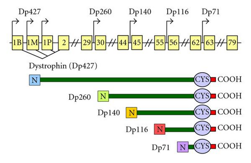
Dystrophin structure is organized in four main domains. The N-terminal domain (246 amino-acids) similar to α-actinin contains several actin-binding sites and a calmodulin-binding site. The central rod domain (2840 aa) has 24 triple-helical spectrinlike repeats thought to give the protein a flexible structure, with several irregular short and long segments, and four proline-rich end regions. The cysteine-rich domain (280 aa) participates in the critical interaction of dystrophin with the transmembraneous DGC component, β-dystroglycan. The COOH-terminus (320 aa) is an α-helical coiled coil region that binds to the cytosolic component dystrobrevin and may also modulate interactions with syntrophins and other DGC-associated proteins.


