Insights into the skin microbiome of sickle cell disease leg ulcers
Julia Byeon, Katherine D. Blizinsky, and Anitra Persaud are co-first authors.
Caterina P. Minniti, Vence L. Bonham, and Elizabeth A. Grice are co-senior authors.
Funding information: National Human Genome Research Institute, Grant/Award Number: ZIAHG200394; National Institute of Arthritis and Musculoskeletal and Skin Diseases, Grant/Award Number: R01AR066663; National Institute of Nursing Research, Grant/Award Number: R01NR015639
Abstract
Leg ulcers are estimated to occur in 1%–10% of North American patients with sickle cell disease (SCD). Their pathophysiology remains poorly defined, but as with other chronic wounds, it is hypothesised that the microbial milieu, or microbiome, contributes to their healing and clinical outcomes. This study utilises 16S ribosomal RNA (rRNA) gene sequencing to describe, for the first time, the microbiome of the SCD leg ulcer and its association with clinical factors. In a cross-sectional analysis of 42 ulcers, we recovered microbial profiles similar to other chronic wounds in the predominance of anaerobic bacteria and opportunistic pathogens including Staphylococcus, Corynebacterium, and Finegoldia. Ulcers separated into two clusters: one defined by predominance of Staphylococcus and smaller surface area, and the other displaying a greater diversity of taxa and larger surface area. We also find that the relative abundance of Porphyromonas is negatively associated with haemoglobin levels, a key clinical severity indicator for SCD, and that Finegoldia relative abundance is negatively associated with CD19+ B cell count. Finally, ratios of Corynebacterium:Lactobacillus and Staphylococcus:Lactobacillus are elevated in the intact skin of individuals with a history of SCD leg ulcers, while the ratio of Lactobacillus:Bacillus is elevated in that of individuals without a history of ulcers. Investigations of the skin microbiome in relation to SCD ulcer pathophysiology can inform clinical guidelines for this poorly understood chronic wound, as well as enhance broader understanding about the role of the skin microbiome in delayed wound healing.
Abbreviations
-
- SCD
-
- sickle cell disease
-
- Faith's PD
-
- Faith's phylogenetic diversity
-
- Hx
-
- history
1 INTRODUCTION
In the United States, chronic wounds impact millions of patients annually and have been recognised as a major public health concern since the early 2000s.1 Over the past two decades, a growing body of literature on their pathophysiology has built an evidence base that informs debridement, antimicrobial, and dressing strategies for a variety of wound types, including diabetic foot ulcers, venous ulcers, pressure injuries, and arterial ulcers.2
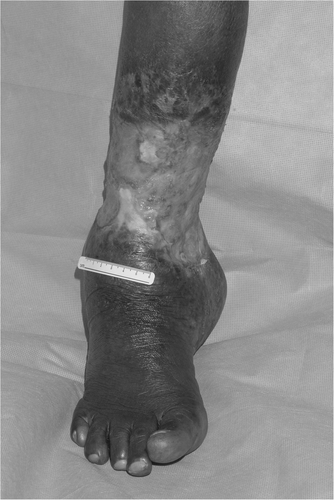
While there is a continuing need to improve and refine treatment guidelines for these most common chronic wounds, the phenotype and pathophysiology of one type of ulcer, the sickle cell disease (SCD) leg ulcer, lacks significant investigation and is poorly understood3 (Figure 1). SCD is a group of genetic blood disorders in which a mutation in the beta-globin gene causes haemoglobin to polymerise in its deoxygenated state, leading red blood cells to lyse or block blood flow in vessels.4 Leg ulcers are estimated to occur in 1%–10% of North American SCD patients, with prevalence in other regions of the world ranging from 11% in Ghana to as high as 43% in Brazil.5-7 These wounds are excruciatingly painful, often recalcitrant and recurring, and may result in perceived social stigma.5, 8 Proposed causes include: venous insufficiency9; increased arteriovenous shunting associated with haemolytic anemia10; impaired endothelial function due to the consumption of nitric oxide by cell-free hemoglobin11; and vessel damage from sickled cells, which allows proteins and inflammatory cells to leak into extravascular space.12 However, these hypotheses remain speculative. Without sufficient knowledge of SCD leg ulcer pathophysiology, treatment strategies vary widely between clinicians and are guided largely by anecdotal experience.6, 13
Treatment of SCD ulcers will benefit from research leveraging a variety of approaches, but investigations of the colonising microbiota may prove especially promising. Wound microbes have been shown to play a role in healing processes by colonising wound beds and overstimulating leukocyte activity, including proteolytic functions and reactive oxygen species production, thereby impairing healing.14 In recent years, research in this area has become even more fruitful through the advent of 16S rRNA gene sequencing, which has allowed researchers to more effectively identify and study specific communities of cutaneous microbes.15-17 With this information, microbial therapies could be refined to promote or prevent colonisation by beneficial and deleterious organisms, respectively. The wound microbiome may also prove a useful diagnostic tool by serving as an accessible source of information about the inflammatory state of a wound.18
Despite their potential to inform treatment, microbial studies of SCD ulcers are scant and dated. Some studies have demonstrated that topical antibacterial therapy can reduce SCD ulcer size.19, 20 In others, SCD ulcers were most often colonised by Staphylococcus aureus,20, 21 a known wound pathogen. Each of these studies has been culture-based, limiting their ability to identify taxa which are difficult to culture in laboratory environments. Culture-independent studies utilising 16S rRNA gene sequencing techniques are non-existent.
This paper contributes to the limited collection of studies on SCD leg ulcers, as well as a growing body of literature on chronic wound microbiomes, by describing the SCD leg ulcer microbiome and its association with clinical outcomes. We use 16S rRNA gene sequencing to characterise the bacterial taxa that colonise the wound bed and surrounding skin, and calculate taxonomic associations with wound severity and other clinical measures salient to SCD. In addition to informing further research on the pathophysiology of SCD ulcers, we establish the SCD leg ulcer as an additional topic of investigation, among diabetic ulcers, venous ulcers, pressure injuries, and other more common chronic wounds, that can provide broader insights into the relationship between the skin microbiome and wound healing.
2 MATERIALS AND METHODS
2.1 Study design and participant characteristics
Individuals 18 years or older with SCD (HbSS, HgSC, HbSB0, or HbSB+) were enrolled as a part of the Insights into Microbiome and Environmental Contributions to Sickle Cell Disease and Leg Ulcers Study (“INSIGHTS Study,” ClinicalTrials.gov Identifier: NCT02156102). Participants were recruited across the United States through convenience sampling at SCD-related community events; ads and flyers disseminated within the SCD community; and referral by physicians and current participants. Participants were seen at the National Institutes of Health, Clinical Center in Bethesda, MD and Montefiore Medical Center in New York, NY. The study protocol (NCT02156102) was approved by National Institutes of Health Institutional Review Board and Montefiore Medical Center (IRB# 2015-5616). Written informed consent was obtained from all participants.
One-hundred and nine participants from the INSIGHTS cohort, with a mean age of 41, and 58 of whom were female (53%), were included in the present study. Thirty-two (29%) participants had one or more active SCD leg ulcers, and 77 (71%) had no active ulcers. Of those 77 without active ulcers, 25 (32%) had a history of one or more healed ulcers, and 52 (67%) had no ulcer history. From 109 participants, 272 samples were collected: 42 (15%) were obtained from the ulcer wound bed, 49 (18%) from periwound skin, and 181 (67%) from intact skin. The distribution of patients and samples are shown in Tables 1 and 2.
| Participants (N = 109) | ||
|---|---|---|
| Genotype, N (%) | HbSS | 90 (83%) |
| HgSC | 11 (10%) | |
| HbSB+ | 7 (6%) | |
| HbSB0 | 1 (1%) | |
| Sex, N (% female) | 58 (53%) | |
| Age, mean (range) | 41 (19–71) | |
| Ulcer status, N (%) | Active ulcer | 32 (29%) |
| Not active, but Hx of ulcer(s) | 25 (23%) | |
| Not active, no Hx of ulcer(s) | 52 (48%) | |
| Samples (N = 272) | ||
|---|---|---|
| Sex, N (% female) | 134 (49%) | |
| Sample Site, N, (%) | Wound | 42 (15%) |
| Periwound | 49 (18%) | |
| Intact | 181 (67%) | |
| Intact skin, active ulcer | 26 (10%) | |
| Intact skin, not active but Hx of ulcer(s) | 53 (19%) | |
| Intact skin, not active and no Hx of ulcer(s) | 102 (38%) | |
2.2 Data collection
Medical history and clinical and physical exams were completed and microbiome samples were collected for each participant at the time of visit. Participants were asked to refrain from showering, using soap, or applying lotion to the lower extremities for 24 hours prior to the clinic visit. Microbiome samples were collected from both non-ulcerated and ulcerated skin tissue. If the participant presented with a non-affected leg(s), non-ulcerated tissue samples were obtained from the medial or lateral malleolus. The swab was pre-moistened and rotated over a 1 cm2 area of skin. For participants with ulcers, non-ulcerated tissue samples were also obtained from the periwound area, defined as non-ulcerated skin 1 cm from the wound edge. For ulcerated tissue, the wound bed was cleaned with sterile 0.9% sodium chloride, the swab was pre-moistened, and the sample was obtained using the Levine technique. Levine's technique is different than other swab techniques in that it samples fluid from the deep tissue layers, similar to aspiration of wound fluid, and has high concordance with tissue biopsies for recovering microbial load and diversity.22, 23 This process was repeated for additional ulcers, if the participant had more than one ulcer. If needed, patients were administered systemic pain medication prior to sampling and topical anesthetics were applied after sample collection. All samples were obtained using Catch-All Sample Collection Swabs (discontinued, Epicentre Biotechnologies #QEC89100) and immediately placed in 2.0 mL Safe-Lock Biopur tubes, Eppendorf; #022600044) tubes that contained 300 μl of Yeast Cell Lysis Solution (Epicentre Biotechnologies; #MPY80200). Samples were stored at −80°C until DNA extraction.
2.3 16S rRNA marker gene sequencing and bioinformatics analysis
DNA was extracted from swab specimens as previously described.15 Amplicons were prepared using Invitrogen AccuPrime High Fidelity Taq kit (ThermoFisher #12346094) for PCR and barcoded primers (Hypervariable regions V1-V3, primers 27F and 534R). The Applied Biosystems SequalPrep kit (ThermoFisher #A1051001) was used for PCR product clean-up and normalisation, and the Qiagen MinElute PCR Purification Kit (Qiagen #28004) for pooled PCR product purification. Amplicon libraries were sequenced using an Illumina MiSeq platform and 2×300 bp chemistry.
Sequencing data were processed and analysed using the Quantitative Insights Into Microbial Ecology 2 (QIIME2) pipeline.24 Divisive Amplicon Denoising Algorithm 2 (DADA2),25 implemented as a QIIME2 plug-in, was used for sequence quality filtering, and taxonomic analysis was done using a Naïve Bayes classifier trained on the Greengenes 13_8 99% operational taxonomic units. The annotation for oxygen tolerance of bacterial taxa was obtained from previous research.26 Alpha diversity was quantified using three metrics: Faith's phylogenetic diversity (Faith's PD, the representation of species present on the phylogenetic tree), richness (the number of species present), and Shannon's diversity index (Shannon's H, the distribution of relative abundance). A rooted phylogenetic tree was generated for calculation of diversity metrics including Faith's PD and UniFrac distances: first a multiple sequence alignment was performed using Multiple Alignment using Fast Fourier Transform27 and high variable positions were masked to reduce noise in a resulting phylogenetic tree. A mid-point rooted tree was then generated using FastTree.28 The Shannon index (H) and richness were calculated using the vegan package version 2.5-6 (https://CRAN.R-project.org/package=vegan) in R (version 3.6.0).
2.4 Statistical analysis
Linear mixed models with patient ID as random effect were used to test the associations between alpha diversity, relative abundances and ratios of taxa, and study groups. Relative abundances and ratios of taxa were log-transformed for these analyses. Fisher's exact test was used for tests of association between categorical variables. A permutational multivariate analysis of variance test as implemented by the function adonis in the vegan package 2.5-5 (https://CRAN.R-project.org/package=vegan) was used to test the null hypothesis of no differences in the study group centroids. For cluster analysis, partitioning around medoids (PAM), implemented by the function pam in the R package cluster (version 2.1.0) (https://www.rdocumentation.org/packages/cluster), was used. Spearman's correlation was calculated between the first principal coordinates analysis (PCoA) axis and Staphylococcus relative abundance. p-Values from multiple comparisons were adjusted to control for the false discovery rate using the Benjamini–Hochberg procedure. All statistical analyses were conducted in R (version 3.6.0).
3 RESULTS
3.1 Abundance of aerobes is inversely proportional to proximity to wound skin
We sought to determine if bacterial community diversity was associated with proximity to the wound in participants with active ulcers. Using linear mixed effects models, we calculated three different diversity metrics, Shannon's H, Richness, and Faith's PD were negatively associated with a shift in sample site from intact to periwound or periwound to intact skin (β = −0.65, padj < 0.001; β = −1.84, padj = 0.04; and β = −24.13, padj < 0.001, respectively, Figure 2A). These findings suggest that the ulcer microbiota is distinct from intact skin, with bacterial community diversity lowest in the ulcer.
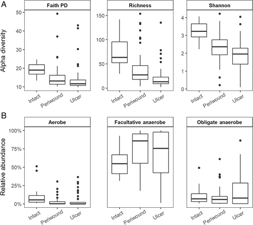
We next assessed whether the drastic reduction in alpha diversity was associated with a change in the fundamental makeup of the bacterial community, in terms of oxygen tolerance. We annotated the bacterial taxa identified in our samples as aerobes, facultative anaerobes, or obligate anaerobes and tested for associations between the relative abundance of these taxa categories and sample site. The relative abundance of aerobic bacteria decreased as the sampling site shifted from intact to periwound, or periwound to wound (β = −2.79, padj < 0.001; Figure 2B). The relative abundance of facultative and obligately anaerobic bacteria did not have a statistically significant association with wound proximity (β = −0.01, padj = 0.92 and β = −0.90, padj = 0.23, respectively, Figure 2B). Thus, the decrease in bacterial diversity observed in the ulcer was accompanied by a decrease in the relative abundance of aerobic bacteria.
3.2 SCD leg ulcer microbiota are heterogeneous, but most often dominated by Staphylococcus
Consistent with our finding that the relative abundance of aerobes decreased with proximity to the wound skin, wound samples were dominated by the presence of facultative and obligate anaerobes (Figure 3A). Of the taxa present, Staphylococcus had the highest mean relative abundance at 52%, of which 80% was identified as S. aureus (Figure 3B; Table S1). This was followed by other facultatively anaerobic bacteria such as Corynebacterium (10%), Streptococcus (4%), Bacillus (2%), and Streptococcaceae, as well as obligately anaerobic bacteria such as Finegoldia (7%), Propionibacterium (3%), Porphyromonas (2%), and Anaerococcus (2%). Of the aerobic bacteria remaining in ulcer samples, Alcaligenes (5%) was the most abundant (Table S1).
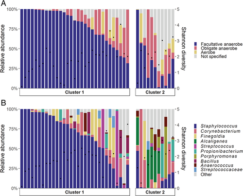
However, the mean relative abundance values were not representative of ulcer samples as a whole, which were highly heterogeneous in the composition, relative abundances, and categorical membership of taxa (Figure 3B). Other than Staphylococcus, which was present in 38 (90%) of ulcer samples, only Corynebacterium and Finegoldia were present in over half the samples (Table S1). Alpha diversity measures were also highly variable: Shannon's H ranged from 0.11 to 4.04 (mean = 1.83), Faith's PD ranged from 9.86 to 42.99 (mean = 14.31), and richness ranged from 2 to 135 (mean = 21.70).
3.3 Ulcers are differentiated by Staphylococcus relative abundance and surface area
Given the heterogeneous taxonomic composition of SCD leg ulcer samples, we pursued a clustering approach to organise samples based on microbiota. We used weighted UniFrac distance to compare ulcer samples based on the relative abundance of bacteria detected, and clustered samples based on an algorithm of PAM. Analysis of silhouette scores indicated that the ulcer samples were best grouped into two clusters (Figures 3 and 4A). The two clusters were separated along the first principal coordinate axis, which accounted for 48% of total variation among samples, primarily by relative abundance of Staphylococcus, which was positively correlated with PCoA Axis 1 (ρ = 0.82, p < 0.001; Figure 4B).
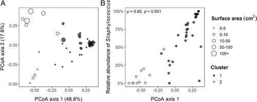
We used mixed effects models to test for differences in the relative abundance of bacterial genera between clusters (Table S2). Cluster 1 was differentiated by high relative abundance of Staphylococcus, specifically S. aureus (β = −11.95, padj < 0.001). Samples in Cluster 2 had a lower relative abundance of Staphylococcus (β = −5.08, padj = 0.03), but greater relative abundances of a variety of bacteria, including Anaerococcus (β = 5.02, padj < 0.001), Porphyromonas (β = 6.20, padj < 0.001), Peptoniphilus (β = 4.83, padj = 0.003), Alcaligenes (β = 6.17, padj < 0.001), and Corynebacterium (β = 5.19, padj = 0.002). Consequently, Shannon's H was higher in Cluster 2 than Cluster 1 (β = 0.683, p = 0.02). Wound size was also greater in Cluster 2 than Cluster 1, as indicated by its greater surface area (β = 4.53, p = 0.02). Other wound measures (depth, % slough, amount of exudate, colour of exudate) were not significantly associated with cluster membership.
3.4 Relative abundances are associated with clinical measures
We tested for associations between wound severity measures (surface area, depth, % slough, amount of exudate, colour of exudate) and alpha diversity, as well as relative abundances of the ten taxa with the highest mean relative abundances across all samples. Consistent with our cluster analysis, surface area was negatively associated at the genus level with the relative abundance of Staphylococcus (β = −0.65, padj < 0.001) but not when considered at the species level (Table S3). Surface area was positively associated with that of Alcaligenes (β = 0.77, padj < 0.001), Anaerococcus (β = 0.75, padj = 0.003), Corynebacterium (β = 0.72, padj = 0.02), and Porphyromonas (β = 0.63, padj = 0.02), all of which were enriched in Cluster 2 (Table S3). Alpha diversity measures were not significantly associated with any wound severity measures.
We also tested for associations between relative abundances of the top ten taxa, alpha diversity measures, and the following clinical variables, which were selected based on their relevance to SCD and previously reported connections to wound healing: levels of c-reactive protein (CRP), albumin, zinc, haemoglobin (Hb), foetal haemoglobin (HbF), creatinine, aspartate aminotransferase (AST), and ferritin; and white blood cell (WBC), neutrophil, natural killer cell, monocyte, reticulocyte, CD19, CD8/3, CD4/3, and platelet counts. Two clinical factors were associated with relative abundance of taxa (Table S3): haemoglobin levels were negatively associated with log relative abundance of Porphyromonas (β = −3.82, padj = 0.001), and CD19+ B cell count was negatively associated with log relative abundance of Finegoldia (β = −0.026, padj = 0.05). Alpha diversity measures were not significantly associated with the clinical variables. We also conducted analyses of associations between reports of ulcer-related pain and the top ten taxa and alpha diversity measures. While we did not find significant associations, these findings were limited by the small number of samples with reported pain scores (only 24/42 ulcer samples had associated pain data).
3.5 Intact skin microbiome differs by ulcer history
To investigate the role that intact skin may play in the formation of leg ulcers, we examined ratios of genera between the intact skin of individuals with and without a history of ulcers. For the purposes of this analysis, individuals with one or more current or healed ulcers were considered to have a history of ulcers. Individuals without a history of ulcers were composed of those who had never had an ulcer. Linear mixed models were used to test for associations between ulcer history and log-transformed ratios of bacterial genera. The ratios of Staphylococcus:Lactobacillus, as well as Corynebacterium:Lactobacillus, were elevated in the intact skin of individuals with ulcer histories (β = 2.132, padj = 0.03 and β = 2.23, padj = 0.03, respectively). Conversely, the mean ratio of Lactobacillus:Bacillus was higher in individuals without a prior history of ulcers, though our results did not indicate a statistically significant difference from a ratio of 1:1 (β = 2.08, padj = 0.06). There were no significant ratio differences between individuals with and without active ulcers.
4 DISCUSSION
In the past decade, novel sequencing technologies have increasingly been used to examine the interplay between skin wounds and microbial colonisation.18 To our knowledge, this is the first study to use 16S rRNA marker gene sequencing to describe the SCD leg ulcer microbiome and its association with measures of wound healing and other clinical factors. We find that the presence of anaerobes and common opportunistic pathogens is a comparable finding between the SCD ulcer microbiome and other chronic wounds. We also report several findings that are unique to this wound type and warrant replication and further exploration.
The SCD ulcer microbiome, as characterised by this study, bears many similarities with that of other chronic wounds. The lower alpha diversity and relative abundance of aerobes in the wound bed relative to intact skin aligns with reports of similarly reduced diversity in diabetic foot ulcer skin,29 as well as the putatively pathogenic nature of anaerobes. Moreover, many of the most abundant taxa in the SCD microbiome, such as Staphylococcus, Corynebacterium, and Finegoldia, are well-established opportunistic pathogens which are routinely found in microbial studies of chronic wounds, including those of venous ulcers, diabetic foot ulcers, and pressure ulcers.16-18, 29-31 Because we sequenced the V1-V3 regions of the 16S rRNA gene, we were able to discern species-level identity of Staphylococcus species for some reads. The majority of Staphylococcus reads were assigned as S. aureus (~80%), with the remainder comprising of coagulase negative species. These findings are consistent with other types of wounds and skin disorders where species-level resolution was possible using amplicon-based sequencing or shotgun metagenomics. Notably, Wolcott et al. found in their study of 2963 chronic wound patients that bacterial compositions did not vary widely between ulcer types.17 Our study adds the SCD ulcer to this collection of chronic wounds whose microbiomes have been found to be similar, and more broadly, suggests that the role of the skin microbiome may be consistent across chronic wounds with distinct pathophysiologic mechanisms.
This study also presents several findings specific to the SCD ulcer that merit further investigation. Our cluster analysis resulted in two groups of ulcer samples: Cluster 1 was characterised by a marked predominance of Staphylococcus and smaller surface area, while Cluster 2 members had significantly greater surface area and microbial diversity. This greater diversity was driven by the increased prevalence and evenness of aerobes such as Corynebacterium and Alcaligenes, and particularly obligate anaerobes, including Anaerococcus, Peptoniphilus, and Porphyromonas.
The presence of these two groups within ulcer samples suggests that microbial community structures vary with wound severity, which in our study was indicated by greater surface area. Similar to our findings, a study of diabetic foot ulcers found that ulcer surface area, depth, and duration were positively associated with alpha diversity measures and relative abundance of anaerobic bacteria, which included Porphyromonas, Anaerococcus, and Peptoniphilus. Shallow ulcers and those of a shorter duration contained greater abundances of Staphylococcus.15 Other studies have also suggested that Staphylococcus may have a predilection for new ulcers over recurrent ones.30 These data, in tandem with our own, collectively suggest that pathologic microbial community structures may evolve with both time and wound severity, with Staphylococcus colonisation predominating in the earlier stages of a wound, and a more diverse set of anaerobic species replacing Staphylococcus as skin and tissue continue to break down over time.
We also assessed the relationships between the ulcer microbiome and clinical factors relevant to SCD and/or wound healing. We note that the great majority of clinical variables were not associated with alpha diversity or relative abundances. However, two associations merit further investigation. First, haemoglobin levels were negatively associated with Porphyromonas relative abundance. Porphyromonas survival may be uniquely related to Hb levels, as some Porphyromonas species require heme and iron for growth and virulence, but lack the mechanisms available to comparable Gram-negative anaerobes to synthesise heme and attain iron from its environment.32 This characteristic of Porphyromonas is especially significant in light of one of SCD's hallmark features: the haemolysis of red blood cells, which can lead to low haemoglobin counts and has previously been linked to SCD ulcer incidence.5, 33 Haemolysis increases the availability of heme in blood and tissue,34 which may provide a favourable environment for Porphyromonas species to scavenge the iron and heme necessary for its survival. We also found that relative abundance of Finegoldia was negatively associated with CD19 counts. The type species of Finegoldia, Finegoldia magna,35 secretes the B-cell superantigen protein L. Protein L has been shown to deplete B cells in both mice and humans,36, 37 and may contribute to the association observed in our study.
Consistent with our findings, obligate anaerobes, including Finegoldia and Porphyromonas, can form co-occurrence networks that predominate the wound microbiome15 and other chronic-wound like disorders, such as hidradenitis suppurativa.38 While their pathogenic roles as polymicrobial communities are not well defined, effective removal of anaerobic bacteria during sharp debridement was associated with better healing outcomes in diabetic foot ulcers.39
Finally, in our analyses of intact skin, we found differences in the ratios of various microbes between individuals with and without histories of ulcers. Specifically, ratios of Corynebacterium:Lactobacillus and Staphylococcus:Lactobacillus were elevated in individuals with a history of SCD leg ulcers, while the ratio of Lactobacillus:Bacillus was elevated in individuals without a history of SCD leg ulcers. Lactobacillus has been implicated in a range of protective mechanisms against cutaneous infection or delayed healing, including immune modulation, promotion of re-epithelialisation, and pathogen clearance.40-42 Conversely, Staphylococcus and Corynebacterium can become opportunistic wound pathogens. Differences between individuals with and without ulcer histories suggest either that the presence of an ulcer induces long-term changes in nearby skin, or that over- or underrepresentation of these taxa can predispose SCD patients to ulcer development. In the latter case, microbial profiles may be useful predictors of future ulcer risk and may inform the development of prophylactic therapies that attenuate, or even, reverse these changes.
This study had some limitations. As a cross-sectional design, this study was not able to assess changes in the skin microbiome over time, although temporal changes have been shown to be important to chronic wound healing.43 Potentially important clinical factors related to the patient were not measured in this preliminary study, including vaso-occlusion and a rigorous assessment of pain. We did not measure important host-level factors related to the ulcer, including the milieu of growth factors, antimicrobial peptides, and cytokines/chemokines that interact with the wound microbiota. We were also unable to control patients' topical dressings prior to their study visit. Moreover, we were limited in our ability to attain species and strain-level resolution of taxa, despite the fact that species and strain-level variation has previously been associated with wound outcomes.39 Nonetheless, the findings of our investigation of the SCD ulcer microbiome may spur subsequent research that advances our understanding of the pathophysiology of this understudied chronic wound. More broadly, this research contributes to a growing body of literature on the shifting dynamics of the skin microbiome in relation to wound healing and clinical biomarkers.
ACKNOWLEDGEMENTS
We would like to acknowledge the individuals living with SCD who generously volunteered to participate in this study. The authors would also like to acknowledge Khadijah Abdallah, MPH; Andrew Crouch, BS; Jacquelyn Meisel, PhD; Joseph Horwinski, BS; and Sabrina Pink, PhD for their laboratory support and assistance in data collection and management for the INSIGHTS study. This work was supported by the Intramural Research Program of the National Human Genome Research Institute, National Institutes of Health (Project Number: ZIAHG200394, Investigator V. L. Bonham); and grants from the National Institute of Nursing Research (R01NR015639 to E. A. Grice) and National Institute of Arthritis, Musculoskeletal, and Skin Diseases (R01AR066663 to E. A. Grice), National Institutes of Health.
CONFLICT OF INTEREST
The authors have no potential financial conflicts of interest.


