A preliminary survey of the histopathological features of skin from the planum nasale and adjacent skin of dogs unaffected by dermatological or respiratory disease
Abstract
enBackground
There is a general belief that immune system cells are present in larger numbers in the planum nasale and adjacent haired skin than in other locations in the dog. However, little published information about the normal histological appearance of the skin of this area exists.
Hypothesis/Objectives
The aim was to obtain information about the normal histological appearance of canine skin for specific anatomical regions of the planum nasale and the haired skin adjacent to the planum nasale.
Animals
Samples from three sites were obtained from the planum nasale and adjacent haired skin of 25 dogs of varying age, breed and sex, with no evidence of dermatological or respiratory disease.
Methods
Samples were analysed to determine and quantify the immune system cells present in the samples. Slides were stained with haematoxylin and eosin or toluidine blue; immunohistochemical stains for CD3 and CD79a were applied.
Results
Immune system cells, including lymphocytes and plasma cells, were either very rare or present in low numbers. The majority of lymphocytes were of T-cell origin, with only infrequent B cells identified. Samples contained numerous melanophages, consistent with pigmentary incontinence, regardless of the presence or absence of inflammatory cells. Mast cells and plasma cells were present in low numbers.
Conclusions
Inflammatory change noted in diagnostic biopsies from this area from dogs with clinical disease is likely to be of pathological significance. However, pigmentary incontinence appears to be common at this site in clinically normal dogs without significant inflammatory cell infiltration and is therefore not necessarily of pathological significance when seen in isolation in this location.
Résumé
frContexte
Il est généralement admis que les cellules du système immunitaire sont présentes en plus grand nombre dans le planum nasal et la peau velue adjacente chez le chien. Cependant, peu de publications existent sur l'apparence histologique normale de la peau de cette zone.
Objectifs
Obtenir des informations sur l'apparence histologique normale de la peau de chien pour les régions anatomiques spécifiques du planum nasal et de la peau velue adjacente.
Sujets
Des échantillons de trois sites ont été obtenus sur le planum nasal et la peau velue adjacente de 25 chiens d'âge, de race et de genre différents, sans maladie apparente cutanée ou respiratoire.
Méthodes
Les échantillons ont été analysés pour déterminer et quantifier les cellules immunitaires présentes dans les prélèvements. Les lames ont été colorées à l'hémalun-éosine et au bleu de toluidine; les colorations immunohistochimiques pour CD3 et CD79 ont été appliquées.
Résultats
Les cellules immunitaires, y compris les lymphocytes et les plasmocytes, étaient soit très rares soit présentes en faible nombre. La majorité des lymphocytes étaient d'origine T avec des cellules B identifiées seulement rarement. Les échantillons contenaient de nombreux mélanophages correspondant à de l'incontinence pigmentaire sans lien avec la présence ou l'absence de cellules inflammatoires. Les mastocytes et plasmocytes étaient présents en faible nombre.
Conclusions
Tout changement inflammatoire, remarqué dans les biopsies de cette zone de chiens présentant des lésions cliniques, doit être considéré comme pathologique. Cependant l'incontinence pigmentaire semble être fréquente sur cette zone, chez les chiens cliniquement sains sans infiltrat cellulaire inflammatoire significatif et n'est donc pas nécessairement pathologique quand observé isolement dans cette région.
Resumen
esIntroducción
hay una creencia generalizada que las células del sistema inmune están presentes en gran cantidad en el plano nasal y en la piel adyacente en el perro. Sin embargo, se ha publicado poca información acerca de la apariencia histológica normal de la piel en esta área.
Objetivos
obtener información acerca de la apariencia histológica normal de la piel canina en regiones anatómicas específicas del plano nasal y de la piel adyacente al plano nasal.
Animales
muestras de tres localizaciones obtenidas del plano nasal y de la piel adyacente en 25 perros de diversa edad, raza y sexo sin evidencia de enfermedades de la piel ni respiratorias.
Métodos
se analizaron muestras para determinar y cuantificar las células del sistema inmunitario presentes en las muestras. Las preparaciones fueron teñidas con hematoxilina-eosina, azul de toluidina, e inmunohistoquímica para CD3 y CD79a.
Resultados
Las células del sistema inmunitarios incluidos linfocitos y células plasmáticas fueron muy raras o presentes en muy bajo número. La mayoría de los linfocitos fueron de origen T, con sólo infrecuentes células de origen B identificadas. Las muestras contenían numerosas melanófagos, consistente con incontinencia pigmentaría, independiente de la presencia de células inflamatorias. Los mastocitos y plasmáticas estaban presentes en bajo número
Conclusiones e importancia clínica
un cambio inflamatorio apreciado en biopsia diagnóstica de esta zona en perros con enfermedad clínica seguramente tenga relevancia patológica. Sin embargo la incontinencia pigmentaría parece ser común en esta localización en perros clínicamente normales sin un número significante de células inflamatorias y por tanto no es necesariamente un hallazgo patológico significativo cuando se ve aislado en esta localización.
Zusammenfassung
deHintergrund
Es wird generell angenommen, dass die Zellen des Immunsystems in größeren Zahlen im Planum nasale und der angrenzenden Haut des Hundes vorkommen. Es gibt jedoch wenig publizierte Information über das normale histologische Erscheinungsbild dieser Körperregion.
Ziele
Information zu gewinnen über das normale histologische Erscheinungsbild der Hundehaut in speziellen Körperregionen wie dem Planum nasale und der behaarten Haut, die an das Planum nasale angrenzt.
Tiere
Es wurden Proben von drei Stellen am Planum nasale und der angrenzenden behaarten Haut von 25 Hunden unterschiedlichen Alters, Rasse und Geschlecht, ohne einer dermatologischen oder respiratorischen Erkrankung genommen.
Methoden
Es wurden Proben analysiert, um die Zellen des Immunsystems in diesen Zellen zu bestimmen und zu quantifizieren. Die Schnitte wurden mit Hämatoxylin und Eosin, sowie mit Toluidinblau angefärbt; es wurden immunhistochemische Färbungen für CD3 und CD79a verwendet.
Ergebnisse
Die Immunzellen, zu denen die Lymphozyten und Plasmazellen gehören waren entweder sehr selten oder nur in sehr geringer Anzahl vorhanden. Die Mehrzahl der Lymphozyten entsprangen den T-Zellen, wobei nur wenige B-Zellen identifiziert werden konnten. Die Proben enthielten zahlreiche Melanophagen, die mit einer Pigmentinkontinenz im Zusammenhang standen, egal ob Entzündungszellen vorhanden waren oder nicht. Mastzellen und Plasmazellen waren in einer geringen Anzahl vorhanden.
Schlussfolgerungen
Entzündliche Veränderungen, die in diagnostischen Biopsien dieser Körperstellen bei Hunden mit klinischer Erkrankung vorkommen, sind wahrscheinlich von pathologischer Bedeutung. Nichtsdestotrotz scheint Pigmentinkontinenz an diesen Stellen auch bei klinisch gesunden Hunden ohne signifikante Entzündungszellinfiltration häufig aufzutreten und ist daher nicht unbedingt von pathologischer Bedeutung, wenn es an diesen Stellen isoliert auftritt.
Abstract
ja

Abstract
zh

Introduction
The skin of the planum nasale of the dog is composed of thick, keratinized epidermis with distinct elevations and grooves.1-3 There are no sweat glands or sebaceous glands in this area.1-3 Aside from these basic observations in veterinary texts detailing the most obvious differences between this region of modified skin and haired skin, there are few reviews of the normal histological appearance of the skin in this anatomical location. Likewise, there is a paucity of published data on the histological features of the junction between haired skin and the modified skin of the planum nasale. It is a common opinion that the haired skin in this location presents a different appearance to haired skin from other anatomical locations, particularly with regard to the cells of the immune system that are present in nondiseased skin. It has been stated that the finding of an interface dermatitis with increased numbers of mononuclear cells in this location is ‘diagnostically weak’.4 However, only one brief survey of the histopathological appearance of skin in these locations from normal dogs has been performed, consisting of five young dogs.5 That study identified increased numbers of inflammatory cells in some samples, and differences in the histological appearance of the skin from different locations within the planum nasale. The aim of the present study was to provide information about the normal histological appearance of the skin of the dog in the specified locations (the planum nasale and the junction between haired skin and the planum nasale), to qualify the cell types present using light microscopy and immunohistochemical techniques, and to quantify the numbers of each cell type present in samples taken from dogs with no signs of skin or respiratory disease.
Materials and methods
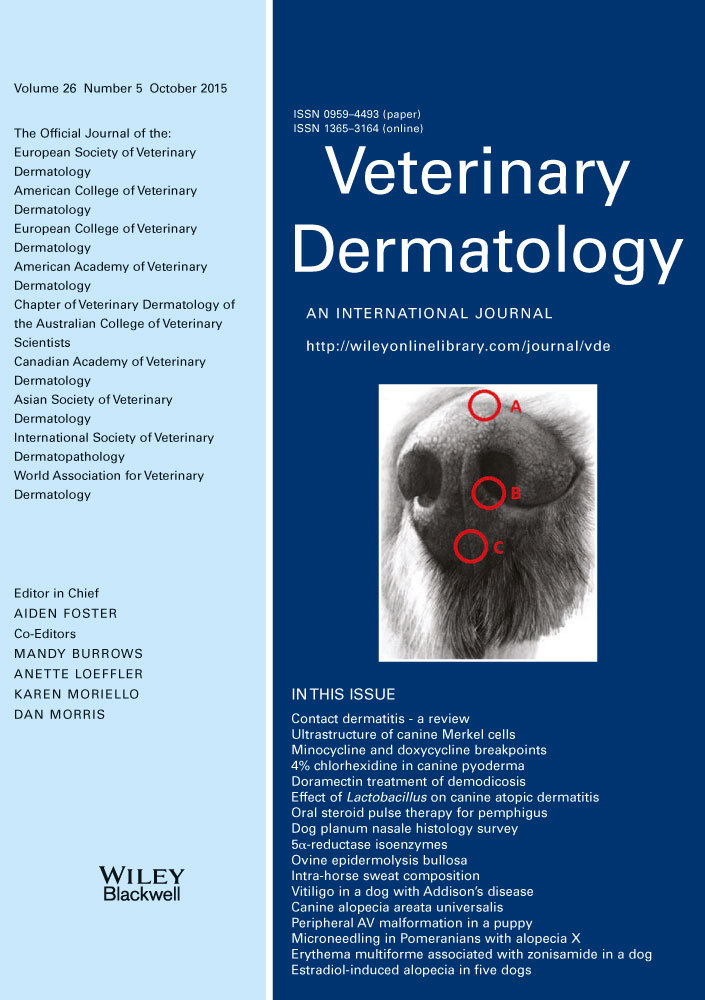
Samples were obtained from 25 dogs with no obvious signs of dermatological or respiratory disease. Dogs used in the study were those presented for euthanasia in the City of Bundaberg, Queensland, Australia, following impounding. The dogs ranged in age from 4 to 84 months of age. There were 10 intact females, three neutered females, 10 intact males and two neutered males. Samples were obtained using a 6 mm biopsy punch from predetermined sites immediately following euthanasia with intravenous sodium pentobarbitone. The sites (see Figure 1) were as follows: the junction between haired skin and the planum nasale on the dorsal aspect of the nose (site A); the planum nasale at the edge of the medial side of the left nare (site B); and the midline rostral portion of the planum nasale incorporating the philtrum (site C).

Samples were placed in 10% neutral buffered formalin solution for routine tissue fixation. The samples were examined macroscopically by the primary investigator, bisected (samples from the junction of haired skin and planum nasale were bisected so that processed samples would include the junction between the skin types) and submitted in labelled tissue cassettes for processing to paraffin wax. After embedding, 5-μm-thick sections were cut and mounted on glass slides, stained according to previously established protocols6 with haematoxylin and eosin for routine histopathology and, separately, with toluidine blue for mast cells, and coverslipped for examination.
Additional 4-μm-thick sections were taken from the paraffin-embedded tissues and mounted on positively charged glass slides (Superfrost Plus; Menzel, Braunschweig, Germany) for immunohistochemistry. Sections were dewaxed and heat-mediated antigen retrieval was performed in a Tris/EDTA buffer, pH 9 (Envision FLEX Target Retrieval Solutions; DAKO, Ely, UK) in a computer-controlled heated water bath (PT Link; DAKO) at 97°C for 30 min. Immunohistochemistry was performed using an automated immunohistochemical staining machine (DAKO Cytomation Autostainer Plus). The tissue sections were incubated with the diluted (Envision FLEX antibody diluent; DAKO) primary antibodies for 30 min at room temperature. Antibodies (DAKO) used were CD3 (rabbit polyclonal anti-human CD3) at a dilution of 1:400 and CD79a, clone HM57 (mouse monoclonal anti-human CD79a) at a dilution of 1:400. Endogenous peroxidase activity was blocked by incubation in hydrogen peroxide solution for 30 min (EnVision FLEX peroxidase-blocking reagent; DAKO). Immunohistochemical staining was detected using a goat anti-mouse/rabbit IgG and horseradish peroxidase-tagged polymer system (EnVisionFLEX/HRP; DAKO). Staining was developed with 3,3′-diaminobenzidine tetrahydrochloride (EnVision FLEX DAB+ chromogen; DAKO) and counterstained with haematoxylin. Negative controls were performed by replacing the primary antibody with antibody dilution buffer.
The haematoxylin and eosin and the toluidine blue samples were examined by both the primary and the secondary investigator. The immunohistochemistry samples were examined by both the primary and the tertiary investigator. For each of the three biopsied sites in each case, 10 separate high-power microscopic fields (using ×10 magnification eyepieces and a ×40 magnification objective lens, corresponding to ×400 magnification, with a calculated field of view 531 μm in diameter) were examined with the aid of a graticule for each of the following four locations: the epidermis, the superficial dermis, the mid-dermis and the deep dermis (see Figure 2). The superficial dermis was defined as the region extending from immediately beneath the epidermis to the upper margin of the sebaceous glands in the sample of junctional haired skin and planum from each individual sampled, and the corresponding level within the planum and philtrum samples from that same individual. The mid-dermis was defined as the region extending from the upper margin of the sebaceous glands to the deepest level containing sebaceous gland lobules in the junctional samples, and at the same level in the corresponding planum and philtrum samples. The deep dermis was defined as the region extending from beneath the sebaceous glands to the subcutis, and the same level in the corresponding planum and philtrum samples for that animal. For each site in every sample, the numbers of polymorphonuclear cells, mast cells, plasma cells, B lymphocytes and T lymphocytes were recorded. In addition, the thickness of the epidermis and the relative thickness, arrangement and features of the stratum corneum in each sample were recorded. Results were collated to provide average cell numbers and average epidermal and keratin thickness for each location.

Results
Polymorphonuclear cells, plasma cells, B lymphocytes and T lymphocytes were all, on average, present in numbers lower than one cell per high-power field (hpf) in all locations. Mast cells were, on average, present in numbers lower than one cell per hpf in the mid-dermis and the deep dermis. No mast cells were identified in the epidermis in any field in any sample. Mast cells were, on average, present at a rate slightly higher than one cell per hpf (1.2253 per hpf) in the superficial dermis.
Epidermal layers consisisted of stratum basale, stratum spinosum and, rarely, stratum granulosum; the stratum granulosum was generally seen only in the junctional haired skin and the stratum corneum.
The stratum corneum in the nasal planum samples was invariably compact and generally orthokeratotic in nature; however, nine cases showed some focal areas of parakeratosis in the absence of inflammation. Where the epidermis was heavily pigmented, the stratum corneum was generally also heavily pigmented. The thickness of the stratum corneum ranged from an average of 3.44 μm in haired skin to an average of 9.4 μm in nonhaired skin.
The thickness of the epidermis ranged from 4.9 to 16.5 cells (averaged over 10 hpf). This equated to an average epidermal thickness of 5.96 μm in haired skin and 14.26 μm in nonhaired skin, not including rete.
Epidermal rete in the nasal planum samples were often irregular in shape and size, but in general tended to reach approximately uniform depth across the sample (see Figure 3).

Epidermal pigmentation ranged from absent through negligible or light to very dark. The pigmentation was also variably confined to the basal layer or spread diffusely throughout the epidermis; however, the majority of cases showed full-thickness pigmentation. Pigment tended to be most marked in the tips of the epidermal rete. Where pigmentation was not diffuse throughout the epidermis, there was occasionally full-thickness pigmentation of the epidermis focally overlying rete. One or two cases showed clumped pigmentation of the basal epidermis, with very darkly pigmented cells present, similar to what is seen in normal colour-dilute epidermis of haired skin.
All samples contained variable numbers of melanophages and, less commonly, free melanin granules within the superficial dermis, consistent with pigmentary incontinence, generally in the absence of observable inflammation (see Figure 4).

Mast cells showed close association with the epidermis in the nonhaired skin of the nasal planum.
Polymorphonuclear cells were present in extremely low numbers in most samples; the majority of those seen were neutrophils. Neutrophils were found in moderate numbers in only one sample. Eosinophils were found as solitary cells in only two samples.
See Table S1 in Supporting information for cell counts and measurements.
Discussion
To the best of the authors' knowledge, there are no systematic surveys of the histopathology of clinically normal dog skin from any location. However, most histopathology textbooks and atlases contain photographic figures and descriptions of the normal histopathological appearance of haired dog skin and the skin of the planum nasale. In this survey, we have identified a number of key differences as well as similarities to what is widely accepted as normal. The key differences from haired skin involved the increased thickness of the epidermis and the stratum corneum, and these changes would appear to be in line with previous observations. The age, breed and sex of the dogs did not appear to have an impact on any of the findings.
In contrast to widely accepted opinion, cells of the immune system do not appear to be present in large numbers in the clinically normal nasal planum and adjacent junctional skin. There was no evidence of lichenoid inflammation or obscuring of the dermo-epidermal junction by cellular infiltrates in any sample. Immune system cells, including lymphocytes and plasma cells, were present, but were either very rare or seen only in low numbers. The presence of these cells may represent low-grade antigenic stimulation in an anatomical location that interacts closely with the environment. Polymorphonuclear cells were present in extremely low numbers in most samples; the majority of those seen were neutrophils, with very rare single eosinophils seen in isolated cases. Neutrophils were found in moderate numbers in only one sample, in the mid- and deep dermis. As this case was such an outlier, it is possible that this individual had sustained a recent injury or microbial infection that was resolving and therefore inapparent at the time of sampling.
Of note was that samples all contained variable numbers of melanophages and, less commonly, free melanin granules, within the superficial dermis, consistent with pigmentary incontinence, generally in the absence of observable inflammation at this location. Pigmentary incontinence is often regarded as a sign of immune-mediated damage to the epidermal melanin unit (such as in lichenoid interface dermatoses), but may also be associated with physical injury or trauma to the epidermis, particularly the basal epidermis.7 It may also be associated with spillover of pigment into the dermis from a hyperpigmented epidermis, which may be associated with a variety of causes, including nonspecific local inflammation. Clinically, it is associated with alterations in visible pigmentation;7 both epidermal hyperpigmentation and, less commonly, hypopigmentation may be seen clinically in association with this histological finding.7 The finding of pigmentary incontinence in the skin of normal dogs and without evidence of significant associated inflammation or true interface change is interesting and challenges the most commonly accepted interpretation of this finding, and may also be responsible for the overdiagnosis of immune-mediated disease at this site due to overinterpretation of background ‘normal’ melanophage infiltration in the nasal planum. Given the almost universal presence of this finding across all the samples examined, it is thought that chronic low-grade injury at this site as a ‘normal’ occurrence gives rise to the finding.
In conclusion, immune system cells are not present in large numbers in this anatomical location in clinically normal dogs. Inflammatory change noted in biopsies from this area is therefore likely to be of pathological significance. However, pigmentary incontinence appears to be common at this site and is therefore not necessarily of pathological significance when seen in isolation in this location. Age, breed and sex do not appear to affect the histological appearance of the skin in this location. Further studies to qualify and quantify other cell types (melanocytes, Langerhan's cells) are required to provide a further evidence base for the normal histological appearance of the skin in both this and other anatomical locations in the dog.