Extracellular matrix in leg basal cell carcinoma: Possible pathogenetic role of chronic venous insufficiency
The main risk factor for the development of basal cell carcinoma (BCC) is ultraviolet radiation (UVR) exposure. However, BCC can also be found in non-exposed skin, so that UVR exposure is not the only risk factor. In the lower extremities, BCC frequency is higher in women, putatively due to greater sun exposure. Chronic venous insufficiency (CVI), which is also more common in women, could be an additional predisposing factor. BCC is locally invasive and the microenvironment around BCC is crucial for tumorigenesis.1 Tumors are characterized by extracellular matrix (ECM) remodeling and stiffening. ECM rigidity in solid tumors is largely mediated by collagen organization. In CVI, the transmission of high venous pressures to dermal microcirculation elicits an inflammatory process and release of cytokines and growth factors, that result in dermal fibrosis and tissue remodeling.2
Second harmonic generation (SHG) is a nonlinear optical microscopy process that allows for studying the supramolecular assembly of collagen in tissues, with a high level of detail.3
In order to investigate a possible relationship between leg BCC development and CVI, we evaluated the SHG texture of dermal collagen fibers in 19 randomly selected biopsies of cancer-free margins of leg BCC. Tumors in patients with predisposing genodermatoses were excluded. The control group consisted of a random sample of leg skin from eight patients in the same age group and sex, obtained for excision of benign lesions.
One 5-μm thick paraffin-embedded section per tumor, containing the BCC and tumor-free margin skin, was stained with hematoxylin and eosin. Histopathological features of venous stasis and solar damage were blindly recorded (Figure 1) in both lesion-free skin groups, as absent/mild or moderate/evident.4 The SHG images were acquired with an inverted Z.1 Axio Observer microscope equipped with a ZeissLSM780 NLO confocal scanning head (Carl Zeiss AG, Jena, Germany). The texture of collagen fibers was blindly investigated by the analysis of four representative regions of interest (42.5 × 42.5 μm), through entropy and anisotropy features, using ImageJ (https://imagej.nih.gov/ij/download.html) software (Figure 2). Entropy and optical anisotropy are high in rough texture and low in homogeneous texture. Statistical procedures were blindly performed using the Statistical Analysis System for Windows 9.4 (SAS Institute, Cary, NC).
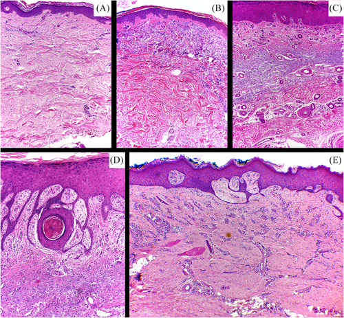
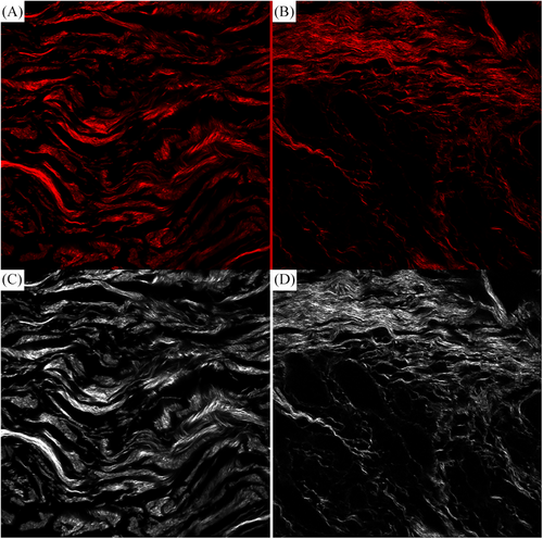
Histological signs of CVI were higher (p-value 0.0267) in the leg BCC specimens than in the control group (Figure 3). We found greater anisotropy and entropy in the control group (p-value < 0.05) than in the vicinity of the BCC, demonstrating collagen remodeling (Figure 4).
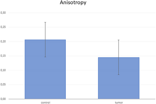
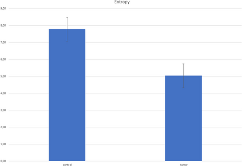
It is known that the compaction of the perineoplastic ECM favors the local invasion of cancer cells and, perhaps, the development of neoplasms. As is, using ex vivo SHG signals generated from multiphoton microscopy in 52 BCC, Sendín-Martín et al. found a more aligned collagen distribution surrounding BCCs nests than in normal skin and benign lesions.5
Summarizing, this study showed, through precise tools, collagen remodeling of the perineoplastic ECM in leg BCC, as described in the dermis in CVI. Additionally, comparative more marked CVI histological signs in the BCC group corroborate the hypothesis that CVI can be a predisposing factor.
Open Research
DATA AVAILABILITY STATEMENT
The data that support the findings of this study are available from the corresponding author upon reasonable request.


