Four fixed points for dermoscope in male androgenetic alopecia: A way to diagnosis and monitor therapy effect
Dear Editor,
Androgenetic alopecia (AGA) is the most common form of alopecia in men. Dermoscope, a modern skin imaging tool, plays an important role in diagnosing alopecia diseases. AGA requires long-term treatment, but not all patients have good response to treatment drugs. Therefore, it‘s crucial to have a method for assessing treatment efficacy, which provides evidence for doctors to determine whether changing treatment drugs is necessary and gives confidence for patients to continue their treatment. However, it has not had unified standard to assess treatment effectiveness. Herein, we present an effective method to assess severity of AGA and evaluate treatment efficacy by dermoscope.
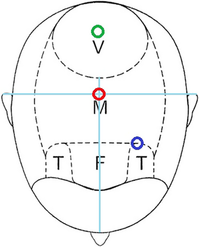
As we all known, the most common features of male AGA comprise a receding hairline at the temples and decreased hair density on the vertex and frontal area of the scalp .1 Temporal angle, baihui acupoint and hair whorl represent the regions of temple, frontal and vertex area of the scalp, respectively. The occipital region is not sensitive to androgen, so there is no serious hair loss. The occipital tuberosity is an important bone marker of occipital region. Therefore, temporal angle, baihui acupoint, and hair whorl represent different hair loss regions in male AGA, while occipital tuberosity is the marker of no hair loss region. Thus, we capture images at these four points using dermoscope to diagnose, evaluate severity and assess treatment effect of AGA. Capture images using dermoscope as follows: (1) Temporal angle: This point is located at the corner of the front hairline (marked by the blue circle in Figure 1). Position the frontal angle at the center of the field of view and capture the image; (2) Baihui acupoint: This point is located at the intersection of the head's midline and the line connecting the tips of the ears (marked by the red circle in Figure 1). After fixing this point, part the hair, then capture the image; (3) Hair whorl: This point is marked with a green circle in Figure 1. Maintain the natural whirlpool of the hair,centering it in the field of view, and then capture the image; (4) Occipital tuberosity: Part the hair at occipital tuberosity, and then capture the image.

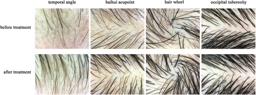
Comparing the dermoscopy images of these four regions can be used to diagnose AGA and assess the severity of this disease in men. Additionally, comparing the dermoscopy images of these four fixed points before and after treatment can be used to evaluate treatment effectiveness (Figure 2). Thus, capturing images at these four points by dermoscopy regularly provides a visual way to assess treatment efficacy.

ACKNOWLEDGMENTS
This work was supported by National High Level Hospital Clinical Research Funding (grant number: 2023-NHLHCRF-YYPP-TS-06) and Elite Medical Professionals Project of China-Japan Friendship Hospital (grant number: ZRJY2021-QM04)
FUNDING INFORMATION
National High Level Hospital Clinical Research Funding, Grant number: 2023-NHLHCRF-YYPP-TS-06; Elite Medical Professionals Project of China-Japan Friendship Hospital, Grant Number: ZRJY2021-QM04
CONFLICT OF INTEREST STATEMENT
The authors declare no conflicts of interest.
Open Research
DATA AVAILABILITY STATEMENT
The data that support the findings of this study are available from the corresponding author upon reasonable request.


