Dermoscopic findings in a patient with papular elastorrhexis
Xigetu He and Qila Sa contributed equally to this work.
Papular elastorrhexis (PE) is an acquired elastic fiber cutaneous disease with rarely clinically and a lower incidence rate. The epidemiological data remain unknown, and the pathogenesis is not completely clear, which is commonly involved in young women.1 Here, we present the clinical, histopathological and dermoscopic findings from a patient with popular elastorrhexis, aimed to explore the dermoscopic findings and its histopathological correlation for providing the noninvasive imaging diagnostic clue and pattern.
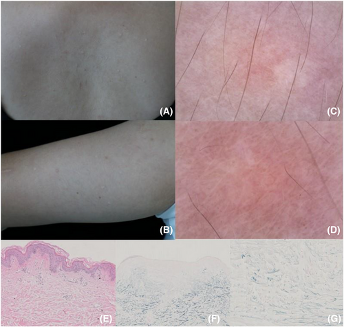
A 24-year-old female was presented to the dermatology clinic with a 1-year history of asymptomatic and multiple cutaneous lesions affecting aesthetics on her trunk and upper arms. Any systemic symptoms, a history of trauma, chronic systemic diseases and similar family history were all denied. Physical examination revealed multiple flat-topped, firm, white and skin-colored round papules about 2–5 mm in diameter on the trunk and upper arms (Figure 1A,B). No positive findings were found in the laboratory examination, that includes the complete blood count, erythrocyte sedimentation rate, C-reactive protein and antinuclear antibodies. A dermoscopy examination was performed, and the imaging showed the central yellow-white unstructured area is surrounded by radiating pigment, linear, rod and other irregular vessels (Figure 1C,D). A skin biopsy was performed, and the specimen obtained from a biopsy of skin lesions was sent for histopathological analysis. In histopathological analysis, the normal epidermis with perivascular inflammatory infiltrate in the superficial dermis (Figure 1E; HE staining ×100) was shown. In further immunohistochemical staining analysis, the focal irregular thickening and homogenization of collagen fibers in the dermis and fragmentation, reduction even the absence of elastic fibers were found (Figure 1F,G; Verhoeff-VanGieson staining, ×100, ×400). The clinical, dermoscopic and histopathological findings were consisting of a diagnosis of PE. The patient refused to accept further examination and treatment and is currently being followed up.

PE is rare may be due to the subtlety of lesions and easy to be ignored or misdiagnosed in clinical practice. Atypical clinical manifestations are often multiple, asymptomatic, firm, circumscribed, oval to round papules with a diameter of about 1–5 millimeters; the etiology and pathogenesis of PE remain unknown; also, it has a stable evolution over the year; reliable treatment option was absent to date; the diagnosis is mainly based on classic clinical manifestations and typical histopathological evidence of reduction even the absence of elastic fibers.1, 2 The dermoscopy examination was considered a potential tool for the non-invasive diagnosis of PE, with unique findings with honeycomb-like reticular pigment surrounded by radial pigment without vessel involvement, was proposed.3 Interestingly, different from the previous report, we found that the vessel structure was involved in our case presented; the findings also support that the vessel involvement may also be the potential dermoscopic clues; the central yellow-white unstructured area was consistent of the reduction and the absence of elastic fibers in histologically, also the vessel involvement may represent the infiltration of inflammatory cells and the perivascular proliferation in super dermis. In conclusion, we proposed the unique dermoscopic findings of PE, which may provide clues and ideas for its diagnosis, and provide a reliable histopathological link and correlation for developing non-invasive diagnosis of PE.
CONFLICT OF INTEREST STATEMENT
The authors declare that there is no conflict of interest that could be perceived as prejudicing the impartiality of the research reported.
FUNDING INFORMATION
The Natural Science Foundation of Inner Mongolia Autonomous Region, Grant Number: 2021ZD15
ETHICS STATEMENT
The written informed consent was obtained from the patient to publish her medical details.
Open Research
DATA AVAILABILITY STATEMENT
Data sharing is not applicable—no new data were generated, or the article describes entirely theoretical research.


