Periodontitis in tonsil cancer patients—A comparative study in accordance with tumour p16 status
Abstract
Objectives
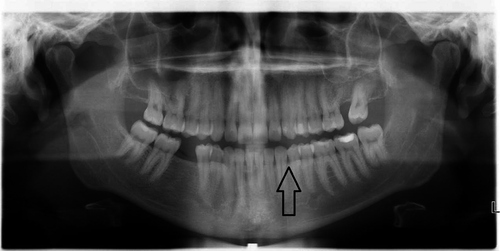
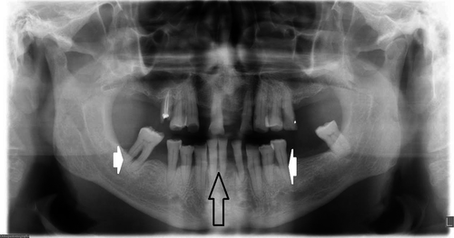
We assessed the periodontal situation radiologically according to tumour p16 status.
Materials and methods
Patients with a diagnosis of tonsillar cancer and availability of a digital panoramic radiograph (DPR) during a 5-year period were included in this retrospective study. The predictor variables were periodontal stability, marginal bone loss, marginal bone loss without periodontal stability and total number of teeth. Periodontal status was compared with p16 status, age, gender, smoking and alcohol use.
Results
Among 115 patients included in the analyses (p16-negative, n = 24; p16-positive, n = 91), smoking (p < .0001), heavy alcohol use (p < .0001) and total number of teeth (p = .0001) were significantly associated with p16 status. Current smoking (OR = 7.3) and heavy alcohol use (OR = 10.1) increased the risk of p16-negative cancer.
Conclusions
Patients with p16-negative tonsillar carcinoma had less teeth than patients with p16-positive tumours. Other periodontal findings were common in both groups without statistical significance. Heavy alcohol use and smoking were the most important risk factors for p16-negative tonsillar carcinoma.
1 INTRODUCTION
Approximately 90% of oropharyngeal squamous cell carcinomas (OPSCCs) develop from the tonsils or base of tongue (D'Souza et al., 2007). Human papillomavirus (HPV) is associated with tonsillar cancer in 70%–73% of patients (Palmer et al., 2014), and the increased incidence of HPV infections is considered to be the reason for increased incidence of tonsillar carcinoma (Park et al., 2013).
OPSCC is nowadays considered as two separate entities according to HPV status (Carpén et al., 2018). The majority of HPV-positive OPSCC patients are men and tend to be younger and to have fewer comorbidities (Carpén et al., 2018; Palmer et al., 2014). In contrast, HPV-negative patients are more often smokers and heavy alcohol consumers (Carpén et al., 2018). HPV status has been shown to be associated with long-term prognosis; HPV-positive oropharyngeal carcinoma patients have better survival rates than patients with HPV-negative carcinoma (Lacau et al., 2017). The immunohistochemical identification of p16 is used as a routine screening method in many clinical settings, as it is easy and cost-effective. This technique does not directly indicate HPV status (El-Naggar, Chan, Grandis, Takata, & Slootweg, 2017; Prigge, Arbyn, von Knebel, & Reuschenbach, 2017), but is a causal and prognostic factor for OPSCC.
Periodontitis is a common oral chronic inflammatory disease (Loesche & Grossman, 2001). It is associated with Gram-negative anaerobic bacteria in the dental biofilm (Loesche & Grossman, 2001). Without treatment, periodontitis leads to the destruction of tissues supporting teeth, clinically detectable as deepened periodontal pockets and alveolar bone loss. This chronic oral disease is usually irreversible and is the main cause for tooth loss among adults (Katarkar et al., 2015).
The severity of periodontitis has been found to increase the mortality from oral and digestive system cancers (Ahn et al., 2012). Recently, a case–control study showed an independent association between periodontitis and oral carcinoma. Patients with periodontitis were 3.7 more likely to have oral cancer than patients without periodontitis (Shin, Choung, Lee, Rhyu, & Kim, 2019). In addition, the periodontal status of patients has been found to be related to tumour HPV status. The risk of periodontal infections in HPV-positive head and neck cancer patients is increased (Tezal et al., 2009, 2012). Based on these previous results, we decided to study the periodontal status of tonsillar carcinoma patients.
We identified the occurrence of periodontal infections and evaluated the characteristics and severity of these findings among p16-positive and p16-negative tonsillar carcinoma patients based on their dental radiographs. We hypothesized that signs of periodontitis are more common and severe in p16-negative patients than in p16-positive patients due to living habits. We designed and performed a retrospective radiological study to determine the occurrence of periodontitis in tonsillar carcinoma patients.
2 MATERIALS AND METHODS
2.1 Patient material
Patient records of all patients (n = 144) with primary tonsillar carcinoma during the 5-year period between January 2013 and December 2017 at the Head and Neck Center, Helsinki University Hospital, Helsinki, Finland, were evaluated. The data search was based on ICD code C09, which indicates “malignant neoplasm of tonsil.” An additional inclusion criterion was the availability of a digital panoramic radiograph (DPR) (Instrumentarium Dental™ Orthopantomograph™ OP200 or Orthopantomograph® OP300) in the Helsinki University Hospital database. Patients with additional malignancy, previous head or neck malignancy, previous radiotherapy in the head or neck region, undefined p16 status, or incomplete information on smoking or alcohol consumption were excluded, as were edentulous patients.
Of the 144 patients with a primary diagnosis of tonsillar cancer, 15 were excluded due to lack of DPR images. An additional 14 patients were excluded due to edentulousness (n = 10), an additional malignancy (n = 2), or previous head or neck malignancy or previous radiotherapy in head or neck region, or both (n = 2). In the remaining 115 patients, requisite data on smoking, alcohol consumption and p16 status were available. Thus, these patients were included in the final analyses.
2.2 Clinical information
Clinical information was collected from the Helsinki University Hospital patient database. The following data were recorded: age, gender, smoking and heavy alcohol use. Age was categorized according to the median age of patients (61 years) as ≥61 or <61 years. Patients were divided into two groups according to their smoking habit: non-smokers (i.e. non-smokers and former smokers who quit ≥5 years ago) or current and former smokers (who quit <5 years ago) (Jerjes et al., 2012). Alcohol use was determined according to the Finnish Current Care Guidelines consumption limits for heavy alcohol use: ≥12 doses (i.e. ≥150 g alcohol) per week for women and ≥23 doses (i.e. ≥287.5 g of alcohol) per week for men as suggested by the Finnish working group (treatment of alcohol abuse: Current Care Guidelines Abstract, 2018).
2.3 P16 ink positivity
The expression of p16INK4A was determined by immunohistochemical (ICH) staining as part of the routine protocol at the Department of Pathology, Helsinki University Central Hospital, on paraffin-embedded formalin-fixed tissue samples. Deparaffinization was performed in xylene, followed by rehydration in a graded ethanol series. Pre-Treatment Module (Lab Vision Corp., UK Ltd.) was used to treat the tissue slides in Tris-HCl buffer (pH 8.5). Endogenous peroxidase was blocked with 0.3% Dako REAL Peroxidase-Blocking Solution. Monoclonal mouse anti-human p16-INK (9517 CINtec Histology Kit, MTM Laboratories) was used as primary antibody. Positive p16 expression was defined as >70% of tumour cells being positive.
2.4 Radiological analyses of periodontal status
All DPR images were reviewed by an experienced oral radiologist with 7 years' experience in maxillofacial radiology (MM-G) twice at 6-month intervals. When the results were in discordance, the more severe status was used in the study.


- Marginal bone loss was determined as present or not present and classified as mild, moderate, or severe.
- Marginal bone loss was defined in accordance with alveolar bone resorption, which was measured at its maximum from the cementoenamel junction to the tooth apex. Marginal bone loss was determined by tertile as follows: maximum bone loss extending to the cervical third (mild), maximum bone loss extending to the middle third (moderate) or maximum bone loss extending to the apical third (severe). The measurement was not performed for unerupted teeth or for those with no remaining supragingival coronal tooth structure.
- Total number of teeth (grouped as 28–32 teeth, 17–27 teeth and 1–16 teeth) was counted.
- The total number of teeth included the third molars and excluded dental implants.
2.5 Study design
P16 status was compared with radiological periodontal status. Explanatory variables were age, gender, smoking and heavy alcohol use.
2.6 Statistical analysis
SPSS 25.0 (IBM Corp, Armonk, NY, USA) was used for statistical analysis. Patient p16 status, clinical data and periodontal status were assessed with chi-square test (χ2 test), two-tailed Mann–Whitney test and multivariate logistic regression analysis. The significance level was set at 0.01.
2.7 Ethical approval
The study was approved by the Internal Review Board of the Head and Neck Center, Helsinki University Central Hospital, Finland (HUS/66/2018).
3 RESULTS
3.1 Patient data
Descriptive statistics of the 115 patients is presented in Table 1. The median age of patients was 61.0 (mean 60.6) years. Over two-thirds of the patients were male (73.9%). The majority of the patients were non-smokers (60.9%) and reported no heavy alcohol use (72.0%). Tumour samples were p16-positive in 91 (79.1%) and p16-negative in 24 patients (20.9%).
| n | % | |
|---|---|---|
| Age (years) | ||
| Range 31–89 | ||
| Mean 60.6 | ||
| Median 61.0 | ||
| <61 | 57 | 49.6 |
| ≥61 | 58 | 50.4 |
| Gender | ||
| Male | 85 | 73.9 |
| Female | 30 | 26.1 |
| Smoking | ||
| Non-smoker | 70 | 60.9 |
| Current smoker | 45 | 39.1 |
| Heavy alcohol use | ||
| Yes | 31 | 27.0 |
| No | 84 | 73.0 |
P16-negative patients were more often current smokers (83.0%) than p16-positive patients (28.0%) (p < .0001) (Table 2). Logistic regression analysis revealed that p16-negative carcinoma was significantly associated with current smoking (p16-negative patients, odds ratio [OR] = 7.261, 95% confidence interval [CI]: 1.778–29.656; p = .006).
| p16-negative (n = 24) | p16-positive (n = 91) | p-value | |||
|---|---|---|---|---|---|
| n | % | n | % | ||
| Age (years) | |||||
| <61 | 9 | 37.5 | 48 | 52.7 | .184 |
| ≥61 | 15 | 62.5 | 43 | 47.3 | |
| Gender | |||||
| Male | 16 | 66.7 | 69 | 75.8 | .363 |
| Female | 8 | 33.3 | 22 | 24.2 | |
| Smoking | |||||
| Non-smoker | 4 | 16.7 | 66 | 72.5 | <.0001 |
| Current smoker | 20 | 83.3 | 25 | 27.5 | |
| Heavy alcohol use | |||||
| Yes | 15 | 62.5 | 16 | 17.6 | <.0001 |
| No | 9 | 37.5 | 75 | 82.4 | |
Heavy alcohol use was more frequently reported among p16-negative patients (62.5%) than among p16-positive patients (17.6%) (p < .0001) (Table 2). Logistic regression analysis revealed that heavy alcohol use was significantly associated with p16-negative cancer (p16-negative patients, OR = 10.053, 95% CI: 2.407–41.982; p = .002).
3.2 Periodontal status
Periodontal stability was present in only 12.5% of patients with p16-negative cancer and in 29.7% of patients with p16-positive cancer. Marginal bone loss was present in 95.5% of p16-negative patients and in 93.4% of p16-positive patients. Most of the p16-positive patients had marginal bone loss categorized as mild (50.5%). The number of p16-negative patients with alveolar bone loss was approximately equal in all categories of marginal bone loss severity (mild 25.0%, moderate 37.5%, severe 33.3%).
A significant association emerged between total number of teeth and p16 status of the tumour (p = .0001) (Table 3). Patients with p16-negative tumours had fewer teeth (1–16 teeth 50.0%, 17–27 teeth 20.8% and 28–32 teeth 29.2%) than patients with p16-positive tumours (1–16 teeth 12.1%, 17–27 teeth 50.5% and 28–32 teeth 37.4%; p = .0001).
| p16-negative (n = 24) | p16-positive (n = 91) | p-value | |||
|---|---|---|---|---|---|
| n | % | n | % | ||
| Periodontal stability | |||||
| Present | 3 | 12.5 | 27 | 29.7 | .088 |
| Not present | 21 | 87.5 | 64 | 70.3 | |
| Marginal bone loss | |||||
| Present | 23 | 95.8 | 85 | 93.4 | .658 |
| Not present | 1 | 4.2 | 6 | 6.6 | |
| Mild | 6 | 25.0 | 46 | 50.5 | .052 |
| Moderate | 9 | 37.5 | 23 | 25.3 | |
| Severe | 8 | 33.3 | 16 | 17.6 | |
| Marginal bone loss without periodontal stability | |||||
| Present | 21 | 87.5 | 63 | 69.2 | .070 |
| Not present | 3 | 12.5 | 28 | 30.8 | |
| Mild | 5 | 20.8 | 31 | 34.1 | .126 |
| Moderate | 9 | 37.5 | 18 | 19.8 | |
| Severe | 7 | 29.2 | 14 | 15.4 | |
| Total number of teeth | |||||
| 28–32 | 7 | 29.2 | 34 | 37.4 | .0001 |
| 17–27 | 5 | 20.8 | 46 | 50.5 | |
| 1–16 | 12 | 50.0 | 11 | 12.1 | |
3.3 P16 status and clinical data
No significant associations were observed between p16 status and age, gender, periodontal stability, marginal bone loss or marginal bone loss without periodontal stability.
In addition, we analysed the correlation coefficients between each variable and the others. There was a significant association between smoking and the following variables: reported history of heavy alcohol use (p = .001), periodontitis of cervical third (p < .001), middle third (p = .005), apical third (p = .002) and total number of teeth (p < .001). Patient age was significantly associated with number of teeth (p = .002).
4 DISCUSSION
The present study assessed radiologically detectable findings of periodontitis in tonsillar carcinoma patients. The hypothesis was that signs of periodontitis are more common and severe in p16-negative patients than in p16-positive patients due to living habits. Our hypothesis was partially confirmed. According to our results, p16-negative patients had significantly fewer teeth than p16-positive patients. However, signs of periodontitis were observed in both patient groups. Abundant smoking and heavy alcohol consumption were significantly more common in patients with p16-negative tumours than in patients with p16-positive tumours, as reported in previous studies as well (Carpén et al., 2018).
Tezal et al. (2009) investigated clinical findings of periodontitis and HPV status in tongue base cancer. Interestingly, in their study, periodontitis was associated with HPV positivity. Similarly, their further studies in 124 head and neck squamous cell carcinoma patients showed that radiological alveolar bone loss is related to HPV infection (Tezal et al., 2012). In addition to oral bacteria, HPV has been found in gingival biopsies of patients with periodontal disease, and the periodontium is suggested to be a reservoir for papillomaviruses (Hormia, Willberg, Ruokonen, & Syrjänen, 2005). Although the present study was conducted on p16 status, our results were contradictory to earlier findings, as we detected tooth loss more often in p16-negative patients. A previous cohort study, including 331 consecutive patients with oropharyngeal cancer, revealed that the periodontal pathogen Treponema denticola was significantly associated with p16-negative oropharyngeal squamous cell carcinoma (Kylmä et al., 2018), supporting our results. The T. denticola chymotrypsin-like protease was also significantly associated with smoking (Kylmä et al., 2018). Thus, the role of periodontitis and the effects of HPV and p16 positivity in tonsillar carcinoma patients (both smokers and non-smokers) remain unknown and warrant further research.
Significant radiological alveolar bone loss in patients with oral cancer has been detected compared with patients without malignant tumours (Moergel et al., 2013). Moreover, proteinases from pathogens associated with severe periodontitis have been detected even at an early stage of tongue cancer (Listyarifah et al., 2018). The type and amount of oral pathogens related to subgingival plaque bacteria are different in normal, paracancerous and cancer tissue (Chang et al., 2019). In view of the established link between periodontitis and OPSCC (Corbella et al., 2018; Shin et al., 2019), periodontitis should be considered as a possible contributor to oral carcinogenesis.
Tooth loss has been observed to elevate the risk of head and neck cancer, other malignancies and neck cancers (Wang, Hu, Gu, Hu, & Wei, 2013; Zeng et al., 2013). Tooth loss, however, is associated with poor oral hygiene (Shin et al., 2019), which in turn is affected by patient diseases and lifestyle. According to our results, tooth loss was more common in p16-negative patients than in p16-positive patients, and alcohol consumption in p16-negative patients was higher than in p16-positive patients. However, smoking as a coefficient variable correlated with tooth loss and severity of marginal bone loss may partly explain the present findings.
We acknowledge that P16 immunohistochemistry, although a valid method according to the WHO, is not a very accurate marker of HPV infection. Our study was not a case–control study, and therefore, p16-positive and p16-negative patient groups were imbalanced in number, with the overall number of p16 patients remaining rather small. Therefore, we could not perform a more reliable subgroup analysis from our data. An additional limitation is that we estimated the periodontal status based on radiological findings alone. However, long-term periodontitis is easily detected radiologically (Rams, Listgarten, & Slots, 2018). Most patients (93.9%) in the present study had radiologically detectable findings of periodontitis, which is higher than the general prevalence of periodontitis in Finland (Heikkinen et al., 2017). A prospective study including both clinical and radiological aspects and healthy controls would provide more specific data for the present aim.
5 CONCLUSIONS
Findings of periodontitis are common in tonsil carcinoma patients. Heavy alcohol use and smoking were found to be strong risk factors for p16-negative tonsillar carcinoma. Patients with 16-negative tumours have fewer teeth, reflecting the poorer oral hygiene in this patient group. However, findings of periodontitis were not independent risk factors for tonsillar carcinoma.
ACKNOWLEDGEMENTS
This work was financially supported by the Helsinki University Hospital Research Fund.
CONFLICTS OF INTEREST
None to declare.
AUTHOR CONTRIBUTIONS
Arvi Keinänen: Data curation, formal analysis, investigation, visualization, writing – original draft. Magdalena Marinescu-Gava: Investigation, methodology, writing – review & editing. Johanna Uittamo: Conceptualization, methodology, project administration, supervision, writing – review & editing. Jaana Hagström: Conceptualization, formal analysis, methodology, validation, writing – review & editing. Emilia Marttila: Data curation, formal analysis, visualization, writing – review & editing. Johanna Snäll: Conceptualization, formal analysis, methodology, project administration, supervision, validation, writing – review & editing.


