The safety and effectiveness of a long-acting transdermal fentanyl solution compared with oxymorphone for the control of postoperative pain in dogs: a randomized, multicentered clinical study
Abstract
A prospective, double-blinded, positive-controlled, multicenter, noninferiority study was conducted to evaluate the safety and effectiveness of transdermal fentanyl solution (TFS) compared with oxymorphone for the control of postoperative pain in dogs. Five hundred and two (502) client-owned dogs were assigned to a single dose of TFS (2.7 mg/kg) applied 2–4 h prior to surgery or oxymorphone hydrochloride (0.22 mg/kg) administered subcutaneously 2–4 h prior to surgery and q6h through 90 h. Pain was evaluated over 4 days by blinded observers using a modified Glasgow composite pain scale, and the a priori criteria for treatment failure was a pain score ≥8 or adverse event necessitating withdrawal. Four TFS- and eight oxymorphone-treated dogs were withdrawn due to lack of pain control. Eighteen oxymorphone-treated, but no TFS-treated dogs were withdrawn due to severe adverse events. The one-sided upper 95% confidence interval of the difference between TFS and oxymorphone treatment failure rates was −5.3%. Adverse events associated with oxymorphone were greater in number and severity compared with TFS. It was concluded that a single administration of TFS was safe and noninferior to repeated injections of oxymorphone for the control of postoperative pain over 4 days at the dose rates of both formulations used in this study.
Introduction
Opioids are generally regarded as an important part of multimodal postoperative analgesia, especially for moderate to severe pain. In human medicine, the use of opioids during and after surgery for most soft tissue and orthopedic surgeries is considered the standard of care and are included in procedure-specific treatment algorithms (Neugebauer et al., 2007). In veterinary medicine, there are limited opioid options to treat moderate to severe pain in conscious, ambulatory dogs beyond the immediate postoperative period, because of inherent limitations of most opioids including poor oral bioavailability and rapid clearance (Garrett & Chandran, 1990; Pascoe, 2000; KuKanich et al., 2005). As a result, extra-label opioid use is primarily limited to single or repeat parenteral injections to treat acute pain or constant rate intravenous infusions and epidural or intrathecal injections delivered during and following anesthesia. The transdermal fentanyl patch approved for use in humans has also been used extra-label in dogs, although safety, consistency and reliability issues exist (Marquardt et al., 1995; Kyles et al., 1996, 1998; Egger et al., 1998, 2007; Robinson et al., 1999; Riviere & Papich, 2001; Welch et al., 2002; Gilbert et al., 2003; Hofmeister & Egger, 2004; Mills et al., 2004; Pettifer & Hosgood, 2004; Janssen Pharmaceutica Products, L.P., 2005; Lafuente et al., 2005; Schmiedt & Bjorling, 2007; Carson et al., 2010).
The recent Food and Drug Administration (FDA) approval of a transdermal fentanyl solution1 (TFS) makes available an approved, long-acting opioid for the control of postoperative pain in dogs and potentially mitigates the disadvantages of oral, parenteral and patch delivered opioids. As a delivery method, the transdermal route has several potential strengths over oral and parenteral administration. These include noninvasive dosing, avoidance of the gastrointestinal tract, and lack of first pass metabolism. A single, rapid drying, small volume (~50 μL/kg), topically applied dose of TFS administered prior to surgery delivers sustained therapeutic plasma fentanyl concentrations over a period of at least 4 days (Freise et al., 2012c,d). Steady, continuous drug delivery avoids the potential side effects associated with repeated postdose peaks in plasma concentrations, as well as end of dose breakthrough pain associated with subanalgesic plasma levels. Additionally, single-dose administration allows for convenience and eliminates compliance concerns (Urquhart, 2000).
The study presented here is a double-blinded, positive-controlled, parallel-arm, multicenter clinical study carried out by recruiting client-owned dogs that presented for orthopedic or various types of soft-tissue surgery from 24 veterinary practices in the United States. The objective of the study was to evaluate the field safety and effectiveness of TFS when administered as a single, topical dose, 2–4 h prior to surgery compared with repeated subcutaneous (SC) injections of oxymorphone hydrochloride administered every six hours.
Materials and Methods
Investigational and control drug
The investigational drug was transdermal fentanyl solution.1 Dogs randomized to TFS were administered a single dose of 2.7 mg/kg (approximately 50 μL/kg) to the dorsal scapular area 2–4 h prior to surgery using the manufacturer provided syringe and applicator tip. The skin over the dorsal scapular area required no specific preparation prior to liquid application. If the hair coat allowed direct contact of the applicator tips to the skin, no specific hair clipping or preparation was necessary, except for thick-coated dogs (e.g., Siberian Huskies) where clipping the application site was necessary. To maintain blinding, the decision to clip the application site was made prior to random assignment to treatment. The dose was applied per the manufacturer's instructions as previously described (Linton et al., 2012). The dog was then restrained for approximately two minutes to prevent removal by shaking or rolling, and no contact was made with the site for five minutes following application to allow full drying of the liquid and fentanyl penetration into the skin. To prevent accidental application of the liquid to humans, treatment administrators wore latex or nitrile gloves, safety glasses, and a laboratory coat while administering the drug to dogs. There were no restrictions for interaction with the application site by professional staff beyond five minutes following topical application.
Oxymorphone hydrochloride injection was the active control drug and is FDA approved for the control of postoperative pain in dogs (New Animal Drug Application [NADA] 030-535). The originally approved product is no longer manufactured, and therefore, a suitable formulation2 approved for use in humans was utilized. To demonstrate substantial evidence of effectiveness of an investigational drug, an active control drug used must be administered at the FDA approved dose in a field study and must be approved for the species and indication for which the investigational drug is being examined (21 CFR 514.117(b)(4)(iii) (2004); FDA-CVM, 2012). Accordingly, dogs randomized to oxymorphone were administered a subcutaneous dose of oxymorphone according to the dosing table in the FDA approved label (Branson & Gross, 2001) (Appendix 1), 2–4 h prior to intubation with additional doses at extubation and then every 6 h through 90 h post-extubation. Injectable oxymorphone has previously been demonstrated to provide postoperative analgesia in dogs when administered repeatedly every six hours (Hardie et al., 1997; Kyles et al., 1998; Bateman et al., 2008).
Inclusion and exclusion criteria
Dogs that qualified for inclusion into the study were client-owned, at least 6 months of age, and weighed >2.7 kg. Pregnant (except for dogs undergoing ovariohysterectomy) or lactating females and males intended for breeding were not eligible for the study. Eligible surgery types included ovariohysterectomy, lateral ear resection, ear crop, cranial or caudal cruciate ligament stabilization, or laparotomy that included one of the following procedures: cystotomy, enterotomy, splenectomy, liver lobectomy or biopsy, kidney removal or biopsy, or tumor removal (including retained testes). Dogs were excluded if clinically relevant medical abnormalities conflicted with the ability of the dog to undergo surgery or other study procedures, if they had a history of seizures, or if they had severe systemic disease (i.e., American Society of Anesthesiologists (ASA) physical status classification score of P3 or greater). Dogs were also excluded if they had recently received corticosteroids or nonsteroidal anti-inflammatory drugs prior to surgery that might interfere with post-operative pain assessments. At no time, other analgesics were allowed other than transdermal fentanyl solution or oxymorphone unless pain intervention was necessary (see Pain assessment and intervention below). All owners were informed of the study procedures and risks and gave signed informed consent to include their dog in the study.
Study procedures and anesthesia
The study was conducted in compliance with the FDA Center for Veterinary Medicine good clinical practice guidance (FDA-CVM, 2001). After meeting eligibility criteria, dogs were hospitalized and randomized to treatment in blocks of two based on clinic and surgery type. The ratio of soft-tissue surgery types was restricted to a maximum of 40% ovariohysterectomies. All clinic personnel were blinded to the identity of the treatment assignments with the exception of the treatment administrator. To maintain blinding, the treatment administrator gave mock injections to all dogs randomized to TFS at the same regimen of oxymorphone injections and was not involved in any post-treatment observations.
A screening physical examination, including ophthalmoscopy and collection of blood samples, for hematology and clinical chemistry was performed for all candidate dogs. Dogs were hospitalized throughout the study from the time of meeting eligibility criteria through discharge 4 days post-surgery. In addition, for dogs assigned to TFS, a single blood sampling time for fentanyl assay was randomly assigned for collection during the 4-day duration of study, and the results are reported elsewhere (Freise et al., 2012a). A presurgical physical examination was performed on the day of surgery. Throughout the study, dogs were observed according to the hospital's standard practice. A termination physical examination, including ophthalmoscopy and collection of blood samples, for hematology and clinical chemistry was conducted prior to discharge.
The anesthetic protocol was restricted in that dogs were limited to being anesthetized using combinations of the following agents according to the investigator's practice standards: (i) premedication: glycopyrrolate, acepromazine, atropine, midazolam, and/or diazepam; (ii) induction: propofol, thiopental, ketamine/diazepam, or tiletamine/zolazepam; and (iii) maintenance: isoflurane or sevoflurane in oxygen (with or without nitrous oxide). Physiological variables including capillary refill time, rectal temperature, pulse rate, and respiratory rate were monitored throughout the anesthetic period and recorded from intubation through extubation at approximately five-minute interval. Additional variables including pulse oximetry, heart rhythm via electrocardiography and blood pressure were recorded if they were monitored according to the hospital's standard procedures.
Pain assessment and intervention
Pain assessments were based on a modification (deletion of section B) of the Glasgow Composite Pain Scale (Holton et al., 2001; Murrell et al., 2008). Assessments were performed by a trained observer prior to treatment, on Day 0 (1-h postextubation ± 30 min, 2, 4, 6, 8, 12 h postextubation ± 1 h), Day 1 (24 h postinitial treatment ± 4 h, then 6–8 h later), Day 2 (48 h postinitial treatment ± 4 h, then 6–8 h later), Day 3 (72 h postinitial treatment ± 4 h, then 6–8 h later) and Day 4 (96 h postinitial treatment ± 4 h). The pain assessments were made by the same pain assessor for each dog. In addition, a sedation score was assigned as determined in Appendix 2. If the sedation score was ≥2 (moderate, profound, or unresponsive), then the dog was considered to be too sedated to adequately assess analgesia and the pain score assessment was not conducted at that time.
At each pain assessment time point, dogs were evaluated for the adequacy of pain control. The a priori criteria for the administration of pain intervention was a composite pain score ≥8 (maximum score of 20) at any time (FDA-CVM, 2007). If pain intervention was necessary, the dog was considered a treatment failure. Any dog removed from the study due to lack of pain control was treated for pain by the Investigator's standard of care and remained at the clinic for safety observations until the scheduled discharge 4 days postsurgery.
Adverse events, opioid reversal, and removal from study
An adverse event was considered to be any observation that was unfavorable and unintended and occurred after the use of the investigational or control veterinary product whether or not considered to be product related (FDA-CVM, 2001). In addition, during anesthesia, physiological variables observed during general anesthesia were considered adverse events if there was a least one excursion outside the normal anesthetic range at any five-minute monitoring interval during the entire duration of anesthesia. At the time of adverse event observation, each was scored as mild, moderate, or severe. Adverse events considered severe included those with an unusual severity, unusual frequency (e.g., repeated vomiting episodes), or a death. In addition, at the time of adverse event observation, additional observations or tests such as physical examination, complete blood count, or serum chemistry were conducted, if necessary, to assign a relationship between adverse events and the investigational or control drug. Adverse events were then classified as unknown, unrelated, possibly related, or related to investigational or control drug. Naloxone,3 a μ-opioid receptor antagonist (Veng-Pedersen et al., 1995) that is FDA approved for use in dogs (NADA 035-825), was to be administered to any dog that showed severe adverse effects consistent with opioid intoxication such as nonresponsive unconsciousness, seizure or marked abdominal breathing. The dose to be administered was 0.04 mg/kg intravenously (IV) or intramuscularly (IM) as an initial dose (Freise et al., 2012b). If clinical reversal was not observed after 2–3 min, administration of naloxone at the same dose was to be repeated. Any dog requiring reversal with naloxone was considered a treatment failure but remained at the clinic until the scheduled study discharge for safety observations.
A dog could be removed at any time if the Investigator determined that an illness, injury, complication, or adverse reaction prohibited the animal from completing the study. At the time of withdrawal, a physical examination, including blood collection for hematology and serum chemistry, was completed. Any dog removed from the study was considered a treatment failure if it was removed for lack of effectiveness or safety reasons and remained at the clinic until the scheduled study discharge on Day 4 for safety assessments.
Statistical analysis
The primary variable for determining effectiveness and safety was the combined treatment failure rate due to lack of pain control (pain score ≥8) or withdrawal from the study due to adverse events or naloxone reversal, and a priori noninferiority margin of difference of 15% was selected based on the treatment failure rate difference between oxymorphone- and placebo-treated human beings following surgery (Gimbel et al., 2005). This human study demonstrated a 32% oxymorphone-placebo difference in failure rate following orthopedic surgery. A failure rate of approximately 19% was significantly different from placebo and to be conservative in this study, the a priori margin of 15% was chosen. Thus, for fentanyl to be considered noninferior to oxymorphone, the one-sided upper 95% confidence interval of the difference between the TFS – oxymorphone treatment failure rates had to be no greater than the noninferiority margin of 15 percentage points (15%). The sample size was selected to achieve a power of at least 80% assuming the true fentanyl failure rate is no more than 5% points higher than this oxymorphone failure rate. A Newcombe-Wilson hybrid score method was used to calculate the confidence interval of the difference in failure rates (Newcombe, 1998). All applicable statistical assumptions of the confidence interval calculations were met.
Secondary variables included treatment failure reason, timewise pain and sedation scores, loss of body weight, and adverse events. A post hoc two-sided Fisher's exact test was used to test difference in treatment failure reason, loss of body weight, and adverse event incidence by treatment. For timewise pain and sedation scores, a two-sided 95% confidence intervals (CI) of the mean difference between the TFS – oxymorphone were constructed. Results that did not contain 0 were considered different (Pickel & Doksum, 2001). For these secondary variables, missing data were omitted from the estimates of the differences and confidence intervals over time. Summary statistics for other data such as age, body weight, breed and adverse events were also calculated. All statistical calculations were conducted using SAS.4
Results
Demographics
A total of five hundred and two (502) dogs were enrolled into the study and were approximately equally divided between TFS (N = 249) and oxymorphone (N = 253). The dose of oxymorphone administered was 0.22 ± 0.079 mg/kg (mean ± SD) Animals enrolled in the study included 200 (39.8%) males and 302 (60.2%) females (Table 1A). The average age of dogs enrolled in both groups was approximately 4 years and ranged from 0.5 to 13 years (Table 1B). The average weight at the time of enrollment was approximately 25 kg and ranged from 2.7 to 59.6 kg (Table 1B). Twenty-seven point nine percent (27.9%) of dogs were crossbred and 72.1% of dogs were purebred. The 10 most common breeds were Labrador Retriever (23.9%), American Pit Bull Terrier (5.6%), Golden Retriever (5.6%), Boxer (4.8%), German Shepard Dog (4.8%), Treeing Walker Coonhound (4.4%), Rottweiler (3.4%), Dachshund (3.2%), Chihuahua (2.2%), and Siberian Husky (2.0%; Table 1C). Two point six percent (2.6% [13/502]) of dogs had the hair clipped at the application site; 3.6% (9/249) of the dogs were treated with TFS and 1.6% (4/253) were treated with oxymorphone. Dogs were equally divided between soft-tissue (40.9%) and orthopedic surgical procedures (50.1%). Of the soft-tissue surgical procedures, the most common were ovariohysterectomy (37.6%), intra-abdominal tumor removal (16.0%), cystotomy (12.8%), liver lobectomy/biopsy (12.4%), and ear crop (9.2%). Of the orthopedic surgeries, the most common cruciate repair procedure was via tibial plateau leveling osteotomy (51.4%), followed by fabellar suture repair (39.4%) and tibial tuberosity advancement (8.8%).
| (A) | ||||||
|---|---|---|---|---|---|---|
| Variable | TFS | Oxymorphone | Total | |||
| N = 249 | N = 253 | N = 502 | ||||
| n | % | n | % | n | % | |
| Gender | ||||||
| Males | 102 | 41.0 | 98 | 38.7 | 200 | 39.8 |
| Females | 147 | 59.0 | 155 | 61.3 | 302 | 60.2 |
| Sex category | ||||||
| Intact males | 42 | 16.9 | 39 | 15.4 | 81 | 16.1 |
| Castrated males | 60 | 24.1 | 59 | 23.3 | 119 | 23.7 |
| Intact females | 60 | 24.1 | 60 | 23.7 | 120 | 23.9 |
| Spayed females | 87 | 34.9 | 95 | 37.5 | 182 | 36.3 |
| (B) | ||
|---|---|---|
| Variable | TFS (N = 249) | Oxymorphone (N = 253) |
| Age (years) | ||
| Mean | 4.2 | 4.3 |
| SD | 3.1 | 3.1 |
| Min | 0.5 | 0.5 |
| Max | 13 | 13 |
| Body weight (kg) | ||
| Mean | 24.7 | 25.9 |
| SD | 13.6 | 13.4 |
| Median | 24.5 | 26.1 |
| Min | 2.7 | 2.7 |
| Max | 56.4 | 59.6 |
| (C) | ||||||
|---|---|---|---|---|---|---|
| Breed | TFS | Oxymorphone | Total | |||
| N = 249 | N = 253 | N = 502 | ||||
| n | % | n | % | n | % | |
| Labrador retriever | 58 | 23.3 | 62 | 24.5 | 120 | 23.9 |
| Golden retriever | 12 | 4.8 | 16 | 6.3 | 28 | 5.6 |
| American pit bull terrier | 11 | 4.4 | 17 | 6.7 | 28 | 5.6 |
| Boxer | 13 | 5.2 | 11 | 4.3 | 24 | 4.8 |
| German shepherd dog | 8 | 3.2 | 16 | 6.3 | 24 | 4.8 |
| Treeing walker coonhound | 9 | 3.6 | 13 | 5.1 | 22 | 4.4 |
| Rottweiler | 7 | 2.8 | 10 | 4.0 | 17 | 3.4 |
| Dachshund | 10 | 4.0 | 6 | 2.4 | 16 | 3.2 |
| Crossbred/mix/no breed stated | 6 | 2.4 | 9 | 3.6 | 15 | 3.0 |
| Chihuahua | 8 | 3.2 | 3 | 1.2 | 11 | 2.2 |
| Australian cattle dog | 7 | 2.8 | 3 | 1.2 | 10 | 2.0 |
| Siberian husky | 5 | 2.0 | 5 | 2.0 | 10 | 2.0 |
| Cocker spaniel | 6 | 2.4 | 3 | 1.2 | 9 | 1.8 |
| Australian shepherd | 8 | 3.2 | 0 | 0.0 | 8 | 1.6 |
| Basset hound | 4 | 1.6 | 4 | 1.6 | 8 | 1.6 |
| Bichon frise | 3 | 1.2 | 5 | 2.0 | 8 | 1.6 |
| Border collie | 2 | 0.8 | 6 | 2.4 | 8 | 1.6 |
| Poodle | 5 | 2.0 | 2 | 0.8 | 7 | 1.4 |
| English pointer | 5 | 2.0 | 1 | 0.4 | 6 | 1.2 |
| Pomeranian | 5 | 2.0 | 1 | 0.4 | 6 | 1.2 |
| Shih tzu | 5 | 2.0 | 1 | 0.4 | 6 | 1.2 |
| American bulldog | 3 | 1.2 | 2 | 0.8 | 5 | 1.0 |
| Shetland sheepdog | 2 | 0.8 | 3 | 1.2 | 5 | 1.0 |
| Doberman pinscher | 3 | 1.2 | 1 | 0.4 | 4 | 0.8 |
| Welsh corgi, pembroke | 1 | 0.4 | 3 | 1.2 | 4 | 0.8 |
| Border terrier | 0 | 0.0 | 4 | 1.6 | 4 | 0.8 |
| Beagle | 2 | 0.8 | 1 | 0.4 | 3 | 0.6 |
| Collie | 2 | 0.8 | 1 | 0.4 | 3 | 0.6 |
| Pug | 2 | 0.8 | 1 | 0.4 | 3 | 0.6 |
| West highland white terrier | 2 | 0.8 | 1 | 0.4 | 3 | 0.6 |
| Samoyed | 1 | 0.4 | 2 | 0.8 | 3 | 0.6 |
| Dalmatian | 0 | 0.0 | 3 | 1.2 | 3 | 0.6 |
| Miniature pinscher | 0 | 0.0 | 3 | 1.2 | 3 | 0.6 |
| Airedale terrier | 2 | 0.8 | 0 | 0.0 | 2 | 0.4 |
| Cane corso italiano | 2 | 0.8 | 0 | 0.0 | 2 | 0.4 |
| Great dane | 2 | 0.8 | 0 | 0.0 | 2 | 0.4 |
| Belgian shepherd dog | 1 | 0.4 | 1 | 0.4 | 2 | 0.4 |
| Brittany spaniel | 1 | 0.4 | 1 | 0.4 | 2 | 0.4 |
| Cavalier King Charles spaniel | 1 | 0.4 | 1 | 0.4 | 2 | 0.4 |
| Chow chow | 1 | 0.4 | 1 | 0.4 | 2 | 0.4 |
| English springer spaniel | 1 | 0.4 | 1 | 0.4 | 2 | 0.4 |
| German shorthaired pointer | 1 | 0.4 | 1 | 0.4 | 2 | 0.4 |
| Great pyrenees | 1 | 0.4 | 1 | 0.4 | 2 | 0.4 |
| Japanese chin | 1 | 0.4 | 1 | 0.4 | 2 | 0.4 |
| Lhasa apso | 1 | 0.4 | 1 | 0.4 | 2 | 0.4 |
| Maltese | 1 | 0.4 | 1 | 0.4 | 2 | 0.4 |
| Newfoundland | 1 | 0.4 | 1 | 0.4 | 2 | 0.4 |
| Weimaraner | 1 | 0.4 | 1 | 0.4 | 2 | 0.4 |
| Bullmastiff | 0 | 0.0 | 2 | 0.8 | 2 | 0.4 |
| English bulldog | 0 | 0.0 | 2 | 0.8 | 2 | 0.4 |
| Louisiana catahoula leopard dog | 0 | 0.0 | 2 | 0.8 | 2 | 0.4 |
| Miniature schnauzer | 0 | 0.0 | 2 | 0.8 | 2 | 0.4 |
| Standard schnauzer | 0 | 0.0 | 2 | 0.8 | 2 | 0.4 |
| Yorkshire terrier | 0 | 0.0 | 2 | 0.8 | 2 | 0.4 |
| American eskimo | 1 | 0.4 | 0 | 0.0 | 1 | 0.2 |
| Antolian shepherd | 1 | 0.4 | 0 | 0.0 | 1 | 0.2 |
| Bouvier des flandres | 1 | 0.4 | 0 | 0.0 | 1 | 0.2 |
| Cairn terrier | 1 | 0.4 | 0 | 0.0 | 1 | 0.2 |
| Dogue de Bordeaux | 1 | 0.4 | 0 | 0.0 | 1 | 0.2 |
| Dutch shepherd | 1 | 0.4 | 0 | 0.0 | 1 | 0.2 |
| English setter | 1 | 0.4 | 0 | 0.0 | 1 | 0.2 |
| Mastiff | 1 | 0.4 | 0 | 0.0 | 1 | 0.2 |
| Mountain cur | 1 | 0.4 | 0 | 0.0 | 1 | 0.2 |
| Portuguese water dog | 1 | 0.4 | 0 | 0.0 | 1 | 0.2 |
| Schipperke | 1 | 0.4 | 0 | 0.0 | 1 | 0.2 |
| Scottish terrier | 1 | 0.4 | 0 | 0.0 | 1 | 0.2 |
| Silky terrier | 1 | 0.4 | 0 | 0.0 | 1 | 0.2 |
| Smooth fox terrier | 1 | 0.4 | 0 | 0.0 | 1 | 0.2 |
| Standard poodle (solid & multi-colored) | 1 | 0.4 | 0 | 0.0 | 1 | 0.2 |
| Whippet | 1 | 0.4 | 0 | 0.0 | 1 | 0.2 |
| Akita | 0 | 0.0 | 1 | 0.4 | 1 | 0.2 |
| Bernese mountain dog | 0 | 0.0 | 1 | 0.4 | 1 | 0.2 |
| Bluetick coonhound | 0 | 0.0 | 1 | 0.4 | 1 | 0.2 |
| Chesapeake bay retriever | 0 | 0.0 | 1 | 0.4 | 1 | 0.2 |
| Keeshond | 0 | 0.0 | 1 | 0.4 | 1 | 0.2 |
| Old English sheepdog | 0 | 0.0 | 1 | 0.4 | 1 | 0.2 |
| Papillon | 0 | 0.0 | 1 | 0.4 | 1 | 0.2 |
| Pharaoh hound | 0 | 0.0 | 1 | 0.4 | 1 | 0.2 |
| Rhodesian ridgeback | 0 | 0.0 | 1 | 0.4 | 1 | 0.2 |
| Russell terrier | 0 | 0.0 | 1 | 0.4 | 1 | 0.2 |
Effectiveness
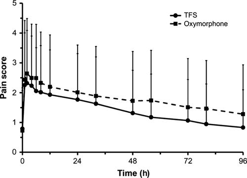
Pain scores were highest 2 h following extubation in both groups where values were 2.32 ± 1.77 (mean ± SD) in TFS- and 2.64 ± 1.85 in oxymorphone-treated dogs (Fig. 1). Pain scores declined throughout the study such that by 4 days poststudy the mean pain scores were 0.83 ± 1.27 in TFS and 1.28 ± 1.65 in oxymorphone-treated dogs. The 95% CI of the mean difference of TFS – oxymorphone pain scores at each pain assessment time contained 0 or was less than 0 throughout the study, suggesting that a single topical dose of TFS provides superior analgesia compared to repeated injections of oxymorphone.

Overall, there were 5 TFS- and 27 oxymorphone-treated dogs that were considered treatment failures due to lack of effectiveness or adverse events (Table 2). Twelve dogs (4 TFS, 8 oxymorphone) were withdrawn due to lack of pain control. The 4 TFS-treated dogs experiencing lack of pain control were withdrawn between 1 and 6 h postextubation with pain scores ranging from 8 to 11. The 8 oxymorphone-treated dogs were withdrawn between Day 0 at 2 hours postextubation and Day 2 with pain scores ranging from 8 to 15. No TFS-treated dogs were withdrawn due to adverse events, and none required reversal with naloxone. Eighteen oxymorphone dogs were withdrawn due to adverse events or were administered naloxone for opioid reversal (Table 3). All but one of the 18 dogs was removed were considered treatment-related. The dog that was removed was not considered as treatment-related but experienced a recurrent prolapsed rectum following surgery to reduce a prolapsed rectum (Table 3). There was no difference in the dose of oxymorphone in dogs withdrawn from the study due to lack of pain control or adverse events compared with those that remained in the study. The most common reasons oxymorphone-treated dogs in the orthopedic surgery population were withdrawn from the study were profound/persistent sedation, hypothermia, bradycardia, bradypnea, vomiting, and anorexia or some combination of these events. Nine of the 18 oxymorphone-treated dogs were reversed with naloxone. One TFS- and 1 oxymorphone-treated dog did not complete the study due to death (see the Adverse events section below).
| Reason | TFS (N = 249) | Oxymorphone (N = 253) |
|---|---|---|
| Safety | ||
| Adverse event | 0 (0.0%)a | 9 (3.6%)b |
| Opioid reversal | 0 (0.0%)a | 9 (3.6%)b |
| Death | 1 (0.4%) | 1 (0.4%) |
| Effectiveness | ||
| Lack of pain control | 4 (1.6%) | 8 (3.2%) |
| Total | 5 (2.0%)a | 27 (10.7%)b |
- Within a Reason, percentages with different a, b superscripts are statistically different (P ≤ 0.05) per a post hoc two-sided Fisher's exact test.
| Signalment | Surgery type | Adverse events | Withdrawal time | Naloxone reversal | Outcome |
|---|---|---|---|---|---|
| 4-year-old spayed Rottweiler | TPLO | Profound sedation, hypothermia, bradycardia and bradypnea | Approximately 9 h following surgery | Yes | Remained at the clinic without incident until the scheduled discharge on Day 4 |
| 9-year-old castrated crossbred Chow | TPLO | Profound sedation, bradycardia, a decreased respiratory rate, and hypothermia | Approximately 7.5 h following surgery | Yes | Remained at the clinic without incident until the scheduled discharge on Day 4 |
| 6-year-old spayed German Shepherd mixed breed | TPLO | Bradycardia, hypothermia, and dyspnea | Approximately 9 h following surgery | Yes | Remained at the clinic without incident until the scheduled discharge on Day 4 |
| 2-year-old spayed Keeshond | TPLO | Tenesmus, colitis, nausea, vomiting, anorexia and fever | Day 1 through Day 3 | No | Resolution by Day 4 and discharged to owner |
| 5-year-old castrated German Shepherd | TPLO | Anorexia, vomiting and nausea | Day 0 through Day 3 | No | Anorexia continued at Day 4 discharge and dog reported normal 1 week following discharge. |
| 6-year-old spayed Boston Terrier | Fabellar suture | Hypothermia and bradycardia | Day 0 through Day 3 | No | Resolution by Day 4 and discharged to owner |
| 3-year-old spayed English Bulldog | TTA | Tachypnea and hypothermia | Approximately 2.5 h following surgery | Yes | Remained at the clinic without incident until the scheduled discharge on Day 4 |
| 11-year-old spayed crossbred Labrador retriever | Lateral ear resection | Ataxia, opisthotonus, clonus, panting and seizure | Day 3 | Yes | Remained at the clinic without incident until the scheduled discharge on Day 4 |
| 5-year-old spayed Cocker Spaniel | Lateral ear resection | Profound sedation | Approximately 12 h following surgery | Yes | Remained at the clinic without incident until the scheduled discharge on Day 4 |
| 6-month-old intact female West Highland White terrier | OVH | Prolapsed rectum | Day 1 | No | Prolapse surgically reduced by purse string; dog treated for intestinal parasites and discharged 7 days following surgery |
| 9-month-old intact female Golden Retriever crossbred | OVH | Persistent excessive sedation, anorexia and vomiting | Approximately 18 h following surgery | No | Remained at the clinic without incident until the scheduled discharge on Day 4 |
| 3-year-old intact female Pit Bull | Liver biopsy | Hypotension and hypothermia | Intra-operative | Yes | Remained at the clinic without incident until the scheduled discharge on Day 4 |
| 6-month-old intact female Cocker Spaniel crossbred | OVH | Persistent sedation and anorexia | Day 2 | No | Remained at the clinic without incident until the scheduled discharge on Day 4 |
| 7-year-old spayed Labrador retriever crossbred | Cystotomy | Sedated, depression and vomiting | Day 2 | No | Remained at the clinic without incident until the scheduled discharge on Day 4 |
| 10-year-old spayed Chihuahua | Cystotomy | Persistent somnolence, vomiting and incision purulent discharge | Day 3 | Yes | Enrofloxacin and amoxicillin/clavulanic acid was continued for 3 weeks following discharge where the dog had fully recovered |
| 2-year-old spayed Bichon Frise | Cystotomy | Persistent vomiting | Day 1 through Day 2 | Yes | Remained at the clinic without incident until the scheduled discharge on Day 4 |
| 4-year-old intact female Boston Terrier | OVH | Persistent vomiting and anorexia | Day 2 | No | Remained at the clinic without incident until the scheduled discharge on Day 4 |
| 3-year-old intact male Beagle | Liver biopsy | Vomiting, lethargy and dehydration | Day 2 | No | Remained at the clinic without incident until the scheduled discharge on Day 4 |
The primary variable for determining effectiveness was a noninferiority evaluation of the TFS and oxymorphone treatment failure rates, which were 2.0% (5/249) and 10.8% (27/251), respectively. The point estimate of the difference between TFS and oxymorphone failure rates was −8.75% and the one-sided upper 95% confidence interval was −5.3%, which was not greater than 15%. Therefore, based on the treatment failure rate difference, TFS was noninferior to oxymorphone at the dose rates of both formulations used in this study'.
Sedation
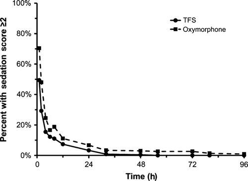
The percentage of oxymorphone-treated dogs with sedation scores ≥2 was higher than the percentage of TFS-treated dogs at all time points (Fig. 2). At 1-h postextubation, 49% and 70% of TFS- and oxymorphone-treated dogs, respectively, had a sedation scores ≥2 and by 12 h this had diminished to 7% and 11% of dogs, respectively. No TFS-treated dogs had a sedation scores ≥2 beyond 48 h. Additionally, the 95% CI of the mean difference of TFS – oxymorphone sedation scores were less than 0 at all time points, suggesting that TFS-treated dogs experienced less sedation than oxymorphone-treated dogs.

Body weight
Over the 4 days of the study, a statistically greater number of TFS-treated dogs lost no body weight (24.3%) compared to those treated with oxymorphone (12.4%). Approximately, equal percentages of TFS- and oxymorphone-treated dogs lost up to 5% of their body weight (51% and 45%, respectively). A statistically greater proportion of oxymorphone-treated dogs lost larger percentages of body weight; 34.5% of oxymorphone-treated dogs lost between 5% and 10% body weight and 8% of oxymorphone-treated dogs lost between 10% and 15% body weight. Twenty one point nine percent (21.9%) of TFS-treated dogs lost between 5% and 10% body weight and 1.6% of TFS-treated dogs lost between 10% and 15% body weight.
Clinical pathology
The mean value for each hematology and clinical chemistry variable was within the reference range prior to treatment and at study completion (Day 4) for both treatment groups. There were no individual excursions outside the normal range that were considered causally related to fentanyl or oxymorphone treatment. There were 5 TFS-treated treated dogs that had a normal amylase at baseline that was elevated at study completion. Of the five dogs with elevated amylase, four underwent TPLO surgery and one had a cryptorchid testes removed. There were no adverse event in any of the five dogs and each completed the study.
Adverse events
Eighty-five percent (211/249) of dogs allotted to TFS and 86.1% (216/251) of dogs allotted to oxymorphone were provided external heat during surgery to support core body temperature. There were no adverse safety events related to heat application at the administration site in TFS-treated dogs. A post hoc statistical analysis of adverse events during anesthesia revealed two events that were significantly different between treatments during this time period (Table 4). Hypothermia affected 28% (71/251) and 9.2% (23/249) of oxymorphone- and TFS-treated dogs during anesthesia, respectively (P < 0.05), while tachycardia occurred in 3.2% (8/251) oxymorphone-treated dogs and 10% (26/249) of TFS-treated dogs (P < 0.05). Tachypnea and bradypnea were the most frequent adverse events during anesthesia, affecting approximately 60% and 45% of dogs in each group, respectively. The remaining adverse events during anesthesia were less frequent and approximately equal between groups. A single oxymorphone-treated dog was reversed with naloxone approximately 30 min into liver lobectomy/biopsy surgery due to severe hypotension and hypothermia. Fluids were also administered, and the blood pressure was immediately increased, and surgery was completed without further incident. No TFS-treated dogs were reversed with naloxone during surgery.
| Adverse event* | TFS(N = 249) | Oxymorphone(N = 251) |
|---|---|---|
| Tachypnea (>20 breaths/min) | 158 (63%) | 151 (60%) |
| Bradypnea (<10 breaths/min) | 116 (47%) | 108 (43%) |
| Hypertension | 37 (15%) | 32 (13%) |
| Hypotension | 32 (13%) | 46 (18%) |
| Hypothermia (<35 °C) | 23 (9.2%)a | 71 (28%)b |
| Tachycardia (>180 beats/min) | 26 (10%)a | 8 (3.2%)b |
| Bradycardia (<50 beats/min) | 9 (3.6%) | 7 (2.8%) |
| Arrhythmia Noted | 2 (0.8%) | 2 (0.8%) |
| Pyrexia (>39.2 °C) | 3 (1.2%) | 1 (0.4%) |
| Oxygen saturation (<85%) | 2 (0.8%) | 3 (1.2%) |
- *Physiological adverse events during general anesthesia were included if there was a least one excursion outside the normal anesthetic range at any 5 min interval during the entire duration of anesthesia.
- Within an Adverse Event, percentages with different a, b superscripts are statistically different (P < 0.05) per a post hoc two-sided Fisher's exact test.
There were a total of 56 individual postoperative adverse events reported in 44 (17.7%) TFS-treated dogs and a total of 228 postoperative adverse events reported in 84 (33.7%) oxymorphone-treated dogs. There was only one severe adverse event in the TFS treatment group, compared with 28 severe adverse events in oxymorphone-treated dogs. All 28 severe adverse events in the oxymorphone-treated dogs occurred in the 19 dogs that were removed from the study (Table 2). Over the first 48 h postoperatively, the most frequent adverse events in TFS-treated dogs were diarrhea ranging from 0.4% to 2%, emesis ranging from 0 to 1.6%, hypothermia ranging from 1.5% to 4.4% and anorexia ranging from 0% to 0.8% (Table 5). The incidence of adverse events in oxymorphone-treated dogs was higher in some categories compared to TFS and persisted throughout the 4-day study period (Table 5). Over the 4-day study period, emesis ranged from 1.6% to 8.7% and hypothermia ranged from 1.4% to 9.5% in oxymorphone-treated dogs.
| Treatment | Adverse event | Day 0 | Day 1 | Day 2 | Day 3 | Day 4 |
|---|---|---|---|---|---|---|
| TFS(n = 249) | Diarrhea | 1 (0.4%) | 5 (2.0%) | 2 (0.8%) | 1 (0.4%) | 0 (0.0%) |
| Emesis | 0 (0.0%)a | 4 (1.6%) | 2 (0.8%)a | 2 (0.8%)a | 0 (0.0%) | |
| Hypothermia | 4 (1.6%)a | 11 (4.4%)a | 0 (0.0%) | 0 (0.0%) | 0 (0.0%) | |
| Pyrexia | 0 (0.0%) | 0 (0.0%) | 1 (0.4%) | 1 (0.4%) | 0 (0.0%) | |
| Anorexia | 0 (0.0%) | 2 (0.8%) | 1 (0.4%) | 0 (0.0%) | 0 (0.0%) | |
| Constipation | 0 (0.0%) | 0 (0.0%) | 0 (0.0%) | 0 (0.0%) | 0 (0.0%) | |
| Hypersalivation | 0 (0.0%) | 0 (0.0%) | 0 (0.0%) | 0 (0.0%) | 0 (0.0%) | |
| Conjunctivitis | 0 (0.0%) | 0 (0.0%) | 0 (0.0%) | 0 (0.0%) | 0 (0.0%) | |
| Death | 0 (0.0%) | 0 (0.0%) | 0 (0.0%) | 1 (0.4%) | 0 (0.0%) | |
| Oxymorphone(n = 253) | Diarrhea | 3 (1.2%) | 3 (1.2%) | 5 (2.0%) | 4 (1.6%) | 0 (0.0%) |
| Emesis | 10 (4.0%)b | 11 (4.4%) | 22 (8.7%)b | 15 (6.0%)b | 4 (1.6%) | |
| Hypothermia | 16 (6.3%)b | 24 (9.5%)b | 4 (1.6%) | 5 (2.0%) | 4 (1.6%) | |
| Pyrexia | 0 (0.0%) | 0 (0.0%) | 0 (0.0%) | 1 (0.4%) | 0 (0.0%) | |
| Anorexia | 0 (0.0%) | 5 (2.0%) | 4 (1.6%) | 2 (0.8%) | 1 (0.4%) | |
| Constipation | 0 (0.0%) | 1 (0.4%) | 0 (0.0%) | 0 (0.0%) | 0 (0.0%) | |
| Hypersalivation | 5 (2.0%) | 1 (0.4%) | 1 (0.4%) | 0 (0.0%) | 1 (0.4%) | |
| Conjunctivitis | 0 (0.0%) | 1 (0.4%) | 0 (0.0%) | 0 (0.0%) | 0 (0.0%) | |
| Death | 0 (0.0%) | 0 (0.0%) | 1 (0.4%) | 0 (0.0%) | 0 (0.0%) |
- Within an Adverse Event and Day, percentages with different a, b superscripts are statistically different (P < 0.05) per a post hoc two-sided Fisher's exact test.
There were two deaths in this study; one each in the fentanyl- and oxymorphone-treated groups. A 13-year-old, castrated, crossbred terrier presented with 4-day history of vomiting and was allotted to fentanyl and underwent gastrotomy surgery to remove a suspected foreign body. No foreign body was identified, but some debris was noted in the caudal esophagus, possibly dog treat fragments. An increased respiratory rate on Days 1 and 2 was considered secondary to pulmonary edema and was treated with furosamide through Day 2. The dog acutely died the morning of Day 3. Necropsy and histopathological evaluation revealed moderate, suppurative pneumonia of bacterial etiology possibly secondary to vomiting and aspiration. An 11-year-old castrated crossbred Labrador retriever was allotted to oxymorphone and underwent splenectomy surgery. On Day 2, the dog was restless and vomited, collapsed and was asystolic. Cardiopulmonary resuscitation was not successful and the dog died. Necropsy revealed a large infarct in the cranial right lung lobe. Histopathology revealed changes consistent with both chronic and acute right-sided heart failure. In both cases, the deaths were judged to be unrelated to investigational or control drug treatment.
Discussion
The results from this study demonstrates that a single dose of TFS applied 2–4 h prior to surgery is safe and effective for the control of pain associated with orthopedic and soft-tissue surgery in dogs and provides analgesia for 4 days. The number of treatment failures due to inadequate pain control were low in both treatment groups indicating that TFS and repeated oxymorphone injections were highly effective in controlling postoperative pain through 4 days postsurgery, a time period demonstrated to result in clinically significant postoperative pain in dogs (Clark et al., 2001; Martinez et al., 2001). As a primary endpoint, the failure rate over 4 days in dogs treated with a single dose of TFS was noninferior to those treated with every 6 h injections of oxymorphone through 90 h. Transdermal fentanyl solution appeared to have a safety advantage compared to oxymorphone at the doses used in this study, with fewer adverse events and adverse events of lesser severity.
A single dose of TFS provides continuous systemic fentanyl delivery over a period of days, and therefore, the control drug choice from an experimental design and regulatory perspective was critical. A placebo control was not deemed ethical because well-studied active controls were available. Oxymorphone hydrochloride injection is an FDA approved opioid for the control of postoperative pain in dogs (NADA 030-535) and was therefore chosen as an active control. Although a Pharmacokinetic/pharmacodynamic (PK/PD) relationship has not been established for oxymorphone at the doses used in this study, repeated injections at a 6-h dosing interval results in effective, long-term opioid exposure (KuKanich et al., 2008) and analgesia in dogs (Hardie et al., 1997; Kyles et al., 1998; Bateman et al., 2008). A noninferiority analysis was chosen instead of a superiority analysis (Weintraub, 2010) because it would not be likely that a full μ-agonist opioid would be superior to another opioid in the same class. However, the one-sided upper 95% confidence bound of the TFS minus oxymorphone treatment failure difference was −5.3%, indicating that on the composite endpoint of treatment failure, TFS was superior over oxymorphone. Similarly, the percentage of subjects that failed due to lack of pain control was higher in oxymorphone-treated dogs (3.2% [8/251]) than in fentanyl-treated dogs (1.6% [4/249]). This difference may be due to steady fentanyl payout with TFS compared with end of dosing interval lack of effectiveness associated with trough oxymorphone concentrations that occur with any opioid when must be repeatedly administered.
The results in this trial were comparable with a similarly designed clinical study conducted in Europe where the positive control was IM buprenorphine (20 μg/kg) administered every 6 h for 90 h instead of SC oxymorphone (Linton et al., 2012). In the European study, the TFS and buprenorphine failure rates were 6.7% (15/223) and 3.6% (8/220), respectively, with a one-sided upper 95% confidence interval of the failure rate difference of 5.6%. Like the current study, the a priori selected margin of the difference was 15% and a single dose of TFS applied 2–4 h prior to surgery was concluded to be noninferior to repeated buprenorphine injections. Thus, TFS has been demonstrated to be safe and effective compared to both repeatedly administered injectable full and partial μ-opioid receptor agonists (e.g., oxymorphone and buprenorphine, respectively).
Furthermore, the results of this current study are consistent with the outcome reported comparing postoperative analgesia of extra-label use of the fentanyl patches to repeated oxymorphone administration in dogs (Kyles et al., 1998). In that comparatively small study (n = 10 per treatment group), a single 50 μg/h fentanyl patch applied 20 h prior to ovariohysterectomy provided comparable analgesia over 24 h to IM 0.05 mg/kg oxymorphone administered presurgery and every 6 h through 18 h postsurgery. A 50 μg/h fentanyl patch had been shown in a previous study (Kyles et al., 1996) in dogs to provide steady-state plasma fentanyl concentrations of 1.60 ng/mL, similar to the average plasma fentanyl concentrations of 1.32 ng/mL achieved in the present clinical trial (Freise et al., 2012a). Average plasma fentanyl concentrations of ≥0.6 ng/mL are generally considered analgesic in dogs (Hofmeister & Egger, 2004).
A marked contrast between TFS and oxymorphone treatments in this study was measures of safety. There were 18 oxymorphone-treated dogs removed from the study due to severe adverse events, nine of which required reversal by naloxone (Table 2). There were no fentanyl-treated dogs removed due to adverse events, and no fentanyl-treated dogs required reversal with naloxone. However, if fentanyl-treated dogs did need to be reversed due to adverse opioid effects, hourly IM administration of naloxone at a dose of 0.04 or 0.16 mg/kg has been demonstrated to provide sustained TFS reversal (Freise et al., 2012b). Alternatively, a constant rate naloxone infusion of 1–4 μg/kg/h has been predicted to provide similar effects. The number and severity of adverse events were also much greater in oxymorphone-treated than fentanyl-treated dogs (Table 5). An explanation for the observed safety difference may be related to the peak and trough drug plasma concentrations that occur after each oxymorphone dosing compared with the steady fentanyl drug concentrations that occur following TFS administration (Freise et al., 2012a,c,d).
Respiratory depression was not a reported adverse event in the TFS treatment group. Unlike in humans, spontaneous respirations are maintained independent of fentanyl concentration in dogs (Bailey et al., 1987; Mathews, 2000). In a study where TFS was administered up to 5-times the recommended dose, mean respiration rates were similar to placebo at the FDA approved dose but decreased slightly in a dose-dependent manner at higher doses to a maximal decrease of approximately 30% (Savides et al., 2012). The present study demonstrates that established tolerance to opioid-induced respiratory depression is not necessary prior to initiating treatment with TFS in dogs.
Along with the desired analgesic effects, sedation is an expected extension of the pharmacological effect of opioids (Gutstein & Akil, 2006). However, excessive sedation was not a key feature of either treatment in this study. At no time was the mean sedation score ≥2 even during the times near extubation where the effects of general anesthesia on sedation scores would be expected to be greatest. Comparatively, oxymorphone resulted in higher mean sedation scores at each time point compared to TFS with some individual dogs having sedation scores ≥2 for the entire 4-day study duration. This too may be the result of steady fentanyl concentrations in contrast to repeated peak oxymorphone concentrations following multiple injections.
In summary, this study demonstrates that a single, small volume of TFS administered 2–4 h prior to surgery provides noninferior efficacy with less adverse events compared with repeated injections of oxymorphone every 6 h over 4 days. It was similar to oxymorphone in behavioral measures of pain over 4 days and exhibited a greater margin of safety with regard to adverse events, sedation and body weight loss. The availability of an FDA approved, long-acting opioid could allow further optimization of postoperative analgesia in dogs.
Footnotes
Acknowledgments
The authors wish to acknowledge the following clinical investigators for recruitment and enrollment of the clinical cases included in this study: LeeAnn Blackford, DVM, DACVS (Blackford Veterinary Surgery Referral, Knoxville, TN); Todd Gauger, DVM (Norway Veterinary Hospital, Norway, ME); Jack Gallagher, DVM, DACVS (Veterinary Surgical Referral Practice, Cary, NC); Samuel Geller, VMD (Quakertown Veterinary Clinic, Quakertown, PA); John Mauterer, DVM, DACVS (Louisiana Veterinary Referral Center, Mandeville, LA); Stephan Ladd, DVM (Hillsboro Animal Hospital, Nashville, TN); David Lukof, VMD (Harleysville Veterinary Hospital, Harleysville, PA); Andrew Pickering, DVM (Wabash Valley Animal Hospital, Terre Haute, IN); Bert Shelley, DVM, MS, DACVS (Bradford Park Veterinary Hospital, Springfield, MO); Fred Williams, DVM (South Texas Veterinary Specialist, San Antonio, TX); John T. Peacock, DVM (MedVet Memphis, Memphis, TN); John Stephan, DVM, MS, DACVS (Indianapolis Veterinary Specialist, Indianapolis, IN); Richard N. Benjamin, DVM (Berkley Dog and Cat Hospital, Berkley, CA); Scott Buzhardt, DVM (The Animal Center West, Zachary, LA); Shane Daigle, DVM (Premier Animal Hospital, Cedar Park, TX); Peter Davis, DVM (Pine Tree Veterinary Hospital, Augusta, ME); Mark Girone, DVM (PetMed, Antioch, TN); Kristy Lively, DVM (Village Veterinary Clinic & Laser Center, Farragut, TN); Mark Marks, DVM (Wakarusa Veterinary Hospital, Lawrence, KS); Wanda Pool, DVM (Deepwood Veterinary Clinic, Centreville, VA); Mike Shelton, DVM (380 West Animal Hospital, McKinney, TX); Roger L. Sifferman, DVM (Bradford Park Veterinary Hospital, Springfield, MO); Joe Wurster, DVM (Colonial Park Animal Hospital, Wichita Falls, TX).
Appendix 1
Food and Drug Administration approved oxymorphone dose (NADA 030-535) used for dogs allotted to the oxymorphone treatment group (Branson & Gross, 2001)
| Body weight | Amount (mg) | Dose (mg/kg) |
|---|---|---|
| 0.9–2.7 | 0.75 | 0.83–0.28 |
| >2.7–6.8 | 1 | 0.37–0.14 |
| >6.8–13.6 | 2 | 0.29–0.15 |
| >13.6–27.2 | 3 | 0.22–0.11 |
| >27.2 | 4 | >0.14 |
Appendix 2
Sedation score scale
0 – No Sedation Present.
1 – Slight Sedation – almost normal; able to stand easily, but appears somewhat fatigued, subdued or somnolent.
2 – Moderate Sedation – able to stand but prefers to be recumbent; sluggish; ataxic or uncoordinated.
3 – Profound Sedation – unable to rise, but can exhibit some awareness of environment; responds to stimuli through body movement; may be lateral or sternal recumbency.
4 – Unresponsive – in a state of coma or semi-coma from which little or no response can be elicited; remains in lateral recumbency.
If the sedation score was ≥2, then the dog was considered to be too sedated to adequately assess analgesia and the pain score assessment was not conducted at that time.


