Lesser vestibular periurethral gland-like inflammation associated with lower urinary tract signs in a female dog
Abstract
Case Description
A 4-year-old female spayed mixed breed dog presented with a 2-year history of painful urination and recurrent hematuria.
Clinical Findings
The dog had a large sensitive bladder, palpation of which was followed by painful urination. Pollakiuria accompanied by vocalization were noted during observation of voiding.
Diagnostics
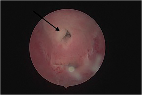
Cystoscopy identified a focal, rounded expansion of epithelial tissue in the right lateral aspect of the urethral papilla containing purulent material consistent with an abscess. A sample submitted for culture yielded growth of Staphylococcus pseudintermedius and Proteus mirabilis.
Treatment and Outcome
Purulent material was expelled by manual pressure during cystourethroscopy. Enrofloxacin (10 mg/kg PO q24h for 42 days) and carprofen (4.4 mg/kg PO q24h for 14 days) were initiated. Clinical signs resolved within 2 days.
Clinical Relevance
Inflammation in the region of the lesser vestibular paraurethral glands should be considered as a differential for female dogs presenting with chronic dysuria.
Abbreviations
-
- BID
-
- twice daily dosing
-
- CFU
-
- colony forming units
-
- MRI
-
- magnetic resonance imaging
-
- USG
-
- urine specific gravity
1 INTRODUCTION
Skene's glands, or lesser vestibular glands, are a subset of paraurethral glands that, when inflamed, have been implicated in the manifestation of acute lower urinary tract symptoms of women.1, 2 The paraurethral glands are homologous to the prostate gland in males. The periurethral glands are located posterolaterally along the course of the urethra within the submucosal layer. The lesser vestibular glands are present only in females and are the distal-most collection of the periurethral glands located inferiorly and laterally to either side of the urethra, adjacent to the urethral meatus in the vestibule. Paraurethral, including lesser vestibular glands, recently were identified in dogs, located caudolaterally and ventrally along the urethra.3 Our purpose was to describe clinical presentation and findings, that were consistent with the condition known as skenitis, or inflammation in the region of the Skene's, or lesser vestibular paraurethral glands in a dog.
2 CASE DESCRIPTION
A 4-year-old female spayed mixed breed dog was presented to NC State Veterinary Hospital with a 2-year history recurrent hematuria along with painful urination characterized by straining and vocalizing. The dog initially presented to the primary care veterinarian for inappropriate urination, lethargy, and decreased appetite. Concentrated urine (USG, 1.050) and proteinuria (1+) were found on urinalysis. A commercial ELISA test (Snap 4Dx, Idexx Laboratories, Westbrook, Maine) was positive for Borrelia burgdorferi; treatment with doxycycline (5 mg/kg PO q12h for 14 days) was initiated. Lethargy and hyporexia improved, but painful urination, recurrent malodorous urine and hematuria persisted. Diagnostic tests performed over a course of 2 years by the primary care veterinarian included repeated urinalyses, urine cultures, abdominal radiographs, abdominal ultrasound examination, and a PCR-based genetic test (Cadet BRAF, Antech Diagnostics, Fountain Valley, California) for detection of BRAF mutation based on poor clinical response to attempted treatment. Therapeutic interventions in the year before presentation included enrofloxacin, amoxicillin-clavulanic acid, cefpodoxime, and feeding a diet for urinary tract disease (Urinary SO, Royal Canin, St. Charles, Missouri). Each antibiotic course was continued for 7 days. No response was noted to these treatments. Other medical history included atopic dermatitis and chronic intermittent vomiting of undetermined etiology.
The dog was bright, alert, and responsive but anxious during examination. Abnormal physical examination findings included vulvar erythema, moderate perivulvar skin folding covering approximately 60% of the vulva, erythematous skin over the nasal planum, periocular, and interdigital regions, as well as dry and crusted ear pinnae. The bladder was large, and immediately after palpation the dog voided a small volume of urine while vocalizing. When taken outside to observe urination, the dog appeared reluctant to urinate, and eventually urinated small volumes multiple times whereas vocalizing. Post-voiding urine residual volume measurements using a 3-D ultrasound device (BladderScan Prime Plus, Verathon, Bothell, Washington) were normal (<1 mL/kg). Results of a CBC and biochemistry profile were within reference range except for mild hypophosphatemia (1.6 mg/dL; reference range [RR], 2.6-5.3) and hypomagnesemia (1.7 mg/dL; RR, 1.9-2.5). Urinalysis of a cystocentesis sample disclosed hyposthenuria (USG 1.007) and bacteriuria (2+) in the absence of pyuria. The remainder of the urinalysis was unremarkable. Abnormalities detected during ultrasound examination limited to the urinary tract included cranioventral bladder wall thickening (0.63 cm in width) suggestive of cystitis and mild left medial iliac lymphadenopathy (0.67 cm).
To further investigate the patient's condition, cystourethroscopy was performed with the dog under general anesthesia. To minimize the likelihood of contamination during the procedure, the external perivulvar area was aseptically prepared, the vestibule was irrigated with betadine solution, and the endoscopist wore sterile gloves. Upon passage of the scope into the urethra, purulent material was released from the right aspect of the urethral papilla. Further examination of this site disclosed a rounded expansion of the epithelial tissue, similar in appearance to an inflamed cyst or abscess (Figure 1). Digital examination identified a small but palpable, firm structure in that location. Direct swabs of the site were collected for bacterial culture. Attempts were made to further drain the structure by passage of a needle through the endoscope's biopsy channel, but, no additional purulent material was expelled, and the structure remained intact. Mild iatrogenic trauma and self-limiting hemorrhage occurred. No other abnormalities were noted in the vestibule. The endoscope was passed through the urethra and into the bladder; no urethral abnormalities were appreciated. The bladder wall appeared diffusely edematous and 2 small sites of hyperemia and mild mucosal hemorrhage were noted. Biopsy samples were obtained from the ventral bladder wall for histopathological examination and aerobic culture. Urine was collected for Ureaplasma culture. Anesthetic recovery was uneventful.

Histopathology of the bladder biopsy sample identified hyperplastic bladder mucosa with multifocal areas of edema within the epithelium. Scattered intraepithelial lymphocytes and a few neutrophils were present. The superficial submucosa was expanded by edema, mild hemorrhage with erythrocyte fragmentation, and scattered macrophages. Mild hemorrhage extended through the mucosa. These findings were suggestive of chronic inflammation in the bladder. Both the direct swab of the periurethral lesion and the culture of bladder wall tissue yielded growth of Staphylococcus pseudintermedius and Proteus mirabilis. Fewer than 10 colony-forming units of each organism were grown from the bladder wall, whereas the direct swab yielded 1+ growth of S. pseudintermedius and <10 colonies of P. mirabilis. Both organisms had a broad antimicrobial susceptibility profile.
These findings were consistent with inflammation in the area of the lesser vestibular paraurethral glands. However, because there was no ultrasonographic or computed tomographic imaging of the lesion itself, it cannot be definitively stated that the gland was involved. A therapeutic plan was made based on typical treatments used in women with skenitis. The dog was discharged with instructions to be given enrofloxacin (10 mg/kg PO q24h) for 42 days and carprofen (4.4 mg/kg PO q24h) for 14 days. The previously prescribed gabapentin was continued. Transition to a hydrolyzed diet was directed given the patient's concurrent dermatologic and gastrointestinal disease. The dog did not urinate until the day after the procedure, at which point straining, vocalization, and hematuria were not noted. Follow-up communication several weeks later indicated complete resolution of the presenting complaints. The owner reported that the dog frequently leaked urine at rest. Because other causes for urinary incontinence were not identified during evaluation, a diagnosis of urethral sphincter mechanism incompetence was made, and the dog was treated with diethylstilbesterol (0.02 mg/kg PO) daily for 5 days and then decreased to twice weekly dosing; urinary incontinence reportedly resolved. Although the patient previously had been treated with enrofloxacin, the duration of treatment was limited to 7 days. We elected to try a 42-day course based on similar recommendations for urinary tract infections associated with prostatitis. A non-steroidal anti-inflammatory drug was prescribed primarily for discomfort, but decreased inflammation associated with the abscess may have facilitated resolution.
3 DISCUSSION
The dog of this report had a constellation of clinical signs, cystoscopic findings, and response to treatment similar to infection and inflammation of the Skene's gland in women. To our knowledge, it is the first report of disease affecting the region of these glands in dogs.
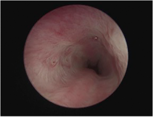
Paraurethral, including lesser vestibular glands, recently were identified in dogs, by histologic evaluation of urethras removed from 25 deceased female dogs (Figure 2).3 Hematoxylin and eosin-stained sections were evaluated for the presence of prostatic gland-like tissue. Other sections were marked with a polyclonal anti-prostate specific antigen antibody. Histopathological findings and immunohistochemical staining consistent with a female prostate-like gland were found in 8 (32%) of the 25 dogs. These findings are suggestive of the presence of the paraurethral, including lesser vestibular glands (ie, Skene's gland), in female dogs.3 Of the included dogs, only 3 were spayed, with 2 of those 3 having presence of the gland. These findings suggest an influence of hormones on the development or regression of this gland, but the number of dogs evaluated is too small to draw clinically relevant conclusions. The dog of this report was spayed at a young age (before 4 months of age) at which time the dog was adopted by the owners.

In women, electron microscopic evaluation of these glands results in findings suggestive of an active secretory function: presence of secretory vacuoles, secretory granules, rough endoplasmic reticulum, developed Golgi complexes, numerous mitochondria, and apical blebs.4 These glands are speculated to support sexual and urinary health by providing lubrication of the urethral opening, contributing to female ejaculation, and exhibiting antimicrobial properties.4, 5
Although the normal function of these glands is likely beneficial, similar to the male prostate gland, both benign and malignant abnormalities of the glands may develop, including cyst or abscess formation secondary to bacterial infection, adenomas, and adenocarcinomas.4, 6 Consistent with what was seen in the dog of this report, when Skene's glands become infected in women, they may become enlarged and tender, a condition known as skenitis. Severe skenitis can progress to urethral obstruction and urinary retention. In women, diagnosis of this condition is based on historical information and physical examination. Although dyspareunia and dysuria are the most common presenting symptoms, others may include pain, presence of a palpable mass, urinary tract infections, or obstructive voiding symptoms.1, 2, 7
The dog of this report had a palpable firm structure that was visualized on the lateral aspect of the urethral papilla. In women, approximately half of Skene's gland abscesses can be palpated and may be visualized as an erythematous painful mass lateral to the urethra. Clear or purulent material may be expressed when pressure is applied to the mass, and spontaneous rupture may be considered a form of conservative management.8 Imaging by ultrasound and magnetic resonance imaging may be necessary to identify and localize the lesion. If rupture is not noted, conservative management with antibiotics is considered the first line of treatment. Surgical intervention is warranted in cases non-responsive to antibiotics. Surgical approaches include cyst removal, marsupialization, puncture, and aspiration. A clear consensus on the preferable approach has not been established.8, 9 The dog of this report had resolution of clinical signs after rupture of the mass during cystoscopy followed by an extended course of antimicrobial treatment. It is possible that the antimicrobial treatment was unnecessary, and the dog would have responded to cystoscopic rupture and drainage of the mass alone. It is also possible that a shorter course of antimicrobial treatment plus drainage would have produced similar results.
In conclusion, inflammation in the region of the paraurethral glands may be considered as an uncommon differential diagnosis for female dogs presenting with chronic dysuria. Abscessation of the lesser vestibular glands may be associated with vocalization during urination, and possibly urine retention. Inflammation of the lesser vestibular glands may be noted cystoscopically with careful observation of the urethral papilla. Continued evaluation of similarly affected dogs is needed to determine if clinical resolution can be obtained by cystoscopic rupture and drainage alone or if antimicrobial treatment is also required.
ACKNOWLEDGMENT
No funding was received for this study.
CONFLICT OF INTEREST DECLARATION
Shelly L. Vaden serves as Associate Editor for the Journal of Veterinary Internal Medicine. She was not involved in review of this manuscript. No other authors declare a conflict of interest.
OFF-LABEL ANTIMICROBIAL DECLARATION
Authors declare no off-label use of antimicrobials.
INSTITUTIONAL ANIMAL CARE AND USE COMMITTEE (IACUC) OR OTHER APPROVAL DECLARATION
Authors declare no IACUC or other approval was needed.
HUMAN ETHICS APPROVAL DECLARATION
Authors declare human ethics approval was not needed for this study.


