Hereditary myotonia in cats associated with a new homozygous missense variant p.Ala331Pro in the muscle chloride channel ClC-1
Abstract
Three-related cats were evaluated for a history of short-strided gait and temporary recumbency after startle. Neurological examination, electromyography (EMG), muscle biopsies, and a chloride voltage-gated channel 1 (CLCN1) molecular study were performed. Clinically, all 3 cats presented myotonia with warm-up phenomenon and myotonic discharges during EMG examination. Muscle biopsies showed normal muscle architecture and variation in the diameter of myofiber size with the presence of numerous hypertrophic fibers. The molecular study revealed a missense variant (c.991G>C, p.Ala331Pro) in exon 9 of the CLCN1 gene, responsible for the first chloride channel extracellular loop. This mutation was screened in 104 control phenotypically normal unrelated cats, and all were wildtype. The alanine at this position is conserved in ClC-1 (chloride channel protein 1) in different species, and 2 mutations at this amino acid position are associated with human myotonia. This is the third CLCN1 mutation described in the literature associated with hereditary myotonia in cats and the first in domestic animals located in an extracellular muscle ClC-1 loop.
Abbreviations
-
- AST
-
- aspartate aminotransferase
-
- CBC
-
- complete blood count
-
- cDNA
-
- ribonucleic acid
-
- CIC1
-
- chloride channel protein 1
-
- CK
-
- creatine kinase
-
- CLCN1
-
- chloride voltage-gated channel
-
- DNA
-
- deoxyribonucleic acid
-
- EDTA
-
- ethylenediamine tetraacetic acid
-
- EMG
-
- electromyography
-
- H&E
-
- hematoxylin and eosin stain
-
- HM
-
- hereditary myotonia
-
- mRNA
-
- messenger ribonucleic acid (mRNA)
-
- PCR
-
- polymerase chain reaction
-
- RNA
-
- ribonucleic acid
-
- SNP
-
- single nucleotide polymorphisms
1 INTRODUCTION
Hereditary nondystrophic myotonia associated with chloride voltage-gated channel 1 (CLCN1) gene mutations occurs in goats,1 dogs,2-6 horses,7 buffalos,8 sheep,9 and pigs.10 Myotonia occurs in cats,11-13 and a mutation associated with this disease in this species is identified.14, 15 Here, we describe a clinical and molecular study in 3 cats with hereditary myotonia.
2 CASE REPORT
All procedures were approved by the Institutional Animal Care and Use Committee (0089/2021—CEUA) of São Paulo State University (UNESP). All methods were performed in accordance with the guidelines and regulations of CEUA. Authorization from the owner of the cats was obtained for the genetic study and images used in this manuscript.
3 HISTORY AND CLINICAL EXAMINATION
Three young mixed-breed cats from the same litter were presented for clinical evaluation with the complaint of stiff gait and recumbency after startle. During the first examination, they were 4 months old: 2 intact males (Cat 1 and Cat 2) and 1 female (Cat 3). The clinical manifestations had been noticed 1 month earlier by the owners who also reported abnormal vocalization patterns and difficulty in cleaning the caudal region of the body (genital area and pelvic limbs). Occurrence of vomiting, regurgitation and difficulty swallowing were denied, as were learning difficulties.
The 3 cats could vocalize only weakly (sometimes just miming meowing), and Cat 2 occasionally kept his tongue exposed and developed transient respiratory stridor during handling. Physical examinations of the 3 cats revealed enlargement of the cervical and proximal appendicular muscles and resistance to opening and then closing the mouth (Video S1). After we forced the cat's mouth open, each presented a delay to close it that became shorter as the forced attempts to open the mouth were repeated (warm-up effect present). These clinical changes were severe in Cat 1, moderate in Cat 2 and mild in Cat 3. Cat 1 and Cat 2 had moderate periodontal disease, gingivitis, and halitosis. Cat 1 also had a broken upper canine. During neurological examination, the 3 cats exhibited normal consciousness and behavior, but they were reluctant to move. They had short steps and stiff gait. All locomotion abnormalities presented phenotypically varying intensities and became less evident with muscle warm-up. When stimulated to jump or exposed to a sudden auditory stimulus, the cats contracted the muscles and then fell into lateral recumbency (startle response), a position from which they only came out seconds later (Video S2). There was also a warm-up effect for these tests. The presence of prolonged blepharospasm and protrusion of the third eyelid in response to digital stimulation of the palpebral fissure were observed (Video S3). No orthopedic abnormalities were identified, and the remaining findings from the physical and neurological examinations, including assessments of posture, postural reaction, spinal reflexes, cranial nerves, and sensation, were all within normal ranges. The cats were re-examined when they were 6 and 11 months old, and all the signs remained stable. The cats were reluctant to climb or descend obstacles. Interestingly, the cats showed an exaggerated reaction to stimulation in the dorsal region of the lumbar region, resulting in nibbling of the paws or surfaces that were supported (Video S4).
Given the observed muscular hypertrophy, improvement of muscle stiffness with exercise (demonstrating a “warm-up” phenomenon), startle response, and absence of other neurological signs, the presence of a skeletal muscle channelopathy was suspected. Specifically, hereditary myotonia associated with CLCN1 abnormality emerged as a highly plausible explanation, particularly in light of the absence of episodic weakness or muscle atrophy. To confirm the diagnosis of hereditary myotonia in these feline cases and to rule out other potential muscular abnormalities (such as myopathy presenting inclusions, nemaline myopathy, or glycogen storage disease), a comprehensive evaluation was conducted, which involved complete blood count (CBC), serum biochemical profile, electromyography, muscle biopsies, and molecular evaluation of CLCN1.
CBC and serum biochemical profile (BS-200E-Mindray, Shenzen, China) results (performed when cats were 11 months old), including creatine kinase (CK) activity (mean 376 IU/L), were within normal limits,16 except for a slight increase in AST. At the time of submission of this report, the 3 cats were 2 years old and had shown no change in their clinical condition. The kittens' mother was 15 years old when she gave birth to the litter, and there is no clear information about the father, as the mother has access to the street and neighbor's cats.
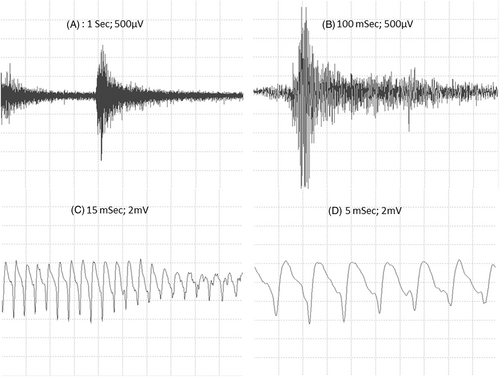
Electromyography (EMG) examinations were performed in all 3 cats in the standing position and after sedation. The proximal muscles of the pelvic and thoracic limbs and tongue were evaluated using concentric needle electrodes with electroneuromyography equipment (Neuromap ENMG/PE, Neurotec, Minas Gerais, Brazil). The tracing of the waveform, frequency, amplitude, and sound produced were documented. All 3 cats presented myotonic discharges and characteristic sounds (Figure 1; Audio S1) during EMG examination.
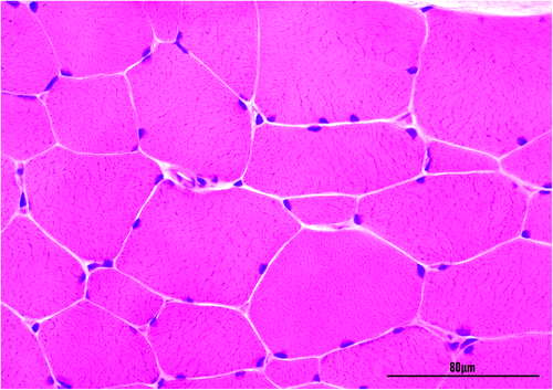
Immediately after the EMG examination and during anesthesia, biceps femoris muscle biopsy sections of Cat 1 and Cat 2 were obtained for histopathological evaluation (fixed in 10% buffered formalin, embedded in wax and stained with hematoxylin and eosin) and molecular analysis (snap frozen in liquid nitrogen). A sample from 1 control animal (with no previous history of neuromuscular abnormalities was also obtained postmortem for molecular study). Muscle biopsies showed a regular muscle architecture and variation in the diameter of myofiber size (ranging from 40 to 125 μm) with the presence of numerous hypertrophic fibers. There were no histological evidence of inflammation, necrosis, fibrosis central nucleation, sarcoplasmic inclusions or splitting fibers (Figure 2).
Blood samples were obtained from all 3 affected animals and stored in ethylenemiamine tetraacetic acid (EDTA) Vacutainer blood collection tubes (Becton Dickinson, São Paulo, São Paulo). These samples were used for CLCN1 exon/intron boundary evaluations and to obtain exon sequences. Genomic DNA was extracted from whole blood using ReliaPrep Blood gDNA Miniprep System (Promega, CA, USA) following the manufacturer's recommendations. Intronic primers previously described14 and other primers were designed (PrimerQuest software—Integrated DNA Technologies, Iowa, USA) to amplify the 23 exons of the Felis catus CLCN1 gene (RefSeq 101 088 564) (Table S1). The PCR (25 μL) contained 2.5 μL of template DNA (about 125 ng), 0.3 μM of each primer, 12.5 μL of PCR Master Mix (Promega, CA, USA), and 8.5 μL of nuclease-free water.
To characterize the full coding sequence of the cat CLCN1 gene, total RNA was obtained from muscle samples (≈30 mg) with SV Total RNA Isolation System (Promega, CA,) following the manufacturer's instructions. The RNA was treated with RQ1 RNase-Free DNase (Promega, CA, USA). First-strand cDNA synthesis was performed with 800 ng of total RNA per 60 μL of reaction volume using an ImProm-II Reverse Transcription System (Promega) and random primers following the manufacturer's instructions. To obtain the feline chlorine channel coding gene sequence, sets of primers were designed (PrimerQuest software—Integrated DNA Technologies) based on the CLCN1 mRNA sequence (NM_001305027.1) (Table S2). All amplicons were analyzed by 1.5% agarose gel electrophoresis, purified, and subjected to Sanger sequencing (Table S1). The obtained sequences and electropherograms were analyzed (Geneious v.2019.1.3; Biomatters, Auckland, New Zealand) and compared with the wildtype feline CLCN1 gene sequence.
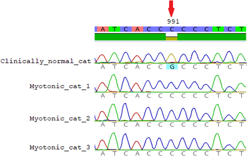
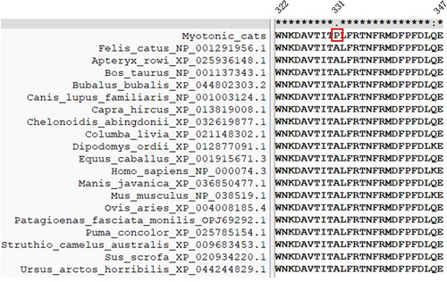
When analyzing the sequences of 23 CLCN1 exons obtained from the 3 affected cats with the Felis catus CLCN1 gene sequence (Genbank, NM01305027.1), we identified 4 variants. All variants were homozygous in the affected cats. Two of them were synonymous variants, c.1182T>G and c.1650G>T, and are located in exons 11 and 15, respectively. The other 2 were missense variants. The c.1855G>A Ala619Thr variant resulting in the substitution of alanine for threonine does not seem relevant, as these modifications are present in cats (NP_001291956.1) and in other species (Southern elephant seal KAF3819440.1 [Mirounga leonina], polar bear XP_040499815.1 [Ursus maritimus], ferret XP_004772118.1 [Mustela putorius furo], dog NP_001003124.1 [Canis lupus familiaris]). Whereas, the variant c.991G>C p.Ala331Pro in exon 9 (Figure 3) of the CLCN1 gene results in an amino acid replacement of alanine with proline (located in the first extracellular loop). The alanine in this position is conserved in ClC-1 in different species (Figure 4).
For variants that resulted in a change to the amino acid sequence, PROVEAN (http://provean.jcvi.org/index.php) was used to determine whether the sequence alteration is expected to significantly affect the protein's function. PROVEAN showed a deleterious score of −4.99 (scores of −2.5 or less are predicted to be deleterious) for p.Ala331Pro. For evaluation of protein stability, MUpro: Prediction of Protein Stability Changes for Single Site Mutations from Sequences (http://mupro.proteomics.ics.uci.edu/) was used, and a delta G = −0.96474552 was obtained, indicating decreased stability. Untrained predictors of protein stability changes upon mutation (https://folding.biofold.org/ddgun/method.html) also indicated decreased stability. Using SAAFEC-SEQ, an online application for calculating folding free energy changes in proteins caused by missense mutations, it was observed that variant p.Ala331Pro destabilizes the protein. The same was observed using PolyPhen-2 (http://genetics.bwh.harvard.edu/pph2/), which indicates that this substitution probably damages the protein function, with a maximum score.
The SNP c.991G>C (p.Ala331Pro) was screened in 104 control phenotypically normal unrelated cats (DNA bank from the clinical veterinary molecular biology laboratory) using primers designed (PrimerQuest software—Integrated DNA Technologies, I) for amplification of the entire exon 9 (Gandolfi_seq_9). The PCR, Sanger sequencing and electropherogram procedures were the same as those previously described. All 104 cats were wildtype for the p.Ala331Pro variant.
4 DISCUSSION
Among the muscular abnormalities, ion channelopathies, particularly chloride channel disorders resulting in hereditary myotonia, were placed at the top of the differential diagnosis list. This was based on signs of muscular hypertrophy associated with warm-up and startle response. Other signs in these 3 cats such as abnormal gait, collapse, dysphonia, and difficult swallowing, are associated with myopathy with oval inclusions,17 nemaline myopathy,18 glycogen storage disease,19 and myopathy with tubulin reactive inclusions20 but were not confirmed during muscle histological evaluation and usually presented muscular weakness and atrophy that were not observed in the cats described here. Abnormalities (such as replacement of muscle fibers by adipose and connective tissue) resulting from muscular dystrophy (Dystrophin-deficient myopathy) were also subsequently excluded through biopsy and there was no need to perform dystrophin immunohistochemistry.
The observed clinical signs of myotonia, associated with the warm-up effect and EMG findings, were consistent with the diagnosis of hereditary myotonia, possibly associated with CLCN1 mutation. Hereditary myotonia was phenotypically characterized in 2 young siblings domestic cats21 and 4 kittens.11 Subsequently, genetic characterization of the disease was performed in 5 affected cats in Canada14 and in a 10-month-old castrated male domestic longhair cat in North Carolina.15 The clinical signs in cats in our study are similar to the 4 previous reports in cats.11, 14, 15, 21
The main clinical signs already described in myotonic cats include stiffness, stilted gait, stiffness reduction during exercise, muscle hypertrophy, blepharospasm, and dysphonia,11, 12, 14, 15 all of which were observed in our study with different intensities, characterizing a phenotypic variation that is described in cases of myotonia. Tongue exposure and transient respiratory stridor were found in 1 of the 3 cats in this study (Cat 2) in the same way as in previous studies,14 but open-mouth breathing was not observed in the cats in our study. Moderate periodontal disease, gingivitis, and halitosis were found in 2 of the 3 cats, as already observed in other myotonic cats.14 Protrusion of the third eyelid is a previously unrecognized clinical sign in cats with hereditary myotonia. Resistance to opening and then closing the mouth is less described, but it has also been found.11 We noted the exaggerated reaction to stimulation in the dorsal region of the lumbar region, which resulted in nibbling of the paws or surfaces that supported the cats, in this study, and this has not been previously described.
The EMG findings were consistent with myotonic discharges as in other 2 reports.14, 15 Serum CK activity was within normal limits, as described in most cats with hereditary myotonia (only 1 animal in a study showed a slight increase14).
The muscle evaluation in this study showed variation of myofiber diameter, hypertrophy of the muscle fibers and no evidence of other abnormalities.22 Overall, histological changes in HM are nonspecific, with occasional degeneration of individual myofibers and moderate diffuse myofiber hypertrophy already described in myotonic cats.11, 12, 14, 21
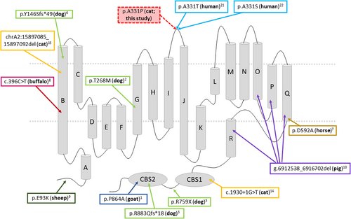
As in this study, all other mutations related to HM in domestic animals are autosomal recessive. The variant observed in this study is different from those observed in other domestic animals with myotonia (Figure 5), such as the first description of the mutation in cats located in the CBS domain14 and a recent mutation described in the first transmembrane domain,15 all of them are present in the ClC-1 protein. It is interesting to note that a mutation in the extracellular loop was detected for the first time in domestic animals. Three large extracellular loops link helices I-J (amino acids 322-347 based on UniProt), helices L-M (amino acids 427-457) and helices N-O (amino acids 499-521).23 As seen in several species of mammals, this region is highly conserved, with 100% similarity. As expected from their location, mutations on extracellular loops are typically less drastic than mutations within helices23; however, some mutations have already been described in this loop and associated with hereditary myotonia in humans (p.V327I24; I329T25; A331T26; A331S27; F337C23). Two mutations have been reported for residue R338*, one nonsense28 and one missense (R338Q).25 Interestingly, in this same position in humans,26 the functional consequences of a novel disease-causing mutation that predicts the substitution of alanine by threonine at position 331 (A331T) by whole-cell patch-clamp recording of recombinant mutant channels has been reported. A331T hClC-1 channels exhibit a novel slow gate that is activated during membrane hyperpolarization and closes at positive potentials. The A331T mutation causes an unprecedented alteration in ClC-1 gating and reveals novel processes defining transitions between open and closed states in ClC chloride channels. Similarly, A331S causes slow gate activation and may contribute to congenital myotonia, with recessive inheritance in humans.27 Although we did not perform patch clamp studies, the mutation in these cats is located in a very conserved region in the I-J extracellular loop; the 2 previous mutations at the same site described in humans and no clinically normal cat presenting this variant indicates that this mutation is likely the cause.
Limitations of this case report include the absence of parental DNA for mutation evaluation, as well as the lack of western blot to confirm protein predicted decreased stability or immunohistochemistry techniques to stain for type 1 and type 2 fibers and localize the voltage-dependent chloride channel, which could have provided additional information for these cases. Furthermore, because of the owner's decision not to pursue treatment for these 3 cats, primarily influenced by economic and daily management factors, pharmacological intervention was not administered.
ACKNOWLEDGMENT
No funding was received for this study.
CONFLICT OF INTEREST DECLARATION
Authors declare no conflict of interest.
OFF-LABEL ANTIMICROBIAL DECLARATION
Authors declare no off-label use of antimicrobials.
INSTITUTIONAL ANIMAL CARE AND USE COMMITTEE (IACUC) OR OTHER APPROVAL DECLARATION
Approved by the IACUC (0089/2021-CEUA) of São Paulo State University (UNESP).
HUMAN ETHICS APPROVAL DECLARATION
Authors declare human ethics approval was not needed for this study.


