Hyponatremia in horses with septic pneumopathy
Abstract
Background
Hyponatremia is common in horses with bacterial pleuropneumonia, but no further characterization of this abnormality has been reported.
Objectives
Describe admission plasma sodium concentration ([Na]) in horses with septic pneumopathy and evaluate any association of plasma [Na] with markers of systemic inflammation.
Animals
Medical records of horses >1 month of age that between 2008 and 2021 had a transtracheal aspirate (TTA) performed, abnormal TTA cytology, positive TTA culture, pulmonary disease on ultrasonography, radiography or both, and plasma [Na] assessed by direct ion-selective-electrode (dISE). Horses with concurrent diarrhea or azotemia were excluded.
Methods
Clinical and clinicopathological variables of interest between hypo- and normonatremic horses were compared. Spearman correlation and Fisher exact tests were used to identify significant associations (P < .05).
Results
Twenty of 35 horses had hyponatremia (median, 132 mmol/L; 25-75th interquartile range [IQR], 129.7-133.1 mmol/L; reference range, 134.2-138.4 mmol/L). A higher proportion of horses with systemic inflammatory response syndrome (SIRS) had hyponatremia (P = .01). Hyponatremic patients had higher mean plasma fibrinogen concentration (461 ± 160.5 mg/dL; P = .01) and higher rectal temperature (38.8 ± 0.7°C; P = .02) than normonatremic horses. Negative correlations were found between plasma [Na] and fibrinogen (P = .001; ρ = −0.57) concentrations and between plasma [Na] and rectal temperature (P = .001; ρ = −0.51). Presence or absence of pleural effusion did not influence severity of hyponatremia. Mean duration of hospitalization was longer (P = .04) in hyponatremic horses (9.8 ± 6.6 days).
Conclusions and Clinical Importance
Hyponatremia at admission is associated with the presence of inflammation, SIRS, and with longer duration of hospitalization.
Abbreviations
-
- ADH
-
- antidiuretic hormone
-
- 95% CI
-
- 95% confidence interval
-
- BUN
-
- blood urea nitrogen
-
- CBC
-
- complete blood count
-
- dISE
-
- direct ion-selective electrode
-
- DOH
-
- duration of hospitalization
-
- EMPF
-
- equine multinodular pulmonary fibrosis
-
- eOsm
-
- effective osmolality
-
- IL-6
-
- interleukin-6
-
- IQR
-
- interquartile range
-
- [Na]
-
- sodium concentration
-
- NSAID
-
- nonsteroidal anti-inflammatory drug
-
- OR
-
- odds ratio
-
- SD
-
- standard deviaiton
-
- SIADH
-
- syndrome of inappropriate ADH release
-
- SIRS
-
- systemic inflammatory response syndrome
-
- TTA
-
- transtracheal aspirate
1 INTRODUCTION
Pulmonary infections are common in adult horses as well as in suckling and weanling foals,1 with inhalation of the infectious causative agent from the nasopharynx or environment being the most common route of lower airway colonization.2-5 Risk factors that have been identified in adult horses for development of septic pneumonia, with or without pleural effusion, include long-distance transportation, prolonged head elevation, viral infections, mechanical ventilation during general anesthesia, esophageal obstruction, and immunosuppression.6-10 In foals transportation and other stressors, such as weaning, high ambient temperature, and overcrowding also play important roles in the development of pneumonia.11, 12 Commonly reported clinicopathologic abnormalities include leukocytosis or leukopenia, neutrophilia, anemia of chronic inflammation, hyperfibrinogenemia, hyperglobulinemia, and azotemia.12, 13
Hyponatremia can affect up to 30% of human patients with pneumonia,14-16 whereas in horses with septic pleuropneumonia it is reported as the second most common biochemistry abnormality.17
In people, hyponatremia associated with respiratory disease has been recognized since at least 1938 in patients affected by tuberculosis,18 and its prevalence can be as high as 46% when specific pathogens are involved.19
The pathogenesis of hyponatremia in patients with pneumonia is not fully understood, but inappropriate secretion of antidiuretic hormone (ADH) secondary to a variety of conditions often has been cited as an underlying dysfunction leading to hyponatremia.20
Patients with pneumonia often suffer from hypoxemia, stress response, and increased concentrations of inflammatory cytokines, which all have been associated with nonosmotic stimulation of ADH release.14, 21-24 Patients with pneumonia also can present with concurrent hypovolemia, which instead represents an appropriate stimulus for ADH secretion.25 Regardless of the stimulus, ADH secretion causes antidiuresis, water retention and, potentially, dilutional hyponatremia.
Although hyponatremia is reported,17 or clinically recognized, to be common in horses with septic pneumopathy, its potential causes, classification, and associations with other variables have not been reported.
The objective of our retrospective study was to describe plasma [Na] at admission in horses with septic pneumopathy and identify potential associations among plasma [Na], presence of systemic inflammatory response syndrome (SIRS), and other clinicopathological variables associated with inflammation. We hypothesized that (a) hyponatremia would be common in this cohort of patients and (b) associations would be present among hyponatremia, markers of inflammation, and SIRS.
2 MATERIALS AND METHODS
2.1 Animals
An electronic medical record database search was performed to identify horses that had a transtracheal aspirate (TTA) performed at the University of Illinois Veterinary Teaching Hospital between 2008 and 2021. Horses in which a TTA was performed were considered to have septic pneumopathy if TTA fluid cytology supported ongoing infection (ie, degenerate neutrophils with evidence of bacterial phagocytosis), or if the presence of degenerate neutrophils, even in the absence of intracellular bacterial organisms, was suggestive of septic inflammation in the context of clinical, hematologic, and imaging findings. All included horses had evidence of pulmonary disease present on ultrasonography (e.g., pulmonary consolidation, atelectasis, abscess-like lesions, variable pleural effusion) or radiography (e.g., alveolar, interstitial, or mixed patterns; presence of pleural effusion). Additionally, horses were included if plasma [Na] had been measured using direct ion-selective electrode (dISE; NOVA Biomedical Analyzer, Waltham, Massachusetts). Horses were excluded if <1 month of age, if, at the time of TTA, diarrhea or azotemia (plasma creatinine concentration >1.7 mg/dL) also was present, or if TTA culture was negative.
2.2 Data collection
Data collected included signalment, presenting complaint, history of nonsteroidal anti-inflammatory drug (NSAID) administration before hospitalization (because of their potential role in hyponatremia development even in the absence of azotemia), vital signs on physical examination at admission (heart rate, respiratory rate, rectal temperature), clinicopathological variables of interest for the aim of the study (plasma [Na] and blood urea nitrogen [BUN], creatinine, glucose, l-lactate, total solids, albumin, fibrinogen concentrations, and white blood cell, neutrophil and band neutrophil counts), TTA fluid cytology and culture results, presence or absence of pleural fluid on admission ultrasonography or radiography or both, duration of hospitalization (DOH) and outcome. Patients were categorized as having SIRS if ≥2 of the following criteria were met at admission (with at least 1 criterion being an abnormal leukogram): heart rate >60 beats/min, respiratory rate >30 breaths/min, rectal temperature >38.6°C, white blood cell count >12 500 cells/μL or <4000 cells/μL, or >10% band neutrophilia.26 Blood gas analyzer variables were collected at admission for all of the included horses and, when available, at the last time point before discharge, euthanasia, or in-hospital sudden death.
Sodium concentration was determined on heparinized blood samples using dISE analysis. Institution reference ranges for equine blood samples determined using a blood gas analyzer were established. Hyponatremia was defined as plasma [Na] < 134.2 mmol/L (reference range, 134.2-138.4 mmol/L)27 and categorized as mild (130-134.1 mmol/L), moderate (125-129 mmol/L), or severe (<125 mmol/L). Once hyponatremia was identified, it was further characterized as hypotonic or nonhypotonic according to effective osmolality (tonicity; eOsm). A reference range for eOsm in horses was generated using 22 blood samples from otherwise healthy horses (>1 month of age) admitted to the University of Illinois Equine Hospital for ophthalmologic conditions. Effective osmolality in control and study populations was calculated by using the formula 2[Na] + glucose/18.28 Blood samples were obtained from these horses as part of routine monitoring for evidence of azotemia before or after initiation of NSAID treatment. Blood samples used for eOsm reference range generation did not show evidence of electrolyte abnormalities.
2.3 Statistical analysis
Sample size was determined by the number of retrieved cases that met inclusion criteria. Data were analyzed using a commercially available software program (GraphPad Prism version 9.0.0; GraphPad Software, San Diego, California). Normality of data was assessed using a Shapiro-Wilk test. Continuous data are presented as mean ± standard deviation (SD) if normally distributed, or as median and 25 to 75th percentile interquartile range (IQR) when not normally distributed. According to data distribution, comparison of clinical and laboratory variables between hyponatremic and normonatremic horses was performed using Mann-Whitney tests (heart rate, respiratory rate, rectal temperature, DOH, plasma [Na]) or unpaired t tests (white blood cell and neutrophil counts, fibrinogen). Spearman correlation analysis was used to investigate the association between plasma [Na] and other clinicopathological variables (rectal temperature, fibrinogen, white blood cell and neutrophil counts). A Fisher exact test was used to assess any relationship between presence or absence of hyponatremia and age (>6 months or <6 months), sex, use of NSAIDs before admission, SIRS status at admission, pleural effusion, and outcome (alive at discharge or dead). The ROUT method29 was used to identify outliers with Q set at 1%. Presence of outliers was assessed for DOH only. Significance was set at <.05.
3 RESULTS
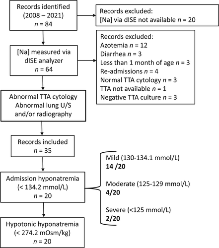
Medical records of 84 horses were retrieved from the electronic search (Figure 1). Of these 84, 20 horses had plasma [Na] evaluated at admission using indirect potentiometry only. Because of substantial differences in analytical technique, these 20 horses were excluded from the study. Fifteen of the remaining 64 horses were excluded because of ongoing gastrointestinal disease (n = 3) or azotemia (n = 12), both potential causes of hyponatremia. Three horses were excluded because they were <1 month of age and because of substantial differences in sodium homeostasis in that age group.30, 31 Four of 46 were readmissions and therefore were not considered new cases. The TTA cytology results did not support underlying inflammation in 3 horses, in 1 instance the TTA was collected but not submitted, and in 3 cases the TTA fluid did not yield any bacterial growth.

Included animals (n = 35) had a median age of 3 years (IQR, 3 months-15 years). Breeds represented were Quarter Horse (14), Standardbred (6), Arabian (5), Paint (3), Thoroughbred (2), Miniature (2), and 1 each of Missouri Fox Trotter and Morgan; breed was not recorded for 1 horse. There were 17 females, 11 castrated males, and 7 intact males.
Presenting complaints included lethargy, fever, nasal discharge, cough, increased respiratory effort or some combination of these. Before referral, 16 horses had received at least 1 NSAID (flunixin meglumine, phenylbutazone, or both). Prior treatment history was not available for 3 horses. Duration of treatment before referral was not available from the medical records.
At admission, hyponatremia was present in 20/35 horses (57%), with a median concentration of 132 mmol/L (IQR, 129.7-133.1 mmol/L). Fourteen of 20 were classified as having mild hyponatremia, 4/20 moderate hyponatremia, and 2/20 severe hyponatremia. When accounting for horses without pleural effusion, hyponatremia was present in 11/25 (44%) of cases. No differences in heart rate (P = .8) or respiratory rate (P = .2) were found between horses with or without hyponatremia, but rectal temperature was significantly higher (P = .02) in hyponatremic (n = 20; 38.8 ± 0.7°C) than in normonatremic (38.1 ± 1°C) horses. A significant correlation between plasma [Na] and rectal temperature was found (P = .001; Spearman ρ = −0.51; 95% confidence interval [CI], −0.72 to −0.2). Age (P = .6), sex (P = .6), or prior use of NSAIDs (P > .9) had no effect on the presence or absence of hyponatremia at admission.
Of the 35 horses that had blood gas analysis performed at admission, 10 (28%) had evidence of pleural effusion. Of the hyponatremic horses at admission, 8/20 (40%) had evidence of pleural effusion. No association (P = .1) was found between presence of pleural effusion and plasma [Na] when accounting for the entire population. No difference was found in the severity of hyponatremia at admission between horses with pleuropneumonia and those with pneumonia (P = .7).
A final measurement of plasma [Na] was available for 15/20 of the hyponatremic horses assessed at admission. Among these, hyponatremia had completely resolved in 5/15 before discharge, 3/15 before euthanasia, and 1/18 before in-hospital sudden death. In the remaining 6 horses, hyponatremia still was present on the last available measurement but had improved compared to the admission result. Specifically, hyponatremia in these 6 horses now was categorized as mild.
The control reference range for effective osmolality was 274.2 to 291 mOsm/kg (mean 282.6 ± 4.2 mOsm/kg). Based on this reference range, all hyponatremic horses were classified at admission as having hypotonic (<274.2 mOsm/kg) hyponatremia (i.e., true hyponatremia). Eight horses with hypotonic hyponatremia had evidence of pleural fluid accumulation, 6 had received drugs before hospitalization that potentially could have caused hyponatremia (≥1 NSAID), 5 had no history of drug administration and no information about previous drug administration was available for 1 horse.
Seventeen of 32 horses met the inclusion criteria for SIRS. In 3 horses there was no CBC available on admission. Of the SIRS positive horses, 14 were hyponatremic. It was significantly more likely for SIRS positive horses to be hyponatremic at admission (P = .01; odds ratio [OR], 9.3; 95% CI, 1.5-38.6).
No difference was found in white blood cell or neutrophil counts between hyponatremic and normonatremic horses.
Fibrinogen concentration (reference range, 125-262 mg/dL) was significantly higher (P = .01) in horses with hyponatremia (n = 17; 461 ± 160.5 mg/dL) compared to those with normal plasma [Na] (n = 12; 321 ± 100.7 mg/dL). A significant negative correlation was found between plasma [Na] and fibrinogen concentration (n = 29; P = .001; Spearman ρ = −0.57; 95% CI, −0.78 to −0.25).
No differences in total solids, albumin, creatinine, BUN, glucose, or l-lactate concentrations were found between hyponatremic and normonatremic horses.
All TTA samples from included horses yielded positive bacterial growth. The most common aerobic isolates were Actinobacillus spp. (11/35; 31%), and Streptococcus equi subsp. zooepidemicus (11/35; 31%), followed by alpha-Streptococcus spp. (9/35; 25%), and R. equi (5/35; 14%). Anaerobes were isolated from 6/35 (17%) submitted TTAs and comprised Bacteroides spp., Fusobacterium necrophorum, Clostridium sordelli, and Prevotella spp. One horse was diagnosed with equine multinodular progressive fibrosis (EMPF) at necropsy.
Median DOH was 7 days (IQR, 4-12 days). No difference (P = .3) was found in DOH between hyponatremic (median, 10 days; IQR, 4-15 days) and normonatremic (median, 6 days; IQR, 4-9 days) patients. When the 2 groups were compared after 2 significant outliers were excluded from the normonatremic group, a significant difference was found (P = .04), with a mean DOH in hyponatremic horses of 9.8 ± 6.6 days, and of 5.6 ± 3.1 days in normonatremic patients. For the first outlier patient, despite the grave prognosis, the owners elected to continue treatment until clinically relevant deterioration ensued, at which point euthanasia was elected. The second outlier patient continued to be hospitalized for PO antimicrobial treatment of R. equi because of client inability to treat the animal at home. These 2 situations led to the prolonged hospitalization observed. Of the 35 horses included in our study, 29 were discharged alive, 4 were euthanized and 2 died in hospital. Both horses that died and 3 that were euthanized were hyponatremic at admission.
4 DISCUSSION
We confirmed that hyponatremia is common in horses with septic pneumopathy, with an overall frequency of 57%, and of 44% when horses with pleural effusion were excluded. The frequency of hyponatremia in our study is lower than previously reported,17 possibly reflecting differences between patient populations (e.g., exclusion of azotemic horses from our study population).
Pseudohyponatremia, a potential cause of falsely decreased plasma [Na], can be associated with specific laboratory techniques32 applied to serum or plasma samples with a decreased water content. A decrease in plasma content of water associated with hyperlipidemia or severe hyperproteinemia creates a phenomenon known as “electrolyte exclusion effect.” While in a previous study17 the technique of sodium analysis was not specified, plasma [Na] was measured in our study using dISE, a technique that is not influenced by the water content of the sample. This method of analysis is recommended to assess plasma [Na] when access to lipid-clearing methods is not available.33
Eight of 20 hyponatremic horses had evidence of pleural effusion on radiography, ultrasonography or both. The development of hyponatremia in horses with pulmonary infection and concurrent pleural effusion likely is multifactorial. The finding of hyponatremia, especially when associated with substantial pleural effusion causing hypovolemia, is not surprising and falls into the category of hypovolemic hyponatremia.25 Substantial third-space loss of water results in ADH release, water retention, and dilutional hyponatremia.34 A major limitation of our study is the inability to retrospectively assess patient plasma volume and the magnitude of pleural fluid accumulation. Furthermore, pleural fluid was not collected, quantified, or classified in many instances, and the assumption that some horses in our study developed hypovolemic hyponatremia remains speculative. Prospective studies would be needed to properly assess decreased circulating volume status and development of hyponatremia.
Twelve of 20 hyponatremic horses had no recorded evidence of pleural effusion at admission, requiring another explanation for hypotonic hyponatremia, such as a nonosmotic release of ADH.22, 24
A main finding of our study is the association between hyponatremia and evidence of SIRS at admission. Hyponatremia development during SIRS has been associated with altered expression of ion channels in nephrons under the influence of pro-inflammatory cytokines35 as well as with nonosmotic ADH release, also secondary to the action of pro-inflammatory mediators.22 To further support the association between hyponatremia and underlying inflammation, we found that both rectal temperature and plasma fibrinogen concentration at admission were increased in hyponatremic patients and negatively correlated with plasma [Na]. Hyponatremia in febrile patients was found to be significantly more common than in nonfebrile patients.37 Fever and endogenous pyrogens are thought to induce nonosmotic release of ADH as a protective mechanism to retain more water and counteract insensible losses caused by the increased body temperature.38, 39
At admission, patients with hyponatremia had higher plasma fibrinogen concentrations compared to those with normonatremia, possibly reflecting more severe or longer duration of inflammation. Fibrinogen is an acute phase protein in horses and is synthesized during inflammation after stimulation by interleukin-6 (IL-6).40 Interleukin-624 and other acute-phase inflammatory cytokines41 have been linked to development of hyponatremia because of their influence on ADH release, potentially leading to a syndrome of inappropriate ADH release (SIADH). Although we did not measure plasma IL-6 concentration in our patients, because of the retrospective nature of the study, it is reasonable to postulate that the significant associations of hyponatremia with SIRS status, rectal temperature, and plasma fibrinogen concentration at admission may be associated with increased pro-inflammatory mediator activity, possibly explaining the hyponatremia observed in the absence of other causes of low plasma [Na].
Additionally, in hyponatremic patients neutrophil count was weakly and inversely correlated with plasma [Na], further supporting a link between inflammation and hyponatremia. Interleukin-6 is a key inflammatory mediator of neutrophil trafficking, and both IL-6 and neutrophil percentages were found to be higher in children with hyponatremia secondary to Kawasaki disease, an acute inflammatory illness.39
In human patients, the use of NSAIDs is considered a rare cause of hyponatremia,42, 43 and we did not find an association between prior usage of NSAIDs and the frequency of hyponatremia at admission. The lack of any significant association between NSAIDs use and hyponatremia in our study population may reflect what is observed in studies of humans or may be related to other factors such as the variable duration of treatment, the dosages used before hospitalization, or small sample size.
Hypoalbuminemia is a common finding in horses with septic pleuropneumonia17 and, in human patients, it has been suggested to be a cause of hyponatremia because of the development of low circulatory volume, which triggers ADH release.44, 45 We did not find a difference in plasma albumin concentration between hyponatremic and normonatremic horses, or any association between sodium and albumin concentrations in our study population.
Hyponatremia at admission is a clinically relevant finding because it is associated with increased incidence of complications, DOH and mortality in people with septic pneumopathy.15, 46 We did not observe a significant influence of hyponatremia on DOH between hyponatremic and normonatremic horses, possibly related to small sample size or early discharge of some patients. Nonetheless, when 2 outliers were removed from the analysis, a significant increase in number of hospitalization days for hyponatremic patients was found.
Reported prognosis for survival in horses with pneumonia and pleuropneumonia has ranged between 43.3% and 95.7%,17, 47-50 and approximately 83% of the horses in our study were alive at the time of discharge. Although both horses that died in-hospital and 3/4 of those that were euthanized were hyponatremic at admission, it is not possible to draw conclusions on the clinical utility of hyponatremia as a predictor of survival or mortality, and larger, multicenter studies are needed.
A single horse was diagnosed with EMPF. In this horse, admission plasma [Na] was 131.4 mmol/L, and before the horse's in-hospital sudden death, its plasma [Na] had normalized. It is unclear if admission plasma [Na] in horses with chronic pulmonary diseases such as EMPF could be a useful predictor for short or long-term mortality.
Our results regarding survival must be interpreted cautiously and are likely to reflect, at least in part, owner's decision to discharge their animals because of financial limitations and against medical advice. Lack of follow-up also limits the ability to assess mortality after discharge in our study population.
The retrospective nature of our study comes with several important limitations. The measurement of plasma [Na] using a blood gas analyzer at admission was inconsistent, which prevented inclusion of all horses retrieved by medical records search. Furthermore, serial plasma [Na] measurement was not standardized throughout hospitalization, but driven by clinician preference, case severity and owners financial limitations. Likewise, for several horses no biochemistry, hematology, and fibrinogen assessment was available at admission.
Several factors impact our ability to draw conclusions regarding other variables that may have influenced plasma [Na], including lack of standardization of serial sodium measurements, incorporation of thoracic imaging, and serial assessment of inflammatory markers.
Although based on data from medical records, none of the horses included in our study suffered from congestive heart failure or chronic liver failure, both potential causes of hypervolemic hyponatremia.51 The retrospective nature of our study, lacking specific functional assessment of the heart and liver, limited our ability to draw firm conclusions. Likewise, intact function of the hypothalamic-pituitary-adrenal axis, dysfunction of which could cause hyponatremia, was not assessed.
A major limitation, which warrants future study, is the lack of urine samples paired with blood samples analysis to assess urine [Na] and osmolality. In human patients with hypotonic hyponatremia, urinary [Na] and osmolality are routinely used to identify impaired renal water excretion.52 The normal response during hypotonic hyponatremia involves suppression of ADH secretion which results in the excretion of low osmolality, gravity urine. Thus, the finding of increased urine osmolality and [Na] in a euvolemic hyponatremic patient without other causes of hyponatremia could indicate the presence of inappropriate ADH secretion.
5 CONCLUSION
In conclusion, we found that hyponatremia was common in horses with septic pneumopathy, even in absence of pleural effusion. Hyponatremia in our study population appeared to be correlated with positive SIRS status, potentially indicating a role of inflammation in the development of hypotonic hyponatremia. The lack of urine osmolality and [Na] limited our ability to further characterize sodium homeostasis. This limitation should encourage clinicians to collect and properly analyze urine samples to determine the underlying cause of hyponatremia in absence of more common causes of low plasma [Na] (e.g., third-space loss of water, kidney disease). When evaluating [Na], dISE methodology should be used to avoid interference from increased plasma protein or triglyceride concentrations, which are common in sick horses.
ACKNOWLEDGMENTS
No funding was received for this study.
CONFLICT OF INTEREST DECLARATION
Authors declare no conflict of interest.
OFF-LABEL ANTIMICROBIAL DECLARATION
Authors declare no off-label use of antimicrobials.
INSTITUTIONAL ANIMAL CARE AND USE COMMITTEE (IACUC) OR OTHER APPROVAL DECLARATION
Authors declare no IACUC or other approval was needed.
HUMAN ETHICS APPROVAL DECLARATION
Authors declare human ethics approval was not needed for this study.


