An investigation of the dynamic relationship between navicular drop and first metatarsophalangeal joint dorsal excursion
Abstract
The modern human foot is a complex biomechanical structure that must act both as a shock absorber and as a propulsive strut during the stance phase of gait. Understanding the ways in which foot segments interact can illuminate the mechanics of foot function in healthy and pathological humans. It has been proposed that increased values of medial longitudinal arch deformation can limit metatarsophalangeal joint excursion via tension in the plantar aponeurosis. However, this model has not been tested directly in a dynamic setting. In this study, we tested the hypothesis that during the stance phase, subtalar pronation (stretching of the plantar aponeurosis and subsequent lowering of the medial longitudinal arch) will negatively affect the amount of first metatarsophalangeal joint excursion occurring at push-off. Vertical descent of the navicular (a proxy for subtalar pronation) and first metatarsophalangeal joint dorsal excursion were measured during steady locomotion over a flat substrate on a novel sample consisting of asymptomatic adult males and females, many of whom are habitually unshod. Least-squares regression analyses indicated that, contrary to the hypothesis, navicular drop did not explain a significant amount of variation in first metatarsophalangeal joint dorsal excursion. These results suggest that, in an asymptomatic subject, the plantar aponeurosis and the associated foot bones can function effectively within the normal range of subtalar pronation that takes place during walking gait. From a clinical standpoint, this study highlights the need for investigating the in vivo kinematic relationship between subtalar pronation and metatarsophalangeal joint dorsiflexion in symptomatic populations, and also the need to explore other factors that may affect the kinematics of asymptomatic feet.
Introduction
The modern human foot has evolved to serve multiple functions during bipedal locomotion including shock absorption, weight transfer, and propulsion. To meet these demands, the foot must be able to function as a flexible appendage at the beginning of stance phase and as a stiff lever at push-off (Scholl, 1920; Morton, 1935; Hicks, 1954; Donatelli, 1996; Bolgla & Malone, 2004; Erdemir et al. 2004; Chi & Schmitt, 2005; Klenerman & Wood, 2006; Lorimer et al. 2006; DiGiovanni & Greisberg, 2007; Aminian & Sangeorzan, 2008; Vereecke & Aerts, 2008; Davids, 2010). Collectively, in vitro and in vivo studies indicate that the plantar aponeurosis plays a key role in enabling the foot to transition from a compliant structure to a rigid strut during gait (Hicks, 1954; Wright & Rennels, 1964; Kitaoka et al. 1994; Aquino & Payne, 1999; Gefen, 2002, 2003; Erdemir et al. 2004; Caravaggi et al. 2009, 2010). The human plantar aponeurosis is an elaborate band composed of longitudinally running collagen and elastic fibers (Wright & Rennels, 1964; Hedrick, 1996; Aquino & Payne, 1999). The main portion of the plantar aponeurosis originates from the medial tuberosity of the calcaneus and, as it courses anteriorly, it divides into five individual digital slips as it approaches the phalanges. Through a complex network, each slip terminates at the base of a proximal phalanx (Bojsen-Møller & Flagstad, 1976; Pontious et al. 1996; Aquino & Payne, 1999; Erdemir et al. 2004; Moraes do Carmo et al. 2008).
Caravaggi et al. (2009, 2010) have demonstrated that the plantar aponeurosis is under varying degrees of tension throughout the entire stance phase of the walking cycle. At heel strike, there is a small increase in tension (relative to the amount measured at rest), which most likely occurs due to active dorsiflexion at the ankle and metatarsophalangeal joints. After the spike in tension at heelstrike, the tension decreases as the ankle becomes plantarflexed and the toes become planted on the ground. From there and into midstance, a slight, gradual increase in tension occurs in the plantar aponeurosis. During this time, the upward-oriented ground reaction force, applied primarily through the heel and forefoot (as shown by plantar pressure measurements) is counteracted by a downward-oriented force originating in the midfoot (Fig. 1a). These opposing forces form a three-point structure that creates tension in the plantar aponeurosis. In response, the plantar aponeurosis elongates, resulting in an initial lowering of the medial longitudinal arch. As the load increases, the plantar aponeurosis will progressively stiffen (Wright & Rennels, 1964) and prevent further deformation of the medial longitudinal arch. In other words, the compliance of the plantar aponeurosis permits some medial longitudinal arch compression in the early parts of stance phase but resists (with contributions from the short and long plantar ligaments) further elongation. This may help prevent further collapse of the medial longitudinal arch to the point at which midfoot joint stability would otherwise be compromised.
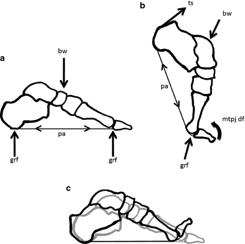
As the stance phase progresses, forward progression of the body in front of the ankle and plantarflexion caused by contraction of the triceps surae lead to lift off of the heel. During this phase, weight is transferred from the rear- to forefoot. As a result, in this latter part of stance the metatarsophalangeal joints are passively dorsiflexed. This leads to tightening of the plantar aponeurosis via dorsal excursion of the proximal phalanx, a pattern classically referred to as the ‘windlass mechanism’ (Hicks, 1954) (Fig. 1b). In this mechanical model, the proximal phalanges, metatarsal heads, and the plantar aponeurosis are treated as a handle, drum, and cable of a simple windlass or winch. With the passive dorsiflexion of the metatarsophalangeal joints, the base of each proximal phalanx (i.e. the handle of the windlass) moves onto the dorsum of the respective metatarsal head (i.e. the drum of the windlass), thus bringing along with it the attached digital slip of plantar aponeurosis (i.e. the cable of the windlass). As a result, each digital slip is wound around the head of the metatarsal and the plantar aponeurosis is stretched in tension (Gefen, 2003; Caravaggi et al. 2009, 2010). Tension in the first digital slip (i.e. the slip attaching to the base of the hallucal proximal phalanx) is the most pronounced compared with the lateral slips (Caravaggi et al. 2009, 2010) corresponding with the relatively larger size of the first metatarsal head (i.e. drum of the windlass) (Bojsen-Møller, 1979; Pontious et al. 1996; Cheng et al. 2008; Christensen & Jennings, 2009) and the first digit's major role as a fulcrum during propulsion (Hetherington et al. 1989; Hopson et al. 1995; Griffin et al. 2010). As the plantar aponeurosis is stretched by the combined contraction of the triceps surae rotating the calcaneus superiorly and the passive metarsophalangeal joint (MTPJ) dorsal excursion pulling the calcaneus distally towards the planted forefoot, the first metatarsal, itself plantarflexes and osseous compression in the midfoot occurs. This compaction of the mid-and forefoot raises the arch (Fig. 1b,c). The forefoot can now serve as a stable lever for propulsion (Hicks, 1954; Aquino & Payne, 1999). In sum, the plantar aponeurosis contributes substantially to foot stability, especially in the midfoot during full foot contact on the substrate (Fig. 1a) and propulsion (Fig. 1b) through passive tension. During full foot contact, the plantar aponeurosis is stretched in tension and as it becomes progressively stiffer, the midfoot is stabilized. During the phase of stance where the calcaneus is lifted from the ground, and the passive dorsiflexion of the MTPJs occurs, the plantar aponeurosis experiences tension and the joints in the midfoot achieve a relatively close-packed position. The resulting midfoot stability allows the forefoot to become a relatively effective lever at push-off, a key characteristic of human bipedalism.
Not only do each of the two events – medial longitudinal arch compression and heel lift combined with MTPJ dorsiflexion – independently stiffen the plantar aponeurosis in different mechanical ways (Fig. 1), both events are also associated with different movements of the subtalar joint. During medial longitudinal arch compression, the subtalar joint moves into pronation (defined as a triplanar joint movement in which the talus inhabits a plantarflexed and adducted position and the calcaneus becomes everted). Later, the subtalar joint supinates (defined as a triplanar joint movement in which the talus inhabits a dorsiflexed and abducted position and the calcaneus becomes inverted) (Donatelli, 1996). As stated by Aquino & Payne (2001), there is a common assumption that excessive subtalar pronation (i.e. subtalar overpronation in which the arch compresses to a relatively high degree) during the stance phase can interfere with the foot's ability to supinate during the latter stance phase, and the latter movement is thought to be essential for stiffening the midfoot in preparation for forefoot propulsion (Fuller, 2000). Although the movements associated with subtalar pronation (and overpronation) at each joint are complex, the overall effect is to lower the medial longitudinal arch. In that sense ‘overpronation’ can be described by the amount of medial longitudinal arch collapse.
Two in vivo studies provide support for the notion that subtalar overpronation can interfere with the timing and amount of subtalar supination and also influence the timing and range of MTPJ1 dorsal excursion. In their study of an asymptomatic sample, Kappel-Bargas et al. (1998) identified two groups (‘immediate’ and ‘delayed’) that significantly differed with regard to the timing of the kinematic markers associated with windlass mechanism (i.e. rise of the medial longitudinal arch relative to the passive dorsiflexion of the MTPJ1). The ‘delayed’ group showed significantly more rearfoot eversion (i.e. calcaneal eversion that takes place during subtalar pronation) during stance and also the medial longitudinal arch rise relative to the dorsiflexion of the MTPJ1 began later compared to the ‘immediate’ group. A more recent dynamic study by Nakamura & Kakurai (2003) also investigated kinematic markers associated with the timing of the movements of the windlass mechanism in an asymptomatic sample. They, too, identified two groups in the sample based on timing of maximum rearfoot eversion during the stance phase. The groups were referred to as ‘early eversion onset’ and ‘late eversion onset’. In addition to exhibiting a delay in timing of rearfoot eversion, the ‘late eversion onset’ group also showed significantly larger measures of both maximum rearfoot eversion and maximum medial longitudinal compression. In general, the ‘later eversion onset’ group showed more medial longitudinal arch compression throughout the entire stance phase. Lastly, and most importantly for the current study, Nakamura & Kakurai (2003) found that the ‘later eversion onset’ individuals showed a significant delay in timing of passive MTPJ1 dorsiflexion compared with the ‘early eversion onset’ subjects. Though passive dorsiflexion occurs later in the ‘later eversion onset’ group, it is possible this timing constraint and the magnitude of subtalar pronation can limit the maximum amount of MTPJ1 dorsal excursion before the foot leaves the ground.
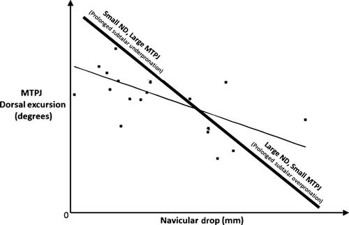
The potential influence of subtalar pronation on maximum MTPJ1 dorsal excursion is highlighted by Paton's (2006) static study on an asymptomatic sample. The author used the measurement of navicular drop or vertical displacement of the navicular tuberosity towards the ground as the proxy for subtalar pronation. For each subject, measurements of the navicular drop and MTPJ1 dorsal excursion were taken independently and during standing (i.e. static stance). Paton (2006) found that navicular drop exhibited a small, but significant, negative correlation with MTPJ1 dorsal excursion (Fig. 2). Based on this outcome, we may predict that if Paton's method was replicated with a sample consisting of individuals characterized as either exhibiting excessive subtalar overpronation and underpronation, excessive subtalar pronators should group near the lower end of the slope line (Fig. 2) because they would exhibit large amounts of navicular drop in association with limited MTPJ1 movement. In contrast, subtalar underpronators should plot near the upper end of the slope line (Fig. 2) because they would exhibit smaller amounts of navicular drop and larger amounts of MTPJ1 dorsal excursion. Under those conditions, one would predict that in a sample with high degrees of variation including subtalar overpronators and underpronators, the result would be a relatively stronger negative correlation between navicular drop and MTPJ dorsal excursion. Before this prediction about subtalar overpronators and underpronators can be tested, the relationship between subtalar pronation and metatarsophalangeal joint excursion needs to be empirically tested under dynamic conditions. We did so here using a sample of habitually shod and minimally shod people walking at self-selected speeds across a flat surface. Rather than relying on traditionally young samples of adults in the United States or Europe who regularly wear athletic and work footwear that may influence foot function even without clear symptoms, we relied on a more age-variant sample of people who use no or minimal footwear in their daily life. This sample provides a valuable perspective on foot function without many confounding factors.
To date, no study has tested the relationship between the amount of subtalar pronation and maximum metatarsophalangeal joint excursion in an asymptomatic population during natural overground walking. Building on what is already known about the role of the plantar aponeurosis during walking gait (Hicks, 1954; Bojsen-Møller & Flagstad, 1976; Bojsen-Møller, 1979; Bojsen-Møller & Lamoreux, 1979; Sarrafian, 1987; Erdemir et al. 2004; Caravaggi et al. 2009, 2010; Christensen & Jennings, 2009) and from studies proposing mechanisms of dysfunction (Kappel-Bargas et al. 1998; Nakamura & Kakurai, 2003; Bolgla & Malone, 2004; Paton, 2006), we hypothesize that there will be a negative relationship between medial longitudinal arch deformation and metatarsophalangeal joint excursion in an asymptomatic foot. Given that dorsal excursion of the first MTPJ1 is essential for normal locomotion and first metatarsals are more susceptible to pathological lesion than the other metatarsals in the foot (Zipfel & Berger, 2007), a better understanding of kinematic variables that may be associated with or affect dynamic metatarsophalangeal joint motion is essential to early identification and treatment of patients suffering from limited push-off capability.
Materials and methods
Sample composition
The sample consisted of asymptomatic adult males (n = 13) and females (n = 13), who were part of an original study led by D'Août et al. (2009). The subjects were native to Bangalore, India, and nearby areas of the region and encompass an age range from 22 to 66 years (mean of 43 years). The sample consists of a mixture of individuals identified as having never worn footwear and those who wore footwear such as sandals, flip flops, or hard-soled shoes but went barefoot when indoors. Permission to include healthy subjects in the sample was obtained by the Ethical Committee of the University of Antwerp (#A0401 granted to K.D.). More specific details of the sample and methods of collection can be found in D'Août et al. (2009). Although somewhat small, this unique study population provides novel insights into foot function by avoiding many of the confounding factors that influence more typical samples of habitually shod subjects from Europe or the United States. Since this is a study of normal variation, it should be noted that this sample is not designed to capture an extreme range of pronation and supination. We believe that this sample will capture a normal range of variation in an asymptomatic sample of men and women. Moreover, previous studies have documented the significant differences in external morphology and kinetics between habitually shod and habitually unshod samples (D'Août et al. 2009) and have shown that habitually unshod individuals are less likely to suffer from foot pathology than their habitually shod counterparts (Kadambande et al. 2006; Zipfel & Berger, 2007). Therefore, our dataset may provide a partial control to some of the confounding factors that restricting footwear may have on the biomechanical variables we measured.
Data collection
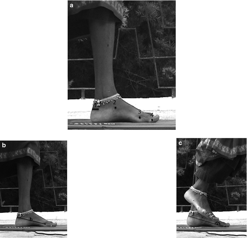
The following anatomical landmarks were indicated with black marker on the right foot of each subject: (i) calcaneal tendon insertion (ii) navicular tuberosity, (iii) head of the first metatarsal, and (iv) head of the hallucal proximal phalanx (Fig. 3a). The height of the navicular marker to the ground was measured for each subject during quiet standing using calipers and was used as a measure of the static medial longitudinal height.
It is worth noting that our calcaneal marker differed slightly from Nielsen et al. (2009). As a result, there was a small but consistent difference in one marker location between our study and that of Nielsen et al. (2009). Nielsen et al. (2009) also calculated navicular drop over a different time range of stance phase as appropriate to their questions and sample. The current study focused on the value of medial longitudinal arch compression at specific instances during the stance phase: (i) at midstance when the navicular drops and (ii) at push-off when the largest amount of MTPJ dorsal excursion occurs. Thus the current study focuses specifically on those points in stance.
Our choice of calcaneal marker was based on reliable correlates discernible from 2D video. Without use of cineradiographic methods the top of the calcaneal tuber and insertion of the calcaneal tendon represent the most distal reliable external reference points on the heel due to compression and movement of the calcaneal fat pad.
Two video cameras (Sony DV, 50 Hz) were set up perpendicular to the runway. The lens of one of the cameras was set at a wide field of view to capture footage of a complete stride so that velocity could be calculated. The second camera, with a high-resolution 3CCD, had a more restricted view to capture a detailed image of the foot during a trial. Each subject was filmed as s/he walked barefoot at a comfortable self-selected velocity across the viewing area (i.e. lateral to camera view). For most subjects, three trials were collected per foot. Trials with the medial side of the right foot facing the camera were used for analysis.
Data analysis
The footage from each trial was de-interlaced in virtualdubmod freeware derived from Avery Lee's virtualdub (http://virtualdubmod.sourceforge.net/) to achieve a full 50-Hz sample. Each trial was imported into DLT dataviewer (DLTdv3, Hedrick, 2008) for digitization of the four anatomical landmarks during the stance phase (see Fig. 3a). Footage was calibrated using the known length of an object (i.e. RSScan pressure pad with a full length of 56 cm) within the field of view of each trial. For each trial, all markers were digitized from the frame showing full foot contact (i.e. when the first ray was completely on the ground) up to the frame before toe-off (i.e. when the foot left the ground).
Studies like this take advantage of populations of people who habitually walk unshod. To facilitate data collection, subjects were encouraged at a comfortable velocity. Such studies avoid use of complex marker systems and multiple cameras for the expediency of converting and collecting data in a short time. However, this does raise legitimate issues of accuracy and precision in data reduction from the original videos. We addressed this in multiple ways include averaging of values and also post hoc filtering of these variables described later in the methods. These two types of methods work to negate any deviations in accuracy and precision during the manual digitizing by one investigator on our team. We also carried out an analysis of both precision and accuracy with three of the authors and one student. We compared results and refined our approach to a position that was most consistent across those collecting the data. We then had one investigator alone do all the digitization following the agreed upon and most consistent approach.
The DLT output file listing the coordinates of the digitized points was imported into matlab (matlab 7.6, The MathWorks Inc., Natick, MA) where the raw navicular height and raw MTPJ angle values were calculated for each frame (Fig. 3b,c). A fourth-order low pass Butterworth filter with a cut-off frequency of 8 Hz was applied to the raw values before calculating the variables navicular drop and MTPJ1 excursion (Fig. 3b,c). Navicular drop and MTPJ1 dorsal excursion values from subjects with more than one trial available were each averaged.
Since subjects were encouraged to walk at a self-selected speed, the influence of velocity (m/s) on navicular drop (in millimeters) and MTPJ1 dorsal excursion (in degrees) was tested for by least-squares regression. The relationship between navicular drop and MTPJ1 dorsal excursion was examined from two perspectives using least-squares regression analysis. MTPJ1 values were regressed against values of both navicular drop and ‘relative navicular drop’. Relative navicular drop represents a proportion of standing medial longitudinal arch height and accounts for differences in medial longitudinal arch height between individuals by providing a measure of comparable proportional deformation – for those with a relatively high medial longitudinal arch an absolutely large navicular drop may represent only a very small relative compressive change.
Results
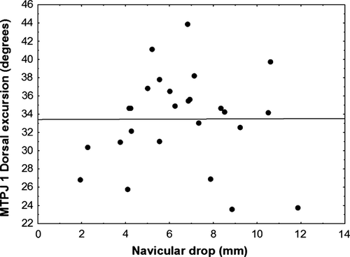
Table 1 lists the summary statistics for each variable. The sample exhibits normal ranges of subtalar pronation because their navicular drop range values are comparable to the normal range reported by Nielsen et al. (2009). Least-square regression testing for the effect of velocity on navicular drop and MTPJ1 dorsal excursion revealed that the coefficients of determination for both variables were small (R2 = 0.14 and 0.0030, respectively) and velocity had no statistically significant effect on the variables (Table 2).
| N | Mean ± SD (minimum to maximum value) | |||||
|---|---|---|---|---|---|---|
| Age (years) | Body mass (kg) | Height (m) | Navicular drop (mm) | MTPJ 1 dorsal excursion (degrees) | Velocity (m s–1) | |
| 26 | 43 ± 13 (22–66) | 58.9 ± 12.3 (41.0–92.0) | 1.61 ± 0.104 (1.44–1.79) | 6.49 ± 2.52 (1.93–11.90) | 33.5 ± 5.08 (23.6–43.9) | 1.05 ± 0.239 (0.584–1.56) |
| Comparison | Slope | Coefficient of determination (R2) | F statistic | P-value |
|---|---|---|---|---|
| Velocity (m/s) vs. navicular drop (mm) | 10.7 − 4.0x | 0.14 | 4.00 | 0.057 |
| Velocity (m/s) vs. MTPJ 1 dorsal excursion (degrees) | 32.3 + 1.1x | 0.003 | 0.070 | 0.79 |
| Navicular drop (mm) vs. MTPJ dorsal excursion (degrees) | 33.0 + 0.0081x | 0 | 0.00039 | 0.99 |
| Relative navicular drop vs. MTPJ dorsal excursion (degrees) | 34.2 − 4.1x | 0.0053 | 0.13 | 0.72 |
Regression analyses of navicular drop vs. MTPJ1 dorsal excursion and of relative navicular drop vs. MTPJ1 dorsal excursion both indicated that navicular drop, a measure of subtalar pronation, does not correlate with or limit first MTPJ1 excursion (see Table 2). These results (Fig. 4) contrast with Paton's (2006) study, which found a significant negative relationship between navicular drop and MTPJ dorsal excursion in a sample of 24 subjects.
Discussion
The results of this study did not support the hypothesis that a negative relationship exists between navicular drop and MTPJ1 dorsal excursion. These results suggest that in asymptomatic individuals walking at a self-selected walking pace, the amount of navicular drop neither correlates with nor directly affects MTPJ1 dorsal excursion. Therefore, subtalar pronation is unlikely to serve as a significant constraint on forefoot leverage capability. These findings that foot function as defined by the windlass mechanism are preserved within normal variation are surprising relative to previous studies and have important functional and clinical implications.
The results of our study indicated that during the stance phase of walking, the hallux's key role of providing leverage for push-off (as measured by MTPJ1 dorsal excursion) is not affected by medial longitudinal arch compression (as measured by navicular drop) in an asymptomatic subject. We only examined walking at a self-selected pace, therefore it is possible to argue that the absence of a relationship between these variables may not persist at higher speeds. In Caravaggi et al.'s (2010) study, the authors sampled subjects at three walking speeds (slow, normal, fast) and found that the foot remained more rigid (i.e. less medial longitudinal arch collapse occurred) at faster walking speeds, and that metatarsophalangeal joint dorsal excursion increased with walking speed. Caravaggi et al. (2010) explain that since the plantar aponeurosis is pre-stretched at heelstrike, and especially so at higher speeds, this extra tension carries over into the later parts of the stance phase and, as a result, less medial longitudinal arch collapse occurs. The authors attribute larger MTPJ dorsal excursions to an increase in velocity because relatively high amounts of dorsal excursion can delay toe-off and therefore allow extra time for the swing foot to move further away from the stance foot, lengthening the stride and increasing subject velocity. Although the authors did not directly test for a relationship between medial longitudinal arch deformation and MTPJ dorsal excursion, their data and interpretation suggest that if there is a relationship between navicular drop and MTPJ dorsal excursion, it should be examined at a variety of speeds.
Collectively, two independent studies suggest that a negative correlation may exist between navicular drop and metatarsophalangeal joint dorsiflexion during running. Dicharry et al.'s (2009) study showed that navicular drop values tend to be larger during running than walking. Together with Griffin's (2009) preliminary data indicating that across individuals the first metatarsophalangeal joint excursion value is significantly smaller during running than walking, these studies suggest that with the transition from walking to running, navicular drop increases, and metatarsophalangeal joint excursion decreases. Greater navicular drop during running is supported by Ker et al.'s (1987) study demonstrating that the foot acts as a spring during running through the elongation and then elastic recoil of the plantar aponeurosis. Thus, larger navicular drop values achieved during running compared with walking can be explained by the greater ground reaction force and tension placed on the plantar aponeurosis causing more compression of the medial longitudinal arch. Because the tension in the plantar aponeurosis is much greater during running than during walking, to avoid the risk of failure, elastic energy stored in the plantar aponeurosis as tension is released earlier in the stance phase before the metatarsophalangeal joints can dorsiflex to a comparable amount measured during walking. Therefore, it is not necessary for a large amount of MTPJ1 dorsiflexion to take place to increase stride length as it does in walking because the elastic recoil of the plantar aponeurosis springs the foot off the ground and into the aerial phase. In this context, it is important to note that in Dicharry et al.'s (2009) study navicular drop values tend to be larger during running than walking, but the differences between these two variables were only statistically significant in the sample classified as hypermobile (i.e. demonstrating a large static navicular drop). If indeed those with more midfoot mobility show an exceptional amount of compliance during the stance phase, the investigation into how this might affect the dynamic function of the windlass mechanism is needed.
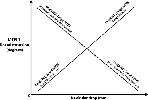
While in vivo studies involving faster-paced locomotion suggest that investigating the relationship between navicular drop and metatarsophalangeal joint excursion requires consideration of velocity, there are also anatomical variables that may need to be identified. In fact, Durrant & Siepert (1993) identify specific variables that can affect the amount of metatarsophalangeal joint excursion during push-off. One critical variable is the presence of a naturally short or inelastic plantar aponeurosis, which has been known to lead to the clinical condition of hallux limitus in which there is limited dorsal excursion of the first proximal phalanx during push-off. Although Durrant & Siepert (1993) discuss the effect of a short or less elastic plantar aponeurosis on MTPJ excursion, we will address the possible effect of a short/inelastic plantar aponeurosis on navicular drop as well as the effect the alternative scenario of having a long or slack plantar aponeurosis would have on the two variables. We predict that during loading of the medial longitudinal arch by body weight in a foot with a short/inelastic plantar aponeurosis, elongation would be minimal and little subtalar pronation would occur, and therefore the navicular drop value would be small. With the initiation of aponeurosis tightening associated with the movements of the windlass, the conservative stretch of the first digital slip would also limit the excursion of the proximal phalanx onto the dorsum of the first metatarsal head. In the second scenario, with a long or intrinsically elastic plantar aponeurosis, the magnitude of force going through on the midfoot during the stance phase of walking may be too minimal for the plantar aponeurosis to reach the critical level of stiffness required to stabilize the midfoot region. As a result, there would be a significant collapse in the medial longitudinal arch, subtalar pronation would be excessive, and the navicular drop value would be large. Then, during heel-up, because the plantar aponeurosis is slack, the proximal phalanx would not be tethered and could move more freely onto the dorsum of the metatarsal head resulting in a large excursion. This latter prediction is supported by data showing that after plantar fasciotomy (transverse surgical cut through the plantar aponeurosis), metatarsophalangeal dorsal excursion increases significantly (Harton et al. 2002).
In Fig. 5, we illustrate our predicted association between navicular drop and metarsophalangeal excursion based on consideration of length/elasticity of the plantar aponeurosis. We predict that if a cohort of individuals with extremely short/inelastic and long/elastic plantar aponeuroses is sampled, the result would be a significant positive correlation between navicular drop and metatarsophalangeal joint excursion. This positive trendline is the opposite of what we would expect for a sample consisting of individuals classified as prolonged overpronators or prolonged underpronators. Together these two trendlines indicate that depending on the cause (subtalar underpronation vs. short/inelastic plantar aponeurosis), an individual exhibiting a small navicular drop during stance phase can either exhibit a small or large amount of MTPJ1 dorsal excursion. The same situation applies to individuals exhibiting a large amount of navicular drop. They can either exhibit a large or small amount of MTPJ1 dorsal excursion depending on the amount of subtalar pronation that takes place before movements of the windlass mechanism are initiated. We are currently not aware of any study that has accounted for potential variation in length or elasticity of the plantar aponeurosis in adults and we do not have this information about our subjects. If the amount and duration of subtalar pronation and the integrity of the plantar aponeurosis influence the relationship between navicular drop and metatarsophalangeal joint excursion in opposite directions, it is understandable that no relationship between navicular drop and metatarsophalangeal joint dorsal excursion during the stance phase of walking will be found in a sample consisting of individuals exhibiting average values of subtalar pronation and assumed to be average values of length/elasticity of the plantar aponeurosis.
Regardless of the potential effects that velocity or length/elasticity of the plantar aponeurosis have on one or both of our variables, our results suggest that normal ranges of subtalar pronation do not appear to interfere, or correlate, with MTPJ1 dorsal excursion, and therefore represent a balanced relationship between medial longitudinal arch deformation and the function of a windlass mechanism in an asymptomatic sample. It is not surprising that we would find a balance in an asymptomatic sample that habitually walk with no or minimal use of light footwear. Previous studies have documented the significant differences in external morphology and kinetics between habitually shod and habitually unshod samples (D'Août et al. 2009) and have shown that habitually unshod individuals are less likely to suffer from foot pathology than their habitually shod counterparts (Kadambande et al. 2006; Zipfel & Berger, 2007). Therefore, our dataset may provide a partial control to some of the confounding factors that restricting footwear may have on the biomechanical variables we measured. This study shows that there is no relationship between navicular drop and metatarsophalangeal joint excursion in a sample of humans that have never worn constricting or very constricting shoes. Thus, the results of this study represent a baseline of normality by which researchers can compare values collected on a sample consisting entirely habitually shod and/or symptomatic samples.
From a clinical perspective, we can conclude that subjects with a normal range of subtalar pronation (i.e. between the extremes of subtalar overpronation and subtalar underpronation) maintain a fully functional windlass mechanism and can effectively accomplish weight transfer and normal propulsion. Hence, those persons with the low and high ranges of subtalar pronation, but still within the normal range, do not need any intervention in their foot mechanics if they are otherwise asymptomatic. Further study including habitually shod and symptomatic subjects would likely reveal clinical relationships and influence treatment modalities to restore the normal balance between medial longitudinal arch compression and MTPJ1 dorsiflexion in patients. Testing these two sample types in a dynamic setting with the simple and portable setup used in this study should not only confirm or disprove that MTPJ dorsal excursion is related to the amount of subtalar pronation, but will also provide clinicians with a simple tool to evaluate the windlass mechanism in patients.
Conclusion
The results of this study contribute to the overall clinical and functional understanding of foot anatomy and show that during the stance phase of walking compression of the foot and thus the stretch of the plantar aponeurosis does not limit the amount of MTPJ1 dorsiflexion in normal, pain-free subjects. Our data are consistent with the null hypothesis that the amount of navicular drop during steady-state walking does not explain a significant amount of variation in first metatarsophalangeal joint dorsal excursion in an asymptomatic sample. This is a counterintuitive result inconsistent with static measures of foot function and provides insight into what is actually ‘required’ mechanically to maintain a stiff foot. Further studies of this pattern during running will elucidate how this balance is maintained at higher speeds, while studies of clinical populations will reveal whether this balance has been lost and treatment is needed for its restoration.
Acknowledgements
The subject data collection for this project was supported by the Fund for Scientific Research - Flanders (project G.0125.05 awarded to K.D.), by the University of Antwerp (BOF grant #1094 awarded to K.D.) and by the Flemish Government through structural support to the Centre for Research and Conservation. We are grateful to Dr. Christine Wall for her statistical expertise and guidance. N.G. is also appreciative of Dr. Bruce Hirsch for reading an earlier draft of the manuscript and also for in-depth conversations about foot anatomy and function. Finally, we extend our appreciation and thanks to the subjects for their participation.


