Hepatobiliary and Pancreatic: Intrahepatic cholangiocarcinoma with intratumoral calcification mimicking hepatolithiasis
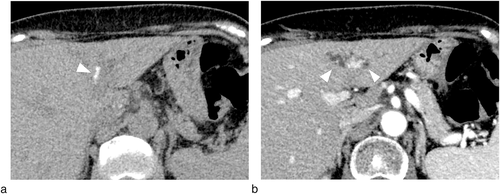
A 68-year-old woman was referred to our hospital for investigation of liver dysfunction and intrahepatic calcification detected by computed tomography (CT). At the time of her referral, CT showed intrahepatic calcification near the umbilical portion of the portal vein (Fig. 1a, white arrowhead) with peripheral bile duct dilatation (Fig. 1b, white arrowhead). Blood tests revealed elevated concentrations of aspartate aminotransferase 32 U/L, alanine aminotransferase 51 U/L, alkaline phosphatase 627 U/L, and γ-glutamyl transpeptidase 327 U/L. Total bilirubin and tumor markers (carcinoembryonic antigen and CA19-9) were within the normal range.

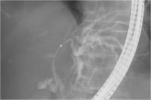
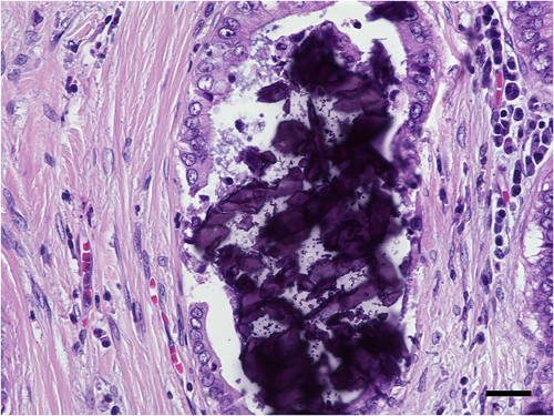
Intrahepatic biliary dilatation caused by hepatolithiasis was first suspected, and endoscopic retrograde cholangiopancreatography was performed. Contrary to our expectations, cholangiography revealed no obvious stones in the bile duct but did reveal a biliary stricture with peripheral biliary dilatation (Fig. 2). Consequently, cholangiocarcinoma was diagnosed by bile duct brush cytology obtained from the stricture. Distant metastasis was not detected, and a left hepatectomy was performed. Postoperative pathologic examination revealed intrahepatic cholangiocarcinoma with intratumoral calcification. Microcalcifications were detected among the cancerous lesions (Fig. 3, scale bar: 30 μm). These results confirmed that the calcification detected by CT was intratumoral calcification.


Intrahepatic calcification occurs in a wide variety of conditions, including inflammation, infection, benign neoplasms, and malignant neoplasms. Calcification is reported in approximately 18% of cholangiocarcinoma cases. Generally, intratumoral calcification can be distinguished from hepatolithiasis by its patchy distribution in the tumor. However, diagnosis may be difficult, as reported previously, that is, calcification distributed in the whole tumor. Conversely, hepatolithiasis is a well-known risk factor of cholangiocarcinoma. Approximately 5.3–12.9% of hepatolithiasis cases are complicated with bile duct cancer. Consequently, when encountering a patient with intrahepatic calcification and peripheral biliary dilatation, cholangiocarcinoma should be carefully ruled out.


