Gallbladder volvulus: A rare cause of acute gallbladder distension
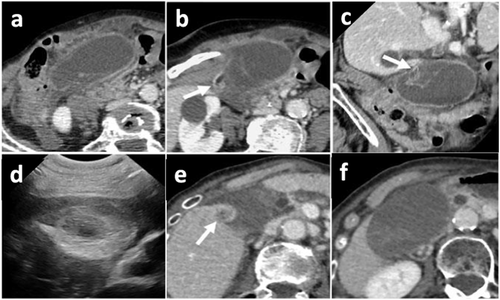
A 90-year-old woman was admitted to the emergency department for severe acute abdominal pain and vomiting. Her medical history included hysterectomy, bilateral ovariectomy, and breast cancer. Physical examination revealed an apyrexic patient with tenderness in the right upper quadrant. Laboratory tests showed increases of the direct bilirubin (22 μmol/L), C-reactive protein (169 mg/L), and neutrophil count (15.5 G/L). Abdominal ultrasonography showed a gallbladder distension with a thickened wall, without any visible stone. Computed tomography (CT) scan of the abdomen was then performed, showing a largely distended gallbladder (Fig. 1a), lying horizontally below its anatomical fossa, a twisted cystic pedicle (arrow, Fig. 1b) above a beak-shaped gallbladder neck (coronal view, arrow, Fig. 1c). No bile duct dilatation nor biliary stone was seen. All CT signs allowed to make the diagnosis of gallbladder volvulus.

Gallbladder volvulus is a rare life-threatening surgical emergency. It may occur at any age, especially in elder skinny women or in infants. Anatomic abnormalities, such as an elongated or absent mesocyst, are the main causes. Clinical presentation is not specific, with symptoms and biologic signs that mimic acute cholecystitis, which is why it was most often diagnosed intraoperatively in previously reported cases. The distended gallbladder can sometimes be felt: “floating gallbladder” sign. When ultrasonography shows a large horizontalized gallbladder with no obstructing stone, gallbladder volvulus must be suspected, and the twisted cystic pedicle has to be searched for. The latter can be seen using ultrasonography and defined as a round or conic hyperechoic mass located in the right side of the gallbladder (Fig. 1d: twisted cystic pedicle on ultrasonography in another patient). In case of doubt, other imaging techniques such as CT scan can be used to increase the preoperative diagnostic accuracy, demonstrating a low-lying distended horizontalized gallbladder, twisted on its cystic mesentery: small “whirlpool sign” (as in midgut volvulus), with a beaked-shape neck: “beak sign,” sometimes a poor enhancement of the gallbladder wall (Fig. 1e and f). This combination of CT scan signs was recently described and allows preoperative diagnosis. Emergency cholecystectomy must then be performed to avoid gallbladder necrosis and perforation. Outcome is often favorable when early diagnosis and surgery are made.
In our case, the patient was operated on a few hours after the CT scan. Outcome was favorable, and the patient is alive at last follow-up 3 years later.


