A multicentre prospective study of post-traumatic endophthalmitis
Abstract.
Purpose: Study the clinical and microbiological characteristics and the prognostic factors of post-traumatic endophthalmitis.
Methods: Seventeen eyes were included between 2004 and 2010, with clinical and microbiological data collected prospectively. Conventional cultures and panbacterial PCR were performed on aqueous and vitreous samples.
Results: Clinical signs of endophthalmitis were observed soon after trauma (1.5 ± 2.5 days). Laceration with an intraocular foreign body (IOFB) was noted in 53% of the patients. At admission, all patients had aqueous humour (71%) and/or vitreous (53%) samples. Fifteen patients (88%) underwent a pars plana vitrectomy. Bacteria were identified in 77% of the cases: Staphylococcus epidermidis (n = 5), Streptococcus (n = 4), Bacillus (n = 2), Pseudomonas stuzeri (n = 1), and Streptococcus salivarius and Gemella haemolysans (multibacterial infection, n = 1). Progression toward phthisis was observed in 35% of the cases; 41% of the patients recuperated visual acuity (VA) ≥20/40. A good final visual prognosis (≥20/40) was significantly associated with initial VA better than light perception (0% versus 70%, p = 0.01) and absence of pupillary fibrin membrane (80% versus 20%, p = 0.05). There was no correlation between visual prognosis and age, the type of laceration (corneal or scleral) or presence of an IOFB. We found a statistical trend toward an association between bacterial virulence and poor final VA.
Conclusion: This series showed that better final VA outcomes were associated with initial VA better than light perception, S. epidermidis or culture-negative cases and absence of retinal detachment during the clinical course.
Introduction
Acute endophthalmitis is a relatively rare but extremely serious complication of ocular trauma. The characteristics of traumatic endophthalmitis have been described most in the US and India more rarely in Europe, and in the Middle East. In these studies, the bacterial spectrum and the associated clinical factors (type of injury, rural environment and time to medical treatment) varied depending on the patients’ geographical location. These ocular infections are for the most part bacterial, occasionally multibacterial (Puliafito et al. 1982; Brinton et al. 1984; Affeldt et al. 1987; Alfaro et al. 1994; Foster et al. 1996; Lieb et al. 2003; Sharma et al. 2003; Essex et al. 2004) and more rarely fungal (Affeldt et al. 1987; Pflugfelder et al. 1988; Boldt et al. 1989; Alfaro et al. 1994; Azad et al. 2003; Lieb et al. 2003; Gupta et al. 2008).
For the past 10 years, microbiological identification of the causal bacteria in endophthalmitis has advanced with the technological progress of molecular biology such as panbacterial PCR (Chiquet et al. 2007, 2008). Vitreoretinal surgery techniques have also been perfected with the appearance of wide-field visualization systems, the high-speed vitrector and high-density silicone oil. The FRENCH INSTITUTIONAL ENDOPHTHALMITIS STUDY (FRIENDS), which has included patients with acute or delayed endophthalmitis since 2004 treated in four French university-affiliated hospital centres, has prospectively collected data from 17 patients with post-traumatic endophthalmitis. This investigation reports the clinical and microbiological characteristics and assesses the visual and anatomical prognosis of these patients.
Material and Methods
Seventeen consecutive patients (17 eyes) presenting acute post-traumatic endophthalmitis managed within the FRIENDS study were included over a 6-year period (May 2004 to May 2010). This study adhered to the recommendations for the conduct of clinical research on human subjects and was approved by the local ethics committee for the protection of humans subjects (CPP Sud-est V, IRB#6705). The inclusion and care procedures followed a standardized protocol (inclusion criteria, clinical and microbiological data, sampling procedure, intravitreous administration of antibiotics and follow-up) (Chiquet et al. 2008).
Clinical data
The patients were included when they presented signs of endophthalmitis occurring within 6 weeks of injury: pain, decline in visual acuity (VA), diffuse conjunctival hyperaemia, chemosis or inflammation of the anterior and posterior segment (diagnoses with biomicroscopy or B-scan ultrasound). An initial assessment form was used to record the patient’s demographic data, the medical and ophthalmological history, the causes and circumstances of the injury, the characteristics and medical and surgical management of the ocular wounds, as well as the results of the clinical and ultrasound examinations at admission. The indication for pars plana vitrectomy (PPV) was left to the clinician’s discretion. The patients were then followed regularly over a minimum period of 6 months.
Microbiological data
At admission, aqueous (AH) and/or vitreous were sampled, followed by an intravitreal injection of vancomycin (1 mg/0.1 ml) and ceftazidime (2 mg/0.1 ml). The ocular sample was used for bacteriological culture on brain–heart infusion broth medium (direct inoculation in the operating room), for mycological culture on Sabouraud medium with added chloramphenicol and genomic analysis with panbacterial polymerase chain reaction (PCR) targeting ribosomal 16S DNA according to previously described procedures (Chiquet et al. 2007, 2008).
Statistical analysis
The quantitative data were expressed in the mean ± standard deviation (SD). The association between the microbiological and clinical data was initially studied using ANOVA for the quantitative data and chi-square analysis (with Yates corrections if necessary) for the qualitative data. The statistical analysis was performed using the statistical package for the social sciences program (SPSS 17.0 for Windows; Chicago, IL, USA) software. Statistical significance was defined as a p-value <0.05.
Results
General data
Table 1. the 17 patients’ initial epidemiological and clinical data are summarized in Table 1. There was a clear majority of males (14/17) and more left-side involvement (11/17). The initial injury was a penetrating scleral laceration in eight eyes (47%) and corneal in nine eyes (53%). An IOFB was present in nine eyes (53%) (for the most part located in the vitreous). The majority of the patients (7/9) underwent surgical extraction of the IOFB, with endophthalmitis systematically occurring before extraction. Two patients initially presented intravitreal haemorrhage and two others retinal detachment (RD).
| Clinical data | Results |
|---|---|
| Gender (M/F) | 14 (82%)/3 (18%) |
| Age (years) | 40 ± 18 |
| Laterality (RE/LE) | 6 (35%)/11 (65%) |
| Interval between trauma and symptoms (days) | 15 ± 25 |
| Interval between first symptoms and presentation to hospital (days) | 12 ± 14 |
| Laceration | |
| Corneal | 9 (53%) |
| Scleral | 8 (47%) |
| Intraocular foreign body (IOFB) | 9 (53%) |
| IOFB size (mm) | 28 ± 1 |
| Metallic IOFB | 8 (89%) |
| IOFB located in the vitreous cavity | 7 (78%) |
| IOFB embedded in the retina | 2 (22%) |
| Surgical removal of the IOFB | 7 (78%) |
| Pain | 12 (70%) |
| Initial visual acuity (iVA) | |
| iVA < 20/400 | 8 (47%) |
| 20/400 ≤ iVA ≤ 20/200 | 1 (6%) |
| 20/200 < iVA ≤ 20/50 | 1 (6%) |
| 20/50 < iVA | 7 (41%) |
| Red reflex loss | 8 (47%) |
| Keratitis | 1 (6%) |
| IOP (mmHg) | 19 ± 5 |
| Anterior chamber flare (SUN classification) (%) | |
| 1+ | 2 (12) |
| 2+ | 2 (12) |
| 3+ | 13 (76) |
| Pupillary fibrin membrane | 10 (59) |
| Hypopyon | 14 (82) |
| <1.5 mm | 9 (53) |
| ≥1.5 mm | 5 (29) |
| Chemosis | 5 (29) |
| Corneal oedema | 8 (47) |
| Posterior synechiae | 4 (23) |
| Anterior chamber examination impossible | 2 (12) |
- IOFB, intraocular foreign body.
The time to the appearance of endophthalmitis symptoms was 1.5 ± 2.5 days after the trauma, and the time to management was 1.2 ± 1.4 days. The main functional signs were VA limited to light perception (LP) in eight eyes (47%). Visual acuity was measured before onset of endophthalmitis in five cases and had decreased to 20/200 or less in the injured eye at this time for four of these five patients (explaining that the decline in vision was not necessarily reported by the patient when endophthalmitis manifested).
B-scan ultrasound performed on 14 patients (82%) systematically showed hyperechogenic vitreous and RD in two cases. All the patients who had IOFB were examined with computed tomography.
Microbiological data
Table 2. each patient had a mean 2.2 ± 1.9 ocular samples taken. Analysis of the AH sampled at admission (12/17 eyes) showed a positive culture in 10% of the cases, and the PCR was positive in 22%. Analysis of the vitreous sampled at admission in 53% of the patients (9/17) showed a positive culture in 43% and a positive PCR in 50%. A second sample of AH or vitreous was taken in some patients (see Table 2).
| Microbiological data | Results |
|---|---|
| Aqueous humour (first sample) | 12/17 (71%) |
| Cultures+ | 1/10 (10%): S epidermidis |
| PCR+ | 2/9 (22%): S epidermidis, S pneumoniae |
| Vitreous from biopsy (first sample) | 9/17 (53%) |
| Cultures+ | 3/7 (43%): S salivarius, S oralis, S haemolyticus |
| PCR+ | 4/8 (50%): 2 S. pneumoniae, 1 S epidermidis, 1 Gemella haemolysans |
| Aqueous humour (second sample) | 6/17 (35%) |
| PCR+ | 0 |
| Cultures+ | 0 |
| Vitreous from biopsy (second sample) | 2/17 (12%) |
| Cultures+ | 0 |
| PCR+ | 2/2 (100%): S. pneumoniae, Bacillus thuringiensis |
| Vitreous from pars plana vitrectomy | 15/17 (88%) |
| Cultures+ | 4/11 (36%): 3 S epidermidis and 1 S Oralis |
| PCR+ | 6/10 (60%): 3 S epidermidis, 1 S. pneumoniae, 1 Pseudomonas stuzeri, 1 Bacillus pumilus |
| Final bacterial identification | 77% (13/17) |
| S epidermidis | 5 (29%) |
| Streptococcus | 4 (24%) |
| Bacillus (Thuringiensis/Pumilus) | 2 (12%) |
| Gram negative bacteria (Pseudomonas stuzeri) | 1 (6%) |
| Polymicrobial infection (S salivarius and Gemella haemolysans) | 1 (6%) |
Pars plana vitrectomy was performed in 15 patients after one or several intravitreal injections. The positivity rate of the culture tested on the vitreous from the vitrectomy was 36%, and the positivity rate of PCR was 60% (p = 0.6). Finally, analysis of the vitreous from the vitrectomy provided a bacteriological diagnosis in seven cases (47%). For six of these seven cases, PPV sample was positive, whereas AH or vitreous biopsy was negative.
In total, the PCR performed in 16 patients (94%) was positive in 62% of the cases and was necessary for five who had negative cultures (29%). Bacterial identification was obtained in 77% of the cases (see Table 2), most often Gram-positive bacteria, with one case of multibacterial infection (S. salivarius + Gemella haemolysans).
Therapeutic data
Fifteen patients (88%) underwent PPV within a mean 7 ± 6.5 days (range 0–22 days), one-third in the first 3 days. Vitrectomy was indicated immediately because of serious anatomic and functional conditions in seven eyes (46%), anatomic or functional aggravation in four eyes (27%) and for removal of the IOFB in four eyes (27%). Five patients underwent delayed PPV, beyond 1 week, the first because of the presence of post-traumatic choroidal detachment and the four others to remove the IOFB after effective treatment of the endophthalmitis with intravitreous injection of antibiotics.
Intraoperative visibility was most often mediocre, with visualization of the retina in seven eyes (46%) after central vitrectomy and absence of retinal visualization during surgery in five eyes (33%). Intra- and perioperative complications were noted in three eyes (20%) (including one case of multiple complications): one cataract (7%), one retinal tear (7%), one macula-off RD (7%) and/or one choroidal haemorrhage (7%).
Clinical course
Table 3. the minimum follow-up was 6 months (mean, 9.7 ± 4.7 months). The main post-operative complications were RD (eight eyes; 47%; time to complication : 7–47 days), isolated retinal tears with no detachment (three eyes; 18%; 8–47 days), epimacular membrane (three eyes; 18%; 90–180 days), intraocular hypertension (three eyes; 18%; 2–12 days), cataract (six eyes; 35%; 1–20 days), corneal oedema (three eyes; 18%; 1st day post-operative) and/or choroidal detachment (three eyes; 18%; 2–30 days). Nine patients presented more than one complication (52%), six of whom progressed toward phthisis (35%).
| Case | Bacteria | Complications (time to onset) (D: day, M: month) | Final visual acuity | Number of surgeries | Number of intravitreal antibiotic injections |
|---|---|---|---|---|---|
| 1 | S epidermidis | Phthisis (M3) | LP− | 1 | 4 |
| 2 | No identification | Epiretinal membrane (M3) | 20/20 | 2 | 4 |
| 3 | S epidermidis | Retinal detachment (M15) | 20/125 | 2 | 3 |
| 4 | S epidermidis | Epiretinal membrane (M3) | 20/20 | 2 | 4 |
| 5 | S epidermidis | Corneal oedema (D1), phthisis (D30) | LP− | 3 + enucleation | 6 |
| 6 | Bacillus thuringiensis | Corneal oedema (D1), retinal detachment (RD) (D10), phthisis (M3) | LP− | 0 | 3 |
| 7 | Streptococcus and Gemella | Retinal detachment with PVR, phthisis (M3) | LP− | 1 | 3 |
| 8 | No identification | Intraocular hypertension (D2), RD with PVR (D7), phthisis (M4) | LP− | 3 + enucleation | 2 |
| 9 | No identification | Intraocular hypertension (D10) | 20/20 | 1 | 2 |
| 10 | Coagulase Negative Staphylococcus | Epiretinal membrane (M6) | 20/20 | 3 | 2 |
| 11 | Streptococcus haemolyticus | Lens opacification (D9) | 20/25 | 1 | 3 |
| 12 | Pseudomonas stuzeri | 0 | 20/30 | 1 | 3 |
| 13 | Streptococcus oralis | Lens opacification (D5), RD with PVR (D25), choroidal detachment (D30), phthisis (M2) | LP− | 2 | 4 |
| 14 | No identification | Lens opacification (D1), choroidal detachment (D5), RD with PVR (D8), intraocular hypertension (D11) | LP− | 2 | 4 |
| 15 | S epidermidis | Choroidal detachment (D2), RD, phthisis (M2) | LP+ | 1 | 2 |
| 16 | Bacillus pumilus | Corneal oedema (D1), cataract (D1), RD with PVR (D10) | 20/200 | 6 | 4 |
| 17 | S. pneumoniae | 0 | 20/20 | 1 | 1 |
- LP, light perception; PVR, proliferative vitreoretinopathy; RD, retinal detachment.
Final VA was ≥20/40 in seven eyes (41.2%). A good final visual prognosis (≥20/40) was significantly associated with the presence of initial VA better than light perception (no cases of final VA ≥20/40 in cases of initial VA limited to LP versus 70% if initial VA was better than light perception, p = 0.01) and absence of pupillary fibrin membrane (80% versus 20%, p = 0.05). There was no significant correlation between visual prognosis and age, the type of laceration (corneal or scleral), presence of IOFB and initial IOP.
After dividing the patients into two groups according to the virulence of their bacterial infection – low (no bacterial identification or Staphylococcus epidermidis) or high (Streptococcus, Bacillus, Pseudomonas) – there was no significant correlation between phthisis and virulence (n = 13; 36% virulent bacterial infection in eyes with absence of phthisis versus 66% virulent bacterial infection in eyes with phthisis). There was a trend toward an association between poor final VA (defined by acuity of hand motion or less) and virulence – 62% of the eyes affected by a virulent bacterium had poor final VA versus 33% in the group of eyes with a less virulent bacterial infection).
Discussion
This prospective study demonstrates that traumatic cases of endophthalmitis are rare in France, for the most part related to monobacterial contamination, that the Staphylococcus and Streptococcus bacteria predominate and that the final visual prognosis, often reserved, is multifactorial, related to the initial traumatic lesions and to bacterial virulence.
Epidemiology
The incidence of endophthalmitis after an open globe injury varies between 4 and 13% depending on the study (Mieler et al. 1990; Verbraeken & Rysselaere 1994; Ariyasu et al. 1995; Thompson et al. 1995; Duch-Samper et al. 1997; Rubsamen et al. 1997; Essex et al. 2004; Zhang et al. 2010). Post-traumatic endophthalmitis accounts for 6.8% of all cases of endophthalmitis included by the FRIENDS group for the study period (17 cases of 251 between 2004 and 2010), a frequency that seems lower than what has been reported in the literature (mean, 25%; range, 17–49%) (Peyman et al. 1980a,b; Puliafito et al. 1982; Brinton et al. 1984; Bohigian & Olk 1986).
Ocular injury (Koo et al. 2005; Ersanli et al. 2006) and consequently post-traumatic endophthalmitis are classically more frequent in young male subjects (Brinton et al. 1984; Affeldt et al. 1987; Thompson et al. 1995; Abu el-Asrar et al. 1999; Essex et al. 2004; Das et al. 2005; Zhang et al. 2010).
Microbiology
The diagnostic yield of conventional culture varies in post-traumatic endophthalmitis, oscillating between 17% and 81% in the literature (Williams et al. 1988; Thompson et al. 1993; Alfaro et al. 1994; Verbraeken & Rysselaere 1994; Duch-Samper et al. 1997; Rubsamen et al. 1997; Kunimoto et al. 1999; Das et al. 2005), and amounts to 48% in this series. If panbacterial PCR is used in AH and/or the vitreous, the microbiological diagnosis increases to reach 77%. Application of PCR techniques to the microbiological diagnosis of post-traumatic endophthalmitis has not been reported to date in other series in the literature. The complementarity of conventional culture and panbacterial PCR, already demonstrated in post-operative endophthalmitis (Chiquet et al. 2008), seems to be confirmed in the context of post-traumatic endophthalmitis. However, the number of subjects remains too low to test the superiority of one of the two techniques.
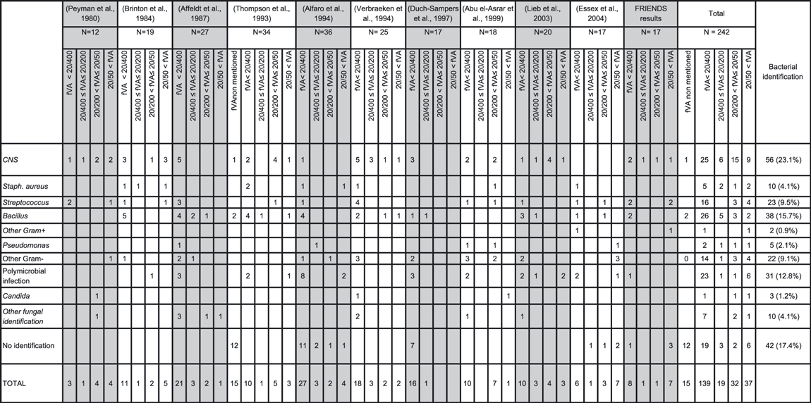
Post-traumatic endophthalmitis is characterized by a different bacterial ecology from post-operative endophthalmitis. Coagulase-negative staphylococci are involved in 45–48% of post-operative cases of endophthalmitis (Johnson et al. 1997; Chiquet et al. 2008) versus only 23% of post-traumatic cases of endophthalmitis (Table 4). The prevalence of streptococci and Gram-negative bacilli is higher in this infection, with an overrepresentation of soil-borne bacteria. Even though there are no other epidemiological data referenced in France on post-traumatic endophthalmitis, the present cohort is probably a relatively representative sample as the FRIENDS study groups four hospital-affiliated centres (population base, 6.6 million) and treats approximately 15–20% of all the post-operative cases of endophthalmitis in France (Chiquet et al. 2008). Comparing this series to the published studies (Table 4), the bacterial ecology in France seems to be characterized by a lower incidence of infection related to Bacillus and multibacterial infections. Bacillus, in particular the cereus and licheniformis species, virulent and destructive strains, is a Gram-positive spore-forming bacillum, aerobic or anaerobic, soil-borne, ubiquitous, often associated with the presence of an IOFB (Brinton et al. 1984; Thompson et al. 1993), which, in terms of frequency, is classically considered the second bacterial aetiology for post-traumatic endophthalmitis after S. epidermidis (Affeldt et al. 1987; Alfaro et al. 1994; Sobaci et al. 2006; Vedantham et al. 2006) and/or streptococci (Kunimoto et al. 1999; Chhabra et al. 2006). In the present series, one of the patients infected by Bacillus presented an IOFB, and the other, a wound with soil. One of the two patients had been infected by Bacillus thuringiensis, a pathogenic agent rarely identified in ocular or periocular infections (Peker et al. 2010), responsible in this patient for a serious and rapidly evolving infection, possibly secondary to the production of toxin and its extreme mobility (Callegan et al. 2005).
Bacteria of the Streptococcus family were the second cause of infection in this series, with VA in 50% of the cases equal to or <20/400 (Table 4). In combining the data from 11 studies (242 cases, Table 4), streptococcal infection accounted for 11% of the causes of post-traumatic endophthalmitis, in which only 21% of the patients presented final VA ≥20/400. The unfavourable prognosis of streptococcal infections has also been reported in post-operative endophthalmitis (Mao et al. 1992; Han et al. 1996; Miller et al. 2004). The virulence and pathogenicity of these bacteria, notably S. pneumoniae, are related to bacterial toxins (pneumolysin) (Ng et al. 1997), the characteristics of the bacterial capsule (preventing phagocytosis) and the components of the wall of streptococci.
Rarely found in post-operative endophthalmitis (1995; Chiquet et al. 2008), multibacterial infection can account for 5–47% of the microbiologically proven cases of post-traumatic endophthalmitis (Brinton et al. 1984; Affeldt et al. 1987; Nobe et al. 1987; Boldt et al. 1989; Alfaro et al. 1994; Abu el-Asrar et al. 1999; Kunimoto et al. 1999; Chhabra et al. 2006) [with the possibility of overrepresentation of Gram-negative bacilli in this context (Kunimoto et al. 1999)].
No cases of fungal endophthalmitis were demonstrated in this study. This is likely related to the limited number of subjects in the study, the small number of plant-related injuries (one scleral wound from a wooden arrow and a plant IOFB) and the lower frequency of fungal infection in our region (4–14% of the cases of traumatic endophthalmitis in other countries) (Affeldt et al. 1987; Abu el-Asrar et al. 1999; Kunimoto et al. 1999; Das et al. 2005; Gupta et al. 2008). As fungal infection symptoms can mimic those of bacterial infection (Imago et al. 2009), culture on specific medium remains recommended (Gupta et al. 2008).
Endophthalmitis with IOFB
In the present series, 53% of the patients presented an IOFB, which is comparable to the data reported for other series (16.4–57.9%) (Brinton et al. 1984; Affeldt et al. 1987; Boldt et al. 1989; Thompson et al. 1993; Alfaro et al. 1994; Verbraeken & Rysselaere 1994; Lieb et al. 2003). The frequency of post-traumatic endophthalmitis in the presence of an IOFB ranges from 6 to 30% (Brinton et al. 1984; Affeldt et al. 1987; Boldt et al. 1989; Thompson et al. 1993; Alfaro et al. 1994; Verbraeken & Rysselaere 1994; Lieb et al. 2003), warranting intravitreous antibiotic prophylaxis (Knox et al. 2004; Soheilian et al. 2007) associated with general antibiotic therapy (Duch-Samper et al. 1998; Mittra & Mieler 1999; Andreoli et al. 2009; Rajpal et al. 2009) during the initial management of the trauma. Injection of intracameral vancomycin has therefore been proposed as an alternative. It should be noted that of the nine patients in this series with an IOFB, eight underwent intravitreous injection of prophylactic antibiotics (vancomycin + ceftazidime) during the initial management of the injury.
Clinical presentation
The precise beginning of post-traumatic endophthalmitis remains very difficult to determine because the clinical and functional signs are usually confused with post-traumatic signs of inflammation. The time to symptom onset is classically short (Brinton et al. 1984), on average 1 day (range, 0–5 days) in our series. In 13 cases (77%), the patient was treated from the start with an injury complicated by endophthalmitis. Initial VA was often low, <20/400 in 86% of the 110 patients in five studies (Peyman et al. 1980a,b; Brinton et al. 1984; Affeldt et al. 1987; Alfaro et al. 1994; Abu el-Asrar et al. 1999). In the series reported herein, the time to treatment was always <24 hr compared to the onset of the first signs of endophthalmitis, except in two cases (≤3 days), which evolved toward ocular phthisis.
Therapeutic management
In this study, 88% of the patients underwent PPV. The indications for this procedure are not standardized, but vitrectomy is classically indicated in the presence of a loss of red reflex, substantial vitreous hyperechogenicity on ultrasound, the presence of an IOFB and/or identification of a virulent bacterium (Reynolds & Flynn 1997; Abu el-Asrar et al. 1999; Mittra & Mieler 1999). The theoretical benefits of PPV are obtaining vitreal samples (Alfaro et al. 1994), reducing the bacterial load and managing the associated lens or retinal injuries (Duch-Samper et al. 1998; Azad et al. 2003). All authors emphasize the difficulty of this surgery performed in conditions of limited visibility (corneal and lens opacity, vitreous haemorrhage) with high risks of intra- and post-operative complications. In our series, intraoperative complications were noted in 20% of the vitrectomies performed. Ultrasound before intravitreal injection or vitrectomy is indispensable so as to identify any associated chorioretinal lesions.
Clinical course, prognosis
The anatomical and functional prognosis is multifactorial (Carter et al. 2010) in this trauma context, with serious initial lesions such as RD (n = 2) or choroidal haemorrhage (n = 1). The precise characterization of the prognostic factors requires large cohorts of patients and multivariate statistical analyses. A recent study from India on 182 cases (Das et al. 2005) showed that final VA <20/60 was significantly associated with five prognostic factors: an IOFB, a needle injury, poor initial VA, reduced fundus visibility and the presence of hyperechogenic membranes on ultrasound.
During the course of treatment, onset of RD is feared because this is a factor of a poor prognosis (Brinton et al. 1984; Affeldt et al. 1987), with a high risk of phthisis. The RD rate after PPV in post-operative endophthalmitis is 10% (Doft et al. 2000), essentially the 1st month after the surgery. The frequency of RD in patients with traumatic endophthalmitis is higher, varying from 17 to 58% (Brinton et al. 1984; Affeldt et al. 1987; Bartz-Schmidt et al. 1996; Abu el-Asrar et al. 1999; Azad et al. 2003; Sobaci et al. 2006). Retinal necrosis secondary to the infection noted histologically in animal models of endophthalmitis (infection of the rabbit with Bacillus, for example) is probably one of the major factors of developing RD in this context (Alfaro et al. 1996). In our series, eight patients (47%) presented RD that had evolved toward phthisis in 75% of the cases (with enucleation in two cases). The PVR rate was 87.5% in our series, more frequent than with post-traumatic RD with IOFB with no endophthalmitis (20–52%) (Jonas et al. 2000; Chiquet et al. 2002).
In the literature, of 10 studies (Table 4) including 225 patients with microbiological data and final VA reported, the latter was ≥20/400 in 38% of the cases (53% in our series). More recently, it was reported that the use of internal tamponade with silicone oil could improve the final prognosis (Azad et al. 2003), with 58% of the patients presenting final VA ≥20/200.
In this series, in nearly half the cases (8/17 eyes), the final prognosis was limited (VA < 20/400), with 7/17 (35%) evolving toward phthisis and two of the seven being enucleated. The literature demonstrates a rate of anatomical loss of the eye (phthisis, evisceration or enucleation) in 6–82% of cases (Brinton et al. 1984; Affeldt et al. 1987; Alfaro et al. 1994; Duch-Samper et al. 1997; Abu el-Asrar et al. 1999; Essex et al. 2004; Al-Omran et al. 2007; Gupta et al. 2008). In a recent publication, the evisceration/enucleation rate was 17% (Zhang et al. 2010), underscoring once again that the prognosis for this infection remains poor. The information initially given to the patient should comply with these results.
In conclusion, this series showed that Gram-positive cocci were the most frequent causative organisms and that better final VA outcomes were associated with better than light perception on initial examination, Staphylococcus epidermidis or culture-negative cases, and absence of RD during the clinical course. The final anatomical and functional prognosis was relatively binary (quite good or very poor), and it was related to microbiological (especially Streptococcus species) and traumatological factors (most particularly occurrence of secondary RD). The diagnosis of post-traumatic endophthalmitis should therefore be made as quickly as possible so as to adapt treatment urgently. Efficient prophylaxis of this complication is needed.
Acknowledgements
Grants: This study was supported by grants from Hospices Civils de Lyon, Bibliothèque Scientifique de l’Association Générale de l’Internat de Lyon, Alcon Laboratories, and Thea Laboratories.


