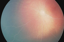
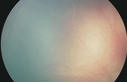
Grade III lipaemia retinalis in a newborn
Editor,
Lipaemia retinalis, first described in 1880 and recently recognized as a paediatric disease, occurs rarely in patients with hyperlipidaemia. In typical cases, ophthalmoscopy evidences creamy-white retinal vessels. Although the picture is graded clinically (Table 1), grade III cases are extremely rare.
| Grade | Intensity | Clinical appearance |
|---|---|---|
| I | Early | White and creamy aspect of peripheral retinal vessels |
| II | Moderate | Creamy-coloured vessels extending toward optic disc |
| III | Marked | Salmon-coloured retina, all vessels having milky aspect |
The first daughter of non-consanguineous parents; spontaneous birth (weight 3430 g) following a full-term uneventful pregnancy. The family history revealed hypercholesterolaemia and hypertrigliceridaemia in the paternal grandfather, grandmother and father, which was moderate, mild and very mild, respectively. The patient underwent phototherapy for jaundice from blood group incompatibility and, a few days after birth, was discharged from hospital in a good clinical condition. However, at 28 days of life, she was admitted to hospital with fever and feeding problems. Breast-fed, and weighing 4045 g, on the first evening of hospitalization the patient had biliary vomiting. Laboratory investigations were performed, but the presence of lypaemic serum precluded blood sample analysis. On transferral to the paediatric intensive care unit for diagnosis and treatment, the clinical examination revealed multiple facial little tip-head xanthomas, and the laboratory investigation a slight increase in leukocytes (17 000 wc/ml), platelets (600 000/ml) and C-reactive protein (114 mg/l). The creamy aspect of the lipaemic serum precluded the determination of coagulation (pro-thrombin and thromboplastin time) and albuminaemia and creatininaemia levels. Triglycerides were 12 380 mg/dl (normal value < 200 mg/dl) and total cholesterol 820 mg/dl (n.v. < 200 mg/dl). Serum chylomicron levels were also elevated markedly. At pancreatic amylase measurement plasma level was normal, as were other bio-humoral values including haemogas. On the suspending of enteral feeding, lipid-free parenteral nutrition, heparin (10 U/kg/hr), ampicillin and gentamycin administration were started; abdominal echography showed a normal liver and pancreas, whereas ophthalmoscopy evidenced pale optic discs and creamy-white arteries and veins, and a salmon-pink retina. The clinical picture was typical of lipaemia retinalis (1, 2). Three days after suspending breast-feeding, parenteral nutrition was started; laboratory examinations showed reduced triglycerides (3750 mg/dl) and total cholesterol (440 mg/dl) levels. All other bio-humoral parameters were normal. Low lipid formula (Basic-F Nutricia) feeding was started and heparin infusion suspended. After another 3 days on the new diet, blood lipids appeared normal, as did the fundus, except for a minimal retinal haemorrhage in the right eye. At follow-up 5 months later, the visual function (Teller Acuity Cards) and ophthalmological findings were normal. Laboratory examinations in the parents confirmed mild hypercholesterolaemia in the father. At DNA analysis, the infant had a familial type 1 lipoprotein–lipases deficit.

Pale optic disc: all vessels (arteries and veins) with creamy aspect; salmon-coloured retina, typical of lipaemia retinalis.

Peripheral retinal vessels with blood column segmentation caused by initial regression of lipaemia (2 days later).
To our knowledge, the only cases of lipaemia retinalis in newborns reported in the literature are in two infants aged 29 days (Hayasaka et al. 1985) and 35 days (Cypel et al. 2008), and in a 77-day-old pre-term infant (Rotchford et al. 1997) in whom lipaemia retinalis was associated with retinopathy of prematurity. Treatments proposed include the administration of a fat-restricted diet, increased protein and carbohydrate, and/or the addition of medium-chain triglycerides and soluble-fat vitamins. Our patient received parenteral nutrition and heparin infusion to lower the risk of small-vessels thrombosis before returning to a diet of artificial skimmed milk. Broad-spectrum antibiotic therapy, started on admission because of the clinical suspicion of sepsis without a laboratory confirmation, was withdrawn on obtaining blood culture results and an improvement in the clinical condition. Prompt interruption of enteral nutrition, intravenous hydratation with no-lipid-balanced solutions, strict monitoring of vital signs and pancreatic enzymes plasma levels are crucial therapeutic strategies. While waiting for normal plasma fat levels to be restored, low-dose heparin infusion may help to prevent small-vessel obstruction.


