Jamming of 23-gauge instruments in the microcannula during vitrectomy for severe vitreous haemorrhage
Editor,
Transconjunctival sutureless vitrectomy has gained popularity in recent years. The use of 25- or 23-gauge instruments has many advantages over conventional 20-gauge vitrectomy, and there have been many reports on the safety and efficacy of 25- or 23-gauge vitrectomy in a variety of vitreoretinal diseases (Fujii et al. 2002; Eckardt 2005). However, complications associated with such small-gauge instruments remain problematic (Inoue et al. 2004; Ooto et al. 2008). Shinoda et al. (2008) reported on the jamming of 25-gauge instruments in the cannula during vitrectomy for vitreous haemorrhage. We experienced the jamming of a 23-gauge endo-illuminator or vitreous cutter in the microcannula during 23-gauge vitrectomy in a patient with severe vitreous opacity.
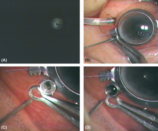
A 47-year-old man was diagnosed with severe vitreous opacity and haemorrhage associated with central retinal vein occlusion. Best corrected visual acuity was 20/20 in the right eye and light perception in the left. Two years previously, the patient had undergone Ahmed valve implantation for neovascular glaucoma in the left eye. A 23-gauge surgical procedure was performed using the DORC two-step system (Dutch Ophthalmic Research Center [DORC] International BV, Zuidland, the Netherlands). Vitreous surgery was carried out using a 23-gauge endo-illuminator and vitreous cutter (DORC International BV) driven by a vitrectomy unit (Associate 2500; DORC International BV). After conventional cataract surgery and subsequent core vitrectomy, peripheral vitrectomy was performed (Fig. 1A). When we attempted to remove the endo-illuminator/vitreous cutter from the microcannula during the peripheral vitrectomy, we found that the instrument was lodged firmly within the microcannula and its removal was likely to pull the cannula out of the sclerotomy site (Fig. 1B). We found that if we stabilized the collar of the microcannula by holding it firmly with a forceps, it was possible to remove the illuminator/vitreous cutter from the microcannula. Thereafter, we identified organized vitreous membranes trapped in the inner tube of the microcannula (Fig. 1C). We removed the entrapped vitreous with a cutter to clear the inside of the cannula and were able to reinsert or remove 23-gauge instruments freely into and out of the microcannula (Fig. 1D). All other procedures were then completed in the usual manner. Intraoperatively and postoperatively, gross and microscopic examinations revealed no specific deformities of or damage to the microcannula or the 23-gauge instruments.

Intraoperative photographs. (A) Dense vitreous opacities are seen during core vitrectomy. (B) The 23-gauge endo-illumination light pipe became jammed in the cannula. The shaft of the light pipe was bent by the force required to remove it from the cannula. (C) Once the light pipe was removed, organized vitreous membranes were seen to be lodged in the inner wall of the cannula. (D) After the entrapped vitreous haemorrhage had been removed with a vitreous cutter, the lumen of the cannula was clear and hollow.
Whereas Shinoda et al. (2008) experienced the jamming of 25-gauge instruments in three (7%) of 45 eyes with vitreous haemorrhage, we experienced jamming in only one of about 50 eyes with vitreous haemorrhage following 23-gauge vitrectomy. By contrast with 25-gauge instruments that are jammed in the microcannula, the jammed 23-gauge cutter or light pipe could be withdrawn from the 23-gauge cannula by the aid of a forceps. Moreover, we did not find any damage to the microcannula or 23-gauge instruments. There are possible explanations for the differences between 25- and 23-gauge instruments in terms of incidence of jamming and recovery of instruments. One explanation may relate to the material of the microcannula. This is made of polyamide in the 25-gauge system, but stainless steel in the 23-gauge system. Therefore, although the 25-gauge plastic cannula is easily damaged, the 23-gauge metal cannula remains firmly intact. Another reason for the difference may concern the inner diameter of the cannula. The 23-gauge microcannula has a larger lumen (0.65 mm) than the 25-gauge (0.50 mm). The microcannula with the larger lumen may be less likely to entrap material inside it.
Although the jamming of 23-gauge instruments during vitrectomy is a rare complication, clinicians should be aware of the possibility of this adverse event when preparing patients with dense vitreous haemorrhage. In order to prevent the occurrence of such a complication, any dense vitreous around or inside the cannula should be carefully removed prior to core vitrectomy.


