Risk of retinal neovascularization in the second eye in patients with proliferative diabetic retinopathy
Abstract.
Purpose: This study aimed to evaluate the risk of proliferative diabetic retinopathy (DR) in the fellow eye of an eye with existing proliferative DR.
Methods: Our DR screening programme database listed 1513 diabetes patients alive at the time of the study. Seventy-six had proliferative DR in one or both eyes.
Results: In 28 of the 76 (37%) diabetes patients, proliferative DR was diagnosed in both eyes at the same examination. Another 28 patients developed proliferative DR in the second eye within 5 years of its diagnosis in the first eye, bringing the total number of diabetes patients with proliferative DR in both eyes at 5 years to 56 (74%). Almost all the diabetes patients eventually developed proliferative DR in the second eye. The median duration of diabetes before the development of proliferative retinopathy was 19 years for type 1 and 14 years for type 2 diabetes.
Conclusions: Proliferative DR is a bilateral disease. Diabetes patients with proliferative DR in one eye are at high risk of developing neovascularization in the second eye and close follow-up is recommended.
Introduction
Diabetic retinopathy (DR) tends to affect both eyes in most patients. It is generally presumed that the onset of proliferative DR in one eye increases the risk of neovascularization in the other, but we are not aware of any study of this risk factor or of the time pattern involved. Several risk factors for the development of proliferative DR have been identified, such as duration of diabetes (Roy et al. 2004), blood glucose (Diabetes Control Complications Trial Research Group 1993; UK Prospective Diabetes Study Group 1998) and arterial blood pressure (Klein et al. 1984a, 1984b). We studied patients in our DR screening programme (Kristinsson 1997; Stefánsson et al. 2000) who developed proliferative DR in at least one eye and report the prognosis for proliferative DR in the second eye.
Materials and Methods
The study was approved by the Icelandic Science Ethics Board. The database of the Icelandic diabetic screening programme listed 1513 diabetes patients alive at the time of the study (Kristinsson 1997; Stefánsson et al. 2000). Of these, 76 had proliferative retinopathy in one or both eyes. Forty-six were male, 41 had type 1 diabetes and 35 had type 2 diabetes mellitus.
The prevalence of type 1 diabetes in Icelandic children under the age of 14 years is 0.57 per 1000 (Helgason et al. 1992). The prevalence of type 2 diabetes in Iceland in people aged 30–79 years is 2.9% (95% confidence interval [CI] 2.5–3.3) for men and 2.1% (95% CI 1.8–2.5) for women (Vilbergsson et al. 1997). From these studies, we have estimated the number of diabetes patients in Iceland to be 4600, of whom 600 have type 1 diabetes and 4000 have type 2 diabetes. Our study group with proliferative retinopathy thus corresponds to 7% of type 1 diabetes patients and 1% of type 2 diabetes patients in Iceland.
All diabetes subjects in this screening programme undergo regular screening visits, which include a complete eye examination, dilated fundus examination and retinopathy staging performed by an ophthalmologist, and fundus photography. The screening programme, documentation and diagnostic criteria have been reported previously (Kristinsson et al. 1994a, 1994b; Kristinsson 1997; Stefánsson et al. 2000; Zoega et al 2005).
From the standardized records, we collected the date of diagnosis of proliferative DR in each eye, the date of diagnosis of diabetes mellitus and the stage of retinopathy in the second eye when the first eye developed neovascularization. We examined medical records, which were available for 51 of the 76 patients, at the Diabetic Clinic at the National University Hospital. We recorded the patients’ glycaemic control (HbA1c), blood pressure and serum cholesterol values and treatment at their last visits, which occurred during the study year. Table 1 shows the clinical data for our study group. Statistical evaluation was performed with Microsoft excel, spss sigma plot 2000 (SPSS Inc., Chicago, IL, USA) and spss sigma stat (SPSS Inc.).
| Type 1 diabetes patients | Type 2 diabetes patients | |
|---|---|---|
| Number of patients | 41 | 35 |
| Male | 29 | 17 |
| Female | 12 | 18 |
| Median age, years, when first diagnosed with proliferative retinopathy (range) | 32 (19–62) | 65 (44–78) |
| Median duration of diabetes, years, when first diagnosed with proliferative retinopathy (range) | 19 (10–39) | 14 (1–30) |
| Median VA in better eye when first diagnosed with proliferative retinopathy (range) | 1.00 (0.25–1.50) | 0.69 (1.0–1.20) |
| Median VA in worse eye when first diagnosed with proliferative retinopathy (range) | 0.90 (HM–1.50) | 0.35 (HM–1.0) |
| Smokers, % | 19 | 24 |
| Patients using insulin, % | 100 | 36 |
| Patients using statins, % | 31 | 40 |
| Patients using anti-hypertensive drugs, % | 92 | 100 |
| Mean HbA1c,*% (SD) | 8.8 (1.62) | 8.1 (1.53) |
| Mean systolic blood pressure, mmHg (SD) | 140 (17.2) | 145 (18.4) |
| Mean diastolic blood pressure, mmHg (SD) | 85 (10.7) | 83 (10.1) |
| Mean serum cholesterol, mmol/l (SD) | 5.14 (0.92) | 5.73 (1.89) |
| Mean HDL, mmol/l (SD) | 1.64 (0.56) | 1.24 (0.47) |
- * Measured with DCA 2000 (Bayer, Germany), reference value < 6.2.
- VA = visual acuity; SD = standard deviation; HDL = high-density lipoprotein.
Results
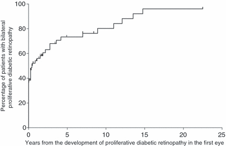
In 28 of the 76 patients (37%), proliferative DR was diagnosed in both eyes at the same visit. An additional 25 patients (33%) developed proliferative DR in the second eye during the follow-up period. Figure 1 presents a Kaplan–Meier graph showing the proportions of patients with proliferative DR in both eyes over the follow-up period. Overall, 28 patients (37%) developed neovascularization at the same time in both eyes and another 28 developed proliferative DR in the second eye within 5 years of its diagnosis in the first eye, bringing the total number of diabetes patients with proliferative DR in both eyes at 5 years to 56 (74%). Gradually, more and more patients developed proliferative DR in the second eye. One patient developed proliferative DR in the second eye 15 years after it appeared in the first eye. Another patient spent 23 years with retinal neovascularization in one eye without developing neovascularization in the second eye. Figure 2 shows a Kaplan–Meier graph comparing patients with type 1 and type 2 diabetes over the time of follow-up. Type 1 diabetes patients seem to have a slightly higher tendency to develop proliferative DR simultaneously in both eyes or within a short time. However, our study group lacks power for statistical evaluation.

Kaplan–Meier graph showing the percentage of second eyes developing proliferative diabetic retinopathy (DR) at various time-points. The interval between the development of proliferative DR in the first and second eyes is given in years. A total of 37% of our patients were diagnosed with proliferative retinopathy in both eyes at the same visit. Within 5 years, the number of patients with neovascularization in the second eye had risen to 74% and virtually all eventually developed bilateral proliferative DR.

Kaplan–Meier graph showing the percentage of second eyes that developed proliferative diabetic retinopathy (DR). The interval between diagnoses in both eyes is given in years. Type 1 diabetes patients are shown in red and type 2 diabetes patients are shown in black. This graph implies that type 1 diabetes patients may have a tendency to develop bilateral proliferative DR sooner than type 2 diabetes patients. However, our study group was too small to support statistical evaluations.
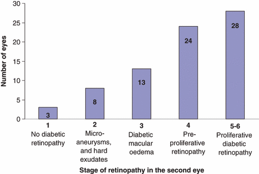
Figure 3 shows the stage of retinopathy in the second eye at the time of the diagnosis of proliferative DR in the first eye. Stage of retinopathy in the second eye did not show a statistically significant correlation with how soon that eye developed proliferative retinopathy. However, the group is small and its statistical power is weak.

Stage of retinopathy in the second eye at the time of diagnosis of proliferative diabetic retinopathy (DR) in the first eye. In all, 28 of our 76 patients already had bilateral disease when diagnosed with neovascularization. Half the remaining 48 patients had developed preproliferative retinopathy in the second eye when neovascularization was diagnosed in the first eye. Only three patients showed no signs of retinopathy in the second eye.
We took a closer look at the 28 patients who were diagnosed with proliferative DR in both eyes at the same visit. Eleven of these 28 patients had been examined less than a year before diagnosis (2–10 months), and proliferative retinopathy most probably developed simultaneously in both eyes in these patients. Eight patients had not attended a screening visit in the year before the diagnosis of bilateral proliferative retinopathy (time since last screening visit: 2–13 years) and nine patients were diagnosed at their first screening programme visit.
Discussion
Proliferative DR presented at the same time in both eyes in about a third of the patient group. Within 5 years of diagnosis of the first eye, another third developed proliferative DR in the other eye. Although the slope representing the development of DR in the second eye is particularly steep in the first 5 years, it continues to rise and most individuals eventually develop proliferative DR in the fellow eye.
Type 2 diabetes patients had been diagnosed with diabetes mellitus for a median of 14 years by the time they developed proliferative DR, whereas type 1 diabetes patients had been diagnosed for a median of 19 years. This difference was statistically significant and may reflect a delay in the diagnosis of type 2 diabetes in the order of 5 years. These results are comparable with those of a previous report by Harris et al. (1992).
Mean HbA1c values for all patients attending the Diabetic Clinic at Reykjavik University Hospital were 7.5% for type 1 diabetes patients and 6.9–7.0% for type 2 diabetes patients. The patients in our proliferative retinopathy group (average HbA1c 8.6%, standard deviation [SD] 1.5) had poorer glucose control than the overall diabetes patient group. We also found the percentage of smokers among the type 2 diabetes patients with proliferative retinopathy (24%) was higher than among all type 2 diabetes patients attending the Diabetic Clinic (13%). Blood glucose control and smoking are known risk factors for developing proliferative DR (Hove et al. 2006; Higgins et al. 2007).
Diabetes patients with proliferative retinopathy in one eye are at high risk of developing neovascularization in the second eye. Patients should be made aware of this and careful follow-up and timely laser treatment may help to prevent loss of vision in the second eye and consequent permanent disability (Bek & Erlandsen 2006; Olafsdóttir et al. 2007).


