Independent and Incremental Value of Severely Enlarged Left Atrium in Risk Stratification of Very Elderly Patients With Chronic Systolic Heart Failure
Abstract
Congest Heart Fail. ****;**:**–**. ©2012 Wiley Periodicals, Inc.
The authors sought to assess the impact on survival of demographic, clinical, and echo-Doppler parameters in patients with chronic heart failure due to left ventricular systolic dysfunction divided according to age groups. This study included 734 patients (age 69±11 years) who were classified into tertiles of age: I (22–66 years), II (67–76 years), and III (77–94 years). Severely enlarged left atrial size was defined as ≥52 mm in men and ≥47 mm in women. Multivariable analysis identified male sex (P=.018) and severely enlarged left atrium (P=.024) as significant correlates of all-cause mortality in the very elderly cohort, while restrictive filling pattern (RFP) (P=.004) and New York Heart Association class III or IV (P=.005) among patients of the first tertile and RFP (P=.028) among patients in the second tertile were independently associated with mortality after 30±21 months of follow-up. At the interactive stepwise model in the very elderly population, a severely enlarged left atrium, added to the model after clinical parameters and ejection fraction, moved the chi-square value from 20.7 to 25.8 (P=.048). RFP emerged as the single best predictor of all-cause mortality in the younger and intermediate ranges, whereas severely enlarged left atrium was the best predictor in the very elderly.
Heart failure (HF) is an expanding syndrome and it is becoming the most important public health problem in medicine.1 The syndrome of chronic HF is a common manifestation of later stages of various cardiovascular diseases.2 Despite the recent achievements in pharmacologic and nonpharmacologic treatments, the morbidity and mortality associated with HF remains high.2–12 Left ventricular (LV) systolic dysfunction is the most common cause of HF13–15 and is associated with reduced quality of life16–19 and poor prognosis.3,12,20 Several previous studies have shown that a number of clinical and echo-Doppler variables can predict major cardiac events, including mortality.21–27 The age of HF patients was also shown as an independent predictor of mortality in most of these studies,25,28–30 even though other studies did not confirm this association between age and mortality in patients with HF.23,26 However, it is not clear which of the echo-Doppler parameters that are commonly recorded in HF patients are most useful to better stratify patients of different age groups, from the younger cohorts to the very elderly. The aim of this study was to assess the impact on survival of demographic, clinical, and echo-Doppler parameters in patients with chronic HF due to LV systolic dysfunction divided according to age groups.
Methods
Study Patients
Consecutive outpatients referred to the Echo Laboratory of the Cardiovascular Unit 2 Tito Giulio Sicca of the Santa Chiara Hospital, Pisa, Italy, from April 2003 to December 2006 were prospectively included in the study. Inclusion criteria were chronic systolic HF, defined as presence of prior or current signs/symptoms of HF, LV ejection fraction (EF) <45%; and sinus rhythm. Patients were excluded if they had primary valvular disease, mechanical valve prosthesis, unstable angina, recent myocardial infarction (within 3 months), or coexisting terminal diseases. Written informed consent was obtained from all patients participating in the study.
Echocardiography
Echo-Doppler examination was performed using an Acuson Sequoia C256 echocardiograph (Siemens, Mountain View, CA) with 2nd-harmonic imaging and a 3.5-MHz transducer. Recordings were made with the patient in the left lateral decubitus position during quiet expiration. Left atrial (LA) size was estimated in parasternal long-axis view from the trailing edge of posterior aortic wall to leading edge of posterior LA wall in systole. LA diameter was qualified as severely enlarged left atrium (≥47 mm for women and ≥52 mm for men) and not severely enlarged.31 LV volumes and EF were calculated from apical 2- and 4-chamber views using the modified Simpson’s method and LV mass index was calculated. LV filling velocities were obtained from the apical 4-chamber view with the sample volume placed by the tips of the mitral valve leaflets. Peak early (E wave) and late (A wave) diastolic LV filling velocities and E-wave deceleration time (EDT) were measured. LV filling pattern was considered restrictive when the EDT <150 ms.32 Mitral and tricuspid regurgitation severities were assessed by color and continuous wave Doppler and were graded as mild, moderate, or severe according to the relative jet area to that of the left or right atrium as well as the flow velocity profile, in line with the recommendations of the American Society of Echocardiography.33 Pulmonary artery systolic pressure (PASP) was measured from the tricuspid retrograde pressure drop using the modified Bernoulli equation and the estimated right atrial pressure.
Follow-Up Data
Patients were followed-up after the baseline Doppler echocardiogram. The end point was all-cause mortality. Follow-up data were obtained by regular visits (from 1- to 6-month intervals as clinically indicated) by telephone calls, from local authority registry, or from hospital records.
Data Analysis
Comparisons between demographic, clinical, and echo-Doppler variables were analyzed by the standard parametric methods. The probability level was P<.05. Demographic, clinical, and echo variables were evaluated for the end point in a univariable Cox proportional hazard model. The variables were dichotomized according to the following threshold value: EF <25%, LA size ≥47 mm for women and ≥52 mm for men, PASP >35 mm Hg, LV end-diastolic volume index >96 mL/m2, and LV end-systolic volume index >42 mL/m2.31,32 Age, body surface area, and heart rate were dichotomized according to their median values. Only variables showing a P value <.1 were tested in multivariable models. Kaplan-Meier curves were constructed and log-rank tests were used to test for differences between survival curves. The likelihood ratio test was used to compare different multivariable Cox models by taking into account the different number of variables used in each model. The log-likelihood and the overall model chi-square (ie, that calculated by testing the hypothesis that all regression coefficients for the variables in the model are identically zero) were calculated for each model.
Results
The study included 734 patients, with a mean age of 69.4±11 years (range 26–94, median 71 years, 23.7% of whom were women). During the follow-up period (30±21 months, median 30 months), 205 (27.9%) patients died.
Comparison Between Age Tertiles
Using one-way analysis of variance test, we found that New York Heart Association (NYHA) class (P=.001), female sex (P=.012), coronary artery disease (P<.001), LV mass index (P=.041), and EDT (P<.001) were significantly increased, whereas body mass index was significantly decreased (P<.001) in age tertiles (Table I). The other clinical and echo-Doppler parameters did not change significantly between age tertiles.
| Variable | First Tertile (n=256) | Second Tertile (n=224) | Third Tertile (n=223) | P Value |
|---|---|---|---|---|
| Age, y | 57±8 | 72±3a | 81±3b,c | <.001 |
| Women, % | 20 | 11d | 30c | .012 |
| BSA | 1.89±0.23 | 1.83±1.17d | 1.76±1.17b,c | <.001 |
| Heart rate, beats per min | 77±15 | 76±15 | 76±13 | NS |
| CAD, % | 47 | 74a | 63b,e | <.001 |
| NYHA class III or IV, % | 35 | 46d | 48f | .011 |
| LVEDV index, mL/m2 | 117±37 | 113±30 | 112±37 | NS |
| LVESV index, mL/m2 | 81±33 | 78±28 | 77±31e,f | NS |
| Ejection fraction, % | 32±8 | 32±8 | 33±9 | NS |
| LV mass index, g/m2 | 157±38 | 155±33 | 163±39e | .041 |
| Left atrial size, mm | 48±6 | 49±6 | 48±6 | NS |
| Mitral regurgitation, %g | 46.8 | 56.9 | 60 | .076 |
| Restrictive mitral flow, % | 46.9 | 38.1 | 29.8 | .001 |
| Sphericity index | 0.49±0.50 | 0.39±0.49 | 0.43±0.50 | NS |
| E-wave deceleration time, ms | 157±50 | 166±57 | 177±58b,e | <.001 |
- Abbreviations: BSA, body surface area; CAD, coronary artery disease; EDV, end-diastolic volume; ESV, end-systolic volume; LV, left ventricular; NS, not significant; NYHA, New York Heart Association. aP<.001 group A vs group B. bP<.001 group A vs group C. cP<.001 group B vs group C. dP<.05 group A vs group B. eP<.05 group B vs group C. fP<.05 group A vs group C. gAny degree.
Predictors of All-Cause Mortality in the Overall Study Group
In the univariable analysis model, a number of measurements predicted cardiac events in the overall study population (Table II), including age, NYHA class, sex, heart rate, ischemic aetiology, LVEF, LV end-diastolic volume index, LV end-systolic volume index, E-wave deceleration time, restrictive LV filling pattern, left atrium diameter, pulmonary artery pressure drop, and LV mass index. However, in the multivariable analysis model, the independent variables that predicted death were presence of NYHA class III or IV (odds ratio [OR], 2.090; 95% confidence interval [CI], 1.425–3.066; P<.001), age (OR, 3.087; 95% CI, 1.985–4.801; P<.001), the presence of restrictive mitral filling (OR, 1.680; 95% CI, 1.081–2.611; P=.021), and LV end-systolic volume index (OR, 1.737; 95% CI, 1.066–2.830; P=.027) (Table III).
| Variable | Odds Ratio (95% CI) | P Value |
|---|---|---|
| Clinical univariable predictors | ||
| NYHA class III or IV | 2.77 (1.98–3.88) | <.001 |
| Age >71 y | 1.04 (1.02–1.05) | <.001 |
| Sex | 0.66 (0.44–0.98) | .041 |
| CAD | 0.67 (0.48–0.94) | .020 |
| BSA >1.70 | 1.02 (0.98–1.06) | .227 |
| Heart rate >80 beats per min | 1.02 (1.01–1.03) | .003 |
| Echo-Doppler univariable predictors | ||
| Severely enlarged left atriuma | 1.79 (1.29–2.48) | .001 |
| LVEF <26% | 2.68 (1.87–3.84) | <.001 |
| LVEDV index >96 mL/m2 | 1.96 (1.38–2.80) | <.001 |
| LVESV index >42 mL/m2 | 2.95 (2.05–4.26) | <.001 |
| Restrictive filling pattern | 2.32 (1.66–3.24) | <.001 |
| PASP >35 mm Hg | 2.19 (1.56–3.06) | <.001 |
| Multivariable predictors | ||
| NYHA class III or IV | 1.55 (1.01–2.40) | <.001 |
| Age >71 y | 3.09 (1.98–4.80) | <.001 |
| Male sex | 1.25 (0.77–1.86) | .020 |
| Restrictive mitral filling | 1.68 (1.08–2.61) | .021 |
| LVESV index >42 mL/m2 | 1.74 (1.07–2.83) | .027 |
| LVEF <26% | 1.60 (0.99–2.57) | .053 |
| CAD etiology | 0.74 (0.49–1.10) | .136 |
| Severely enlarged left atriuma | 1.21 (0.81–1.80) | .348 |
| Heart rate >80 beats per min | 1.00 (0.99–1.02) | .768 |
| PASP >35 mm Hg | 1.31 (0.85–2.03) | .228 |
| LVEDV index >96 mL/m2 | 1.16 (0.72–1.86) | .535 |
- Abbreviations: BSA, body surface area; CAD, coronary artery disease; CI, confidence interval; EDV, end-diastolic volume; EF, ejection fraction; ESV, end-systolic volume; LV, left ventricular; NYHA, New York Heart Association; PASP, pulmonary artery systolic pressure. a≥47 mm for women and ≥52 mm for men.
| Variable | Odds Ratio (95% CI) | P Value |
|---|---|---|
| Univariable predictors in the first tertile | ||
| NYHA class III or IV | 4.90 (2.35–10.19) | <.001 |
| Severely enlarged left atrium | 3.33 (1.68–6.62) | .001 |
| LVEF <26% | 3.97 (1.98–7.95) | <.001 |
| Restrictive filling pattern | 5.69 (2.49–13.02) | <.001 |
| PASP >35 mm Hg | 2.32 (1.16–4.62) | .017 |
| Univariable predictors in the second tertile | ||
| NYHA class III or IV | 1.99 (1.15–3.46) | .014 |
| Severely enlarged left atrium | 1.35 (0.79–2.32) | .277 |
| LVEF <26% | 2.90 (1.59–5.29) | .001 |
| Restrictive filling pattern | 2.81 (1.61–4.93) | <.001 |
| PASP >35 mm Hg | 2.08 (1.19–3.61) | .010 |
| Univariable predictors in the third tertile | ||
| NYHA class III or IV | 2.36 (1.36–4.10) | .002 |
| Severely enlarged left atriuma | 1.90 (1.08–3.34) | .025 |
| LVEF <26% | 3.01 (1.45–6.24) | .003 |
| Restrictive filling pattern | 2.14 (1.19–3.86) | .012 |
| PASP >35 mm Hg | 1.86 (1.08–3.22) | .026 |
- Abbreviations: CI, confidence interval; LVEF, left ventricular ejection fraction; NYHA, New York Heart Association; PASP, pulmonary artery systolic pressure. a≥47 mm for women and ≥52 mm for men.
Predictors of All-Cause Mortality in Age Tertiles
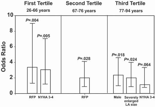
In the first age tertile (age range, 26–66 years), LVEF <26%, presence of restrictive filling pattern, NYHA class III or IV (P<.001, for all), severely enlarged left atrium (P=.001), and PASP >35 mm Hg (P=.017) were univariable predictors of all-cause mortality (Table III). NYHA class III or IV and the presence of restrictive mitral filling resulted as independent predictors of death (Table IV and Figure 1).
| Variable | Odds Ratio (95% CI) | P Value |
|---|---|---|
| Multivariable predictors in the first tertile | ||
| Restrictive mitral filling | 3.75 (1.53–9.22) | .004 |
| NYHA class III or IV | 3.14 (1.41–7.02) | .005 |
| Male sex | 1.37 (0.82–2.23) | .434 |
| Coronary artery disease aetiology | 1.29 (0.61–2.73) | .498 |
| Multivariable predictors in the second tertile | ||
| Restrictive mitral filling | 2.08 (1.08–3.99) | .028 |
| Coronary artery disease aetiology | 2.11 (0.62–7.27) | .234 |
| LVEF <26% | 1.98 (0.99–3.93) | .052 |
| Male sex | 1.51 (0.74–3.05) | .255 |
| PASP >35 mm Hg | 1.34 (0.68–2.65) | .393 |
| NYHA class III or IV | 1.18 (0.60–2.28) | .633 |
| Multivariable predictors in the third tertile | ||
| Male sex | 2.25 (1.15–4.39) | .018 |
| Severely enlarged left atriuma | 2.07 (1.10–3.88) | .024 |
| NYHA class >II | 1.82 (0.96–3.44) | .064 |
| Restrictive mitral filling | 1.65 (0.85–3.20) | .135 |
| LVEF <26% | 1.78 (0.80–3.96) | .157 |
| PASP >35 mm Hg | 1.35 (0.72–2.54) | .350 |
| CAD etiology | 1.13 (0.61–2.10) | .694 |
- Abbreviations: CAD, coronary artery disease; CI, confidence interval; LVEF, left ventricular ejection fraction; NYHA, New York Heart Association; PASP, pulmonary artery systolic pressure. a≥47 mm for women and ≥52 mm for men.

Histograms showing the odds ratio of variables independently related to all-cause morality in the three age groups. RFP indicates restrictive filling pressure; NYHA, New York Heart Association; LA, left atrium.
In the second age tertile (age range, 67–76 years), LVEF <26% (P=.001), presence of restrictive filling pattern (P<.001), NYHA class III or IV (P=.014), severely enlarged left atrium (P=.001), and PASP >35 mm Hg (P=.010) were univariable predictors of death (Table III). The presence of restrictive mitral filling was the only independent predictor of death (Table IV and Figure 1).
In the third age tertile (age range, 77–94 years), NYHA class III or IV (P=.002), LVEF <26% (P=.003), presence of restrictive filling pattern (P=.012), severely enlarged left atrium (P=.025), and PASP >35 mm Hg (P=.026) were univariable predictors of death (Table IV). The independent correlates of survival in the third tertile are reported in Table IV and Figure 1.
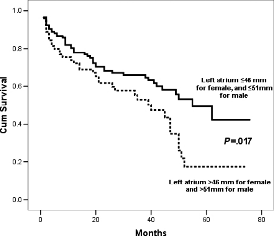
The survival rate was 72.1% in the overall study population, 84% in the first tertile, 70.5% in the second tertile, and 60.3% in the third tertile. On Kaplan-Meier survival analysis, patients with NYHA class III or IV HF exhibited a significantly worse survival rate than those with NYHA class I or II in the first and second tertiles (17.7% vs 6.3%; log-rank, 20.6; P<.001; and 43.7% vs 21.6%; log-rank, 23.4; P<.001). In the third tertile, a worse survival rate was observed in patients with an enlarged left atrium, >46 mm for women and >51 mm for men (50% vs 34.5%, log-rank: 5.73, P=.017, Figure 2).

Survival free of death predicted by severely enlarged left atrium in the third age tertile (log-rank: 5.73, P=.017).
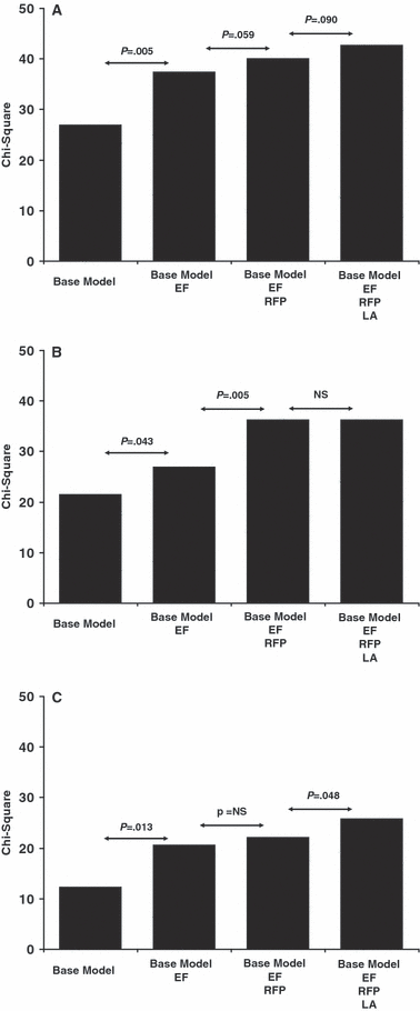
Finally, Cox proportional hazard models using the likelihood ratio chi-square statistics were assessed to evaluate whether an enlarged left atrium was additive to demographic, clinical, and instrumental findings in the prediction of death at follow-up. The base model included age, sex, etiology, NYHA class, and systolic blood pressure. This model was compared with models including EF (base model+EF), restrictive filling (base model+EF+diastolic dysfunction), and severely enlarged left atrium (base model+EF+diastolic dysfunction+severely enlarged left atrium). In the overall study population, EF added prognostic value to the base model (73.5–100.5, P<.0001), and the presence of restrictive filling moved the chi-square value up to 110.2 (P=.001). As far as the three tertiles are concerned (Figure 3), the incremental analysis showed that a severely enlarged left atrium significantly contributed to the prediction of the outcome in the very elderly population (P=.048).

The incremental analysis in the three age tertiles in patients with systolic heart failure. EF indicates ejection fraction; RFP, restrictive filling pressure; LA, left atrium; NS, not significant.
Discussion
The main findings of our study were that predictors of all-cause mortality in patients with chronic systolic HF change with age and that in the very elderly, severely enlarged LA size has independent and incremental value in risk stratification of all-cause mortality.
To our knowledge, our study is the first to assess the effect of age on the echo-Doppler predictors of all-cause mortality in patients with chronic systolic HF. Prior studies have shown that several echo-Doppler parameters play an important role in predicting cardiac events, including death, in patients with HF.21–27 However, there is no consensual statement on which of these parameters is the best predictor of outcome. The results of previous studies were often contradictory regarding the role of echo-Doppler measurements to risk-stratify patients with systolic HF.23,25,26,28–30 LV restrictive filling has been shown to be one of the most powerful predictors of mortality in systolic HF patients,21–23,34 and there was a more significant relationship with the severity of HF as compared with LVEF.21,22,35 The findings of our study also showed that the restrictive filling pattern is a powerful independent predictive factor of mortality in the younger cohorts of patients with chronic systolic HF. Nevertheless, restrictive filling did not predict mortality in older patients (older than 77 years).
It has been shown that LA volume provides powerful prognostic information incrementally and independently of maximal oxygen consumption and LVEF in patients with systolic HF.36 The Valsartan in Acute Myocardial Infarction (VALIANT) Echo study also showed that LA size was an independent predictor of death and hospitalization in patients with poor systolic function after acute myocardial infarction.37 A meta-analysis of 18 prospective studies in HF patients showed that LA dilatation is associated with much worse prognosis in patients with HF.38 Our results showed that echo-Doppler predictors of survival in systolic HF patients change with age. In particular, a severely enlarged left atrium was independently associated with negative outcome in the very elderly HF patients. A possible explanation is that mitral flow patterns do not accurately reflect an increased LV filling pressure with advancing age. An impaired relaxation pattern despite a higher-than-normal LV filling pressure has been observed in elderly patients with LV hypertrophy.39
Study Limitations
A number of study limitations should be addressed. First, LA size was measured using the M-mode approach, whereas most recent studies suggested that LA volume index is best to ascertain prognosis in patients with HF.40–42 The contribution of several comorbidities, including chronic obstructive lung disease, chronic kidney disease, and anemia, was not considered. Finally, the evaluation of LV diastolic function was obtained only through the analysis of the mitral flow pattern, since data on tissue Doppler imaging were not available in all the study patients.
Conclusions
Predictors of all-cause mortality changed with age, and LV restrictive filling pattern emerged as the single best predictor in the younger and intermediate age ranges, whereas severely enlarged left atrium was the best predictor in the very elderly. Severely enlarged left atrium also has an incremental role in prediction of death from all causes in the very elderly cohort.


