Amelioration of Airway Stenosis in Rabbit Models by Photodynamic Therapy with Talaporfin Sodium (NPe6)
Abstract
It is difficult to treat patients with acquired airway stenosis, and the quality of life of such patients is therefore lowered. We have suggested the application of photodynamic therapy (PDT) as a new treatment for airway stenosis and have determined the efficacy of PDT in animal disease models using a second-generation photosensitizer with reduced photosensitivity. An airway stenosis rabbit model induced by scraping of the tracheal mucosa was administered NPe6 (5 mg kg−1), and the stenotic lesion was irradiated with 670 nm light emitted from a cylindrical diffuser tip at 60 J cm−2 under bronchoscopic monitoring. PDT using NPe6 improved airway stenosis (P = 0.043) and respiratory stridor. A significant prolongation of survival time was seen in the PDT-treated animals compared to that in the untreated animals (P = 0.025) and 44% of the treated animals achieved long-term survival (>60 days). In conclusion, PDT using NPe6 is effective for improvement in airway stenosis.
Introduction
Due to advances in management techniques for artificial respiration such as prolonged endotracheal intubation (1), the life-saving rate of ultra/extreme low birth weight babies with respiratory failure has increased. However, a significant increase in tracheal and subglottic stenosis among cases of prolonged endotracheal intubation (2) has also been observed, and airway stenosis in newborn babies with prolonged intubation has been reported to be 0.7–4% (3–5). Adult cases are not negligible: catheter indwelling after tracheotomy or tracheal intubation for respiratory management often causes granulation formation, leading to tracheal stenosis. Airway stenosis also develops for other reasons (e.g. trauma, airway burn, systemic diseases, etc.) (6). For the treatment of airway stenosis, surgical resection, balloon dilatation, laser vaporization and stent implantation using a bronchoscope have been performed. However, an effective treatment method with a high cure rate and few side effects has not yet been established (7). Although tracheal resection/anastomosis has been shown to have a relatively good outcome (8,9), postoperative infection, wound-healing failure and restenosis caused by increased granulation (10) occasionally occur. In addition, for stress-intolerant cases, including low weight/immature babies, elderly adults and patients requiring respiratory management, surgical methods are highly invasive and easily induce severe complications. Although balloon dilatation, laser vaporization and stent implantation using a bronchoscope are therapies with low levels of invasiveness, they can cause complications such as laceration/perforation of the trachea and displacement or fracture of stents, thus resulting in an unsatisfactory outcome (11,12).
We have proposed the application of photodynamic therapy (PDT) for the treatment of airway stenosis and have provided significant evidence of its usefulness (13–15). PDT is a therapeutic modality for solid tumors as well as proliferative diseases in which the cytocidal effects of singlet oxygen that is generated from photochemical reaction of photosensitizers (16,17) are utilized, inducing an antitumor effect in tumor cells and endothelial cells of tumor vessels. Using airway stenosis animal models (13), we have confirmed that a photosensitizer (Photofrin) selectively accumulates in granulation tissue (14). Moreover, PDT with Photofrin significantly improves airway stenosis and prolongs survival time of the treated animals (15). However, Photofrin causes an unavoidable side effect (skin photosensitivity) when exposed to sunshine (18). This compels Photofrin-administered patients to rest in a dark room for 3–4 weeks to prevent skin photosensitivity. On the other hand, mono-l-aspartyl chlorin e6 (Talaporfin sodium, NPe6, Laserphyrin®; Meiji Seika, Tokyo, Japan), which has recently been approved for clinical use in Japan, is water-soluble and is washed out from the body faster than Photofrin, causing little skin photosensitivity (19,20). The aim of this study was to establish PDT for airway stenosis with reduced side effects, and we therefore investigated the effect of PDT using NPe6. NPe6-PDT for airway stenosis rabbit models was carried out and the therapeutic effect was evaluated by bronchoscopic and pathologic observations.
Materials and Methods
Animals. Male Japanese white rabbits (Tokyo Laboratory Animal Science, Tokyo, Japan) weighing 2.5–3.5 kg were used for the experiment. The rabbits were intramuscularly anesthetized with 35 mg kg−1 ketamine and 5 mg kg−1 xylazine (21). To enhance analgesia, 2 mL of 1% lidocaine hydrochloride was injected into the subcutaneous area of the anterior neck. During anesthesia and procedures, all rabbits breathed spontaneously. Animals were maintained under a light environment that had a light–dark cycle every 12 h. All animal procedures were conducted in accordance with guidelines published in the Guide for the Care and Use of Laboratory Animals (DHEW publication NIH 85-23, revised 1996, Office of Science and Health Reports, DRR/NIH, Bethesda, MD). The study protocol was approved by the Ethics Committee for Laboratory Animals of the National Defense Medical College, Tokorozawa, Japan.
Preparation of airway stenosis models. Rabbit models of airway stenosis were prepared by the previously reported method (13). Briefly, after making a midline skin incision in the anterior neck, the larynx and trachea were exposed. The trachea was incised transversely along the tracheal cartilage with an incised length of two-thirds of the circumference. The incision point was located 2–3 cm caudal to the bottom edge of the cricoid cartilage. A nylon brush was inserted at the incision point into the trachea in the direction toward the mouth. The tracheal mucosa was then circumferentially scraped by pushing and pulling the brush several times. The incised trachea was closed using 6-0 monofilament polypropylene thread. Muscle, subcutis and skin were closed with continuous suture with each layer using 3-0 polyamide thread (Nylon).
PDT. Twenty-six rabbit airway stenosis models were separated into two groups: a treatment group (nine rabbits) and a nontreatment group (17 rabbits). NPe6 (5 mg kg−1) was intravenously administered 4 days after scraping the trachea. At 24 h after administration of NPe6 (5 days after scraping), the tracheal lesion was observed under anesthesia by a bronchoscope with an external diameter of 2.8 mm (BF Type XP60; Olympus, Tokyo, Japan). Immediately after the bronchoscopic observation, an optical fiber for irradiation equipped with a cylindrical diffuser tip (external diameter = 0.25 mm, tip length = 20 mm) (22) was inserted via an additional channel of the bronchoscope and intraluminal irradiation was carried out, resulting in circumferential irradiation of the stenotic lesion. A 670-nm diode laser (HLD2000MT 7A; High Power Device, North Brunswick, NJ) was used as a light source. The lesion was irradiated for 10 min at 400 mW, corresponding to a dosage of approximately 100 mW cm−2 and 60 J cm−2 for a rabbit tracheal diameter of 6–7 mm. For the nontreatment group, neither NPe6 administration nor light irradiation was performed after scraping the trachea.
The degree of airway stenosis under a bronchoscope was categorized into four groups (I: <25%; II: 25–50%; III: 50–75%; IV: ≥75%). The trachea of the dead rabbits was removed and fixed in 10% formalin solution. The whole trachea was cross-sectionally sliced at a thickness of 3 mm. The sliced trachea was paraffin-embedded, thin-sectioned and stained with hematoxylin–eosin.
Statistical analysis. Data are expressed as mean ± SD. The data were analyzed by the Mann–Whitney U-test. A survival curve was obtained by the Kaplan–Meier method and analyzed by the log-rank test. P < 0.05 was considered statistically significant.
Results
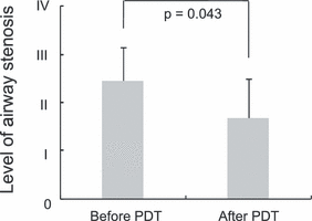
At 5 days after tracheal mucosa scraping, designated as Day 0, all of the animals showed tracheal stenosis, which was confirmed by bronchoscope (Table 1). No significant difference was found between the weights of rabbits in the treatment group and nontreatment group (P = 0.08) on Day −1 (4 days after scraping). A significant difference was also not found between the two groups in respiratory stridor (P = 0.77) and degree of stenosis (P = 0.18) immediately before PDT on Day 0. On Day −1, animals in the treatment group were administered NPe6. On Day 0, tracheal lesions of animals in the treatment group were irradiated by keeping a cylindrical diffuser tip placed at the center of the stenotic lesion followed by bronchoscopic inspection of the tracheal lumen. For the treatment group, unusual events were not seen immediately after PDT. Six (75%) of the eight rabbits that had respiratory stridor before PDT showed improvement in the symptom after PDT (Table 1). In addition, five (62.5%) of the eight rabbits with stenosis degree of II or more showed improvement in airway stenosis (P = 0.043) (Table 1, Fig. 1). Figure 2A shows a typical time course of tracheal stenosis after PDT. The granulation tissue-predominant stenotic lesion was ameliorated on Day 5 compared with the lesion before PDT, although white plaque was seen in a part of the treated lesion. On Day 14, the stenotic lesion had almost disappeared in this case. On the other hand, in the nontreatment group, tracheal stenosis gradually worsened over time (Fig. 2B), and respiratory stridor tended to be evident. Aside from the stenotic lesion, no evidence of redness/edema after PDT was seen on the nonscraped part of the tracheal mucosa located on the trachea caudal to the trachea incision line (data not shown).
| Stridor | Degree of stenosis† | |||
|---|---|---|---|---|
| 5 days after scraping (Day 0)* | After PDT*‡ | Immediately before PDT | After PDT‡ | |
| Treatment group (PDT) | − | − | II | I |
| ± | − | III | I | |
| ++ | ± | III | I | |
| + | ± | II | II | |
| ± | − | I | I | |
| ++ | − | III | II | |
| ++ | ++ | III | III | |
| + | + | III | III | |
| + | − | II | I | |
| Nontreatment group (control) | + | III | ||
| ++ | III | |||
| ± | III | |||
| ± | III | |||
| + | III | |||
| ++ | II | |||
| ± | II | |||
| ++ | III | |||
| + | III | |||
| + | III | |||
| + | IV | |||
| ± | III | |||
| ± | IV | |||
| − | II | |||
| ++ | II | |||
| ++ | III | |||
| − | II | |||
- PDT, photodynamic therapy. *−, no stridor; ±, mild in an excited state; +, mild; ++,marked. †Degree of tracheal stenosis by bronchoscope: I, <25%; II, 25–50%; III, 50–75%; IV, ≥75%. ‡At the best condition after PDT.

Improvement in airway stenosis by PDT. Improvement in the degree of airway stenosis was seen in PDT-treated rabbits.

Bronchoscopic views of intratracheal icavities of a PDT-treated rabbit (A) and an untreated rabbit (B).
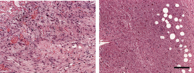
Pathohistologically, fibroblasts and collagen fibers had formed thick granulation tissue on the scraped lesion of untreated animals (Fig. 3, left), but in the treatment group granulation tissue formation was reduced in the scraped lesion, which showed few fibroblasts and collagen fibers, and the remaining granulation tissue was patchy in distribution over the lesion. In addition, the PDT-treated animals showed coagulation necrosis and vacuole formations had developed in some areas (Fig. 3, right).

Cross-sectioned views of the trachea in an untreated rabbit that died on Day 7 (left) and in a rabbit in the PDT group that died on Day 9 (right). Scale bar = 0.2 mm.
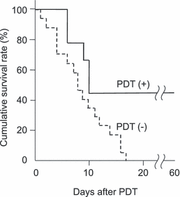
Survival times in the treatment group and nontreatment group are shown in Fig. 4. Even after Day 60, four rabbits (44%) were alive in the treatment group. The average survival time was 31.2 ± 8.6 days (mean ± SD) for the treatment group and 8.7 ± 1.2 days for the nontreatment group, and the median survival time point in the treatment and control groups were Day 10 and Day 8, respectively, indicating a significant improvement in life expectancy by PDT (P = 0.025).

Overall survival curves. There was a significant difference between PDT-treated rabbits and untreated rabbits (P = 0.025).
Discussion
Although PDT has already been regarded as one of the established therapies for solid tumors, it has been drawing attention as effectiveness even in proliferative diseases such as age-related macular degeneration (23) or atherosclerosis (24) has been reported. We also showed the effectiveness of PDT in one of the proliferative diseases, airway stenosis: PDT using Photofrin improved airway stenosis in an animal model (15). In this study, PDT using NPe6, which is water-soluble and has less incidence of photosensitivity, showed reduction in granulation stenosis and respiratory stridor in airway stenosis models, resulting in an increase in survival time of the PDT-treated animals.
This study showed results equivalent to those previously reported for Photofrin-PDT (15). Prolongation of survival time was seen in the animals treated with NPe6-PDT: four out of nine treated rabbits survived >60 days after PDT, while only two out of twelve rabbits treated with Photofrin-PDT in our previous study survived >60 days after PDT (15). However, the median survival time point was longer in the animals treated with Photofrin-PDT (Day 13) when compared with that in the animals treated with NPe6-PDT (Day 10). Damage to the normal tracheal mucosa was not remarkable in the rabbits that received NPe6-PDT. As the irradiation method used in this study was exactly the same as that used in our previous study for animals that received Photofrin-PDT (15), the nonscraped part of the tracheal mucosa located on the trachea caudal to the trachea incision line must have been inevitably exposed to light. However, temporal redness or edematous changes in the nonlesion light-exposed portion were not evident in rabbits that received NPe6-PDT, while those were seen in rabbits that received Photofrin-PDT (15). The cause of pathological differences might have been due to the difference in tissue distribution pattern of each photosensitizer, but a further study is required.
Although the standard treatment of airway stenosis is surgical resection, this methodology has serious invasiveness and high risks for cases such as low weight/immature babies and elderly diseased adults. It is therefore more desirable to treat such patients as conservatively as possible. Balloon dilatation, laser vaporization and stent implantation using a bronchoscope have been used as conservative therapeutic modalities with less invasiveness for the treatment of airway stenosis. However, there have been only a few successful cases without complications in which these therapeutic modalities were used (8,11,12). Considering the current conditions for treatment for airway stenosis and the results of this study, NPe6-PDT seems to be very promising as a new treatment for airway stenosis.
Conclusion
We examined the therapeutic effects of PDT with NPe6 on airway stenosis using rabbit models. NPe6-PDT improved airway stenosis and prolonged survival time of the treated animals in comparison with those of the untreated animals.
Acknowledgments
Acknowledgements— The authors thank Meiji Seika Ltd. for the gift of NPe6. This work was supported in part by Kawano Masanori Memorial Foundation for Promotion of Pediatrics and by The Japanese Foundation for Research and Promotion of Endoscopy.


