Immature stages of Glyphocassis spilota (Gorham) (Coleoptera: Chrysomelidae: Cassidinae) from Korea
Abstract
Detailed morphological descriptions and illustrations of the immature stages of Glyphocassis spilota (Gorham) are presented for the first time. Taxonomic remarks and notes on its biology are also provided.
Introduction
The genus Glyphocassis Spaeth contains three species, and is distributed in China, Japan, India and Vietnam, with only one species known in Korea (Gressitt & Kimoto 1963; Seeno & Wilcox 1982; Kimoto & Takizawa 1994). The cassidine larvae have characteristic features: they possess a supra-anal process to which is attached successive molted skins in the course of larval growth, and generally, accumulated feces. This structure, called the “parasol”, serves as a protective and concealing device against natural enemies (Gressitt 1952). Very little is known about the immature stages of this genus: only the larvae and pupae of Glyphocassis trilineata (Hope) have been briefly described and illustrated by Zaitsev (1988, 1992). The purpose of the present paper is to describe and illustrate the immature stages of Glyphocassis spilota (Gorham), with notes on their biology.
Materials and methods
Immature stages of Glyphocassis spilota (Gorham) were reared on the leaves of Calystegia hederacea Wall. in the laboratory. Materials used in the present study were preserved in 70% ethyl alcohol. Larvae and pupae were cleared in 10% KOH solution for 30 minutes, rinsed in water, and then dissected under a stereoscopic microscope (SZX12; Olympus, Japan). For morphological study of the microscopic structures, the relevant parts were mounted on slides and observed through a compound microscope (SZ4045; Olympus). Slide mounting procedures were carried out according to LeSage (1984), and the terminology follows Anderson (1947) and Takizawa (1980).
Taxonomic account
Subfamily Cassidinae Erichson, 1847.
Genus Glyphocassis Spaeth, 1914.
Glyphocassis spilota (Gorham, 1885)
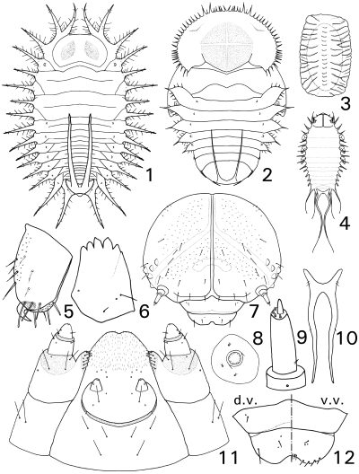
Egg (Fig. 3)

Immature stages of Glyphocassis spilota (Gorham). 1 Last instar larva (d.v.); 2 pupa (d.v.); 3 egg (d.v.); 4 first instar larva (d.v.); 5 leg (tibia, l.v.); 6 mandible (d.v.); 7 head (d.v.); 8 spiracle (d.v.); 9 antenna (d.v.); 10 supra-anal process (d.v.); 11 lower mouth parts (v.v.); 12 clypeus and labrum (d.v. and v.v.). d.v., dorsal view; l.v., lateral view; v.v., ventral view.
Length 1.3 mm, width 0.6 mm (n = 15). Dark yellow, flat and oval, covered with a semi-transparent egg-case.
Egg case. Length 1.7 mm, width 0.9 mm. Dark brown, flat and rectangular. Streaked, consists of two layers of membranes.
First instar larva (Fig. 4)
Similar to mature larva except for following characters: Body length 1.9 mm, width 0.8 mm, head width 0.4 mm (n = 5), pale yellow, flat and long obovate. Head well exposed. Relative length of lateral projections: fifteenth–sixteenth > first–tenth > eleventh–fourteenth; supra-anal processes twice as long as sixteenth lateral projections.
Last instar larva (Figs 1, 5–12)
Body length 4.8 mm, width 2.5 mm; head width 0.9 mm (n = 10). Body yellow, flat and obovate, with 16 pairs of lateral projections and a pair of supra-anal processes; lateral projections covered with many spinules. Club-shaped setae on dorsum. Spiracles elevated, pale brown.
Head (Fig. 7). Hypognathous, rounded, brown, well sclerotized, completely concealed by pronotum in dorsal view. Many spinules present centrally, subtriangular shape. Coronal suture and endocarina distinct; frontal suture indistinct. Frons depressed centrally, with eight pairs of setae; epistomal suture well developed. Five stemmata present on each side. Antennae (Fig. 9) two-segmented, segment 1 with one sensillum, segment 2 long, with one seta laterally, a conical sensory papilla and four setae at apex. Clypeus (Fig. 12) trapeziform, with two setae and two sensilla; labrum somewhat notched at anterior margin, with four setae and four sensilla; epipharynx with seven pairs of setae anteriorly, a pair of setae and two pairs of sensilla centrally. Mandible (Fig. 6) palmate, with six distal teeth, exterior face with two mandibular setae and two sensilla. Maxillary palp two-segmented; palpifer with three setae and two sensilla; stipes with two setae; galea with eight setae and one sensillum; cardo absent. Labial palp (Fig. 11) one-segmented, with one sensillum; ligula bifurcated, with numerous micro-setae; prementum with two pairs of setae and six pairs of sensilla; postmentum with three pairs of setae.
Thorax. Pronotum with four pairs of lateral projections, with a pair of dark patches centrally. Mesothoracic spiracles elevated, annuliform, situated on epipleural anterior parts (EPa). Legs rather short and stout; tibia (Fig. 5) with 14 setae (six pairs not pointed at apex); claw strongly curved, hook-like without seta; pulvillus absent.
Abdomen. Abdomen with a pair of lateral projections on each segment. Abdominal spiracles (Fig. 8) elevated, present on segments 1–8, similar to mesothoracic spiracle but smaller. Supra-anal process (Fig. 10) long, slender and setiferous.
Pupa (Fig. 2)
Body length 4.4 mm, width 3.0 mm (n = 10). Dark brown with dorsum largely light brown, glabrous, flat and oval.
Head. Longer than wide, directed backwards, completely concealed by pronotum in dorsal view.
Thorax. Pronotum with approximately 60 marginal spinules, of which two pairs on the anterior margin are distinctly longer and stouter than the others, cross shape present centrally.
Abdomen. Abdominal segments 1–5 each with a leaf-like lateral projection that ends apically in a long spinule; segments 6–8 each with a spinule-like process ventro-marginally. Apical processes of ninth abdominal segment long, slender, reaching fourth abdominal segment. Spiracles elevated, dark brown, first to third each as high as wide, fourth and fifth each higher than wide.
Material examined
Thirty exs., Andong, Gyeongbuk Province, 12.vii.2004, H.W. Cho; 5 exs., Andong, Gyeongbuk Province, 16.vi.2005, H.W. Cho.
Distribution
Korea, Japan, China.
Host plants
Calystegia hederacea Wall., Calystegia japonica (Thunb.) Chois.
Remarks
This species is easily distinguished from the other known cassidine larvae by the following characters: head with many spinules centrally, subtriangular shape; excretions on the supra-anal process are triangular in shape, without dense rod-like or filamentous projections.
Biology
Adults appear in mid-July and oviposit in late July. Eggs are laid singly on the undersides of leaves in an egg-case with feces. The mature larva bears a flat and large triangular mass of feces on the cast skins of the four preceding larval instars, and pupates with all the cast skins and feces. The total life cycle from egg to adult ranged from 30 to 35 days in the laboratory.
Acknowledgments
We thank Dr Lech Borowiec (Wroclaw University, Wroclaw, Poland) for providing valuable published material. This study was supported by a Korea Institute of Environmental Science and Technology Ministry and Environment Grant (KIEST 052-041-029, 2005).


