RADIOGRAPHIC AND SCINTIGRAPHIC EVALUATION OF THE THIRD CARPAL BONE OF CONTROL HORSES AND HORSES WITH CARPAL LAMENESS
Abstract
We compared the radiographic and scintigraphic findings in the third carpal bone of horses performing different work disciplines and investigated their relationship with lameness. Horses had undergone carpal radiography including acquisition of a dorsoproximal-dorsodistal oblique (DPr-DDiO) image of the distal row of carpal bones and/or scintigraphic examination of the carpi. Cause of lameness, breed, age, and work discipline were recorded. Increased opacity in the third carpal bone was graded, ratio of radiopharmaceutical uptake calculated objectively, and increased radiopharmaceutical uptake graded subjectively. Relationships between radiographic, scintigraphic, and clinical findings were assessed statistically. Increased opacity in the third carpal bone (P = 0.003) and ratio of radiopharmaceutical uptake (P = 0.015) were associated with the work discipline. Increased opacity in the third carpal bone was associated with both increased radiopharmaceutical uptake grade (P = 0.002; rs = 0.59) and ratio of radiopharmaceutical uptake (P = 0.013; rs = 0.46). Increased radiopharmaceutical uptake and increased opacity in the third carpal bone were not always observed concurrently. Lameness related to the middle carpal joint was associated with increased opacity (P < 0.001), ratio of radiopharmaceutical uptake (P = 0.037), and increased radiopharmaceutical uptake grade (P < 0.001). Radiographic and scintigraphic abnormalities were observed in horses performing all disciplines, indicating that high‑speed exercise may not be the only factor determining the development of osseous disease in the third carpal bone. Both increased opacity and increased radiopharmaceutical uptake were more likely to be seen in horses with lameness related to the middle carpal joint than in horses with other sources of pain.
Introduction
There is wide range of anatomic variation and pathologic conditions of the equine carpus.1, 2 The frequency of occurrence of carpal injuries is higher in racehorses compared with horses from other sports disciplines,2-4 although carpal injuries have also been documented in sports horses.5-8 Carpal lameness in racehorses is believed to be a consequence of carpal hyperextension occurring during high-speed exercise, which predisposes to injury of the dorsal aspect of the carpus, including the third carpal bone.3, 4, 9, 10 Race training results in an adaptive process in the third carpal bone, which is manifested radiographically by increased opacity in the radial facet of the third carpal bone 4, 10-12 and increased radiopharmaceutical uptake.11 Differentiation between this normal adaptive process and a pathologic condition causing lameness is not always clearly defined.4, 10-13 There are limited data on the association between radiographic and scintigraphic findings in the equine third carpal bone and their association with lameness in horses not performing high-speed exercise.5-8, 11
Our aims were (1) to document the radiographic and scintigraphic findings in the third carpal bone of horses of different breeds, work disciplines, and ages; (2) to compare radiographic and scintigraphic findings; and (3) to investigate their relationship with lameness. We hypothesized that radiographic and scintigraphic abnormalities would be found in horses of all work disciplines, but with higher frequency in racehorses; there would be a positive relationship between the severity of radiographic findings and the degree of increased radiopharmaceutical uptake; radiographic and scintigraphic grades of the third carpal bone would be highest in horses with middle carpal joint pain.
Material and Methods
Radiographic records from the Animal Health Trust (AHT) between January 1997 and November 2010 and from Rossdales Equine Hospital in 2009 were searched for horses with lameness or poor performance that had undergone unilateral or bilateral carpal radiography. The imaging examination had to have included a flexed dorsoproximal-dorsodistal oblique (DPr-DDiO) projection of the distal row of carpal bones and/or a scintigraphic examination of the carpi. One hundred and fifty-three lame or poorly performing horses were included in the study; 83 had a DPr-DDiO image of the distal row of carpal bones, 100 had scintigraphic examination of the carpi. Thirty horses (15 in group MC; 15 in group L) had both a DPr-DDiO image of the distal row of carpal bones and a scintigraphic examination of the carpi.
Age, gender, breed (Thoroughbred, Thoroughbred-cross [including Thoroughbred cross Warmblood], Warmblood, and other breeds) and work discipline (racehorses [high-speeed exercise], horses performing moderate-speed exercise [eventing, endurance, western performance] and low-speed exercise [show jumping, dressage, general purpose]) were recorded. Horses were divided into four age groups: <6 years; 6–8 years; 9–11 years; > 11 years.
Horses were grouped based upon the source of pain causing lameness, usually confirmed using local analgesia: Group MC = horses with lameness related to the middle carpal joint, determined by the presence of effusion or substantial improvement in lameness after intraarticular analgesia of the middle carpal joint; group L = horses with lameness unrelated to the middle carpal joint. Scintigraphic data of horses in group L were used as controls and compared with those of horses in group MC for the investigation of the clinical significance of the scintigraphic features. To validate the use of scintigraphic data of the horses in group L as controls, scintigraphic data of these horses were compared with those of seven clinically sound horses in full work (group S). There were 100 horses in group L and 53 in group MC. Breeds, work disciplines, and age groups of horses in groups MC and L are summarized in Table 1.
| Group MC | Group L | |
|---|---|---|
| Age (years) | ||
| 6 | 28 | 28 |
| 6–8 | 9 | 30 |
| 9–11 | 9 | 22 |
| >11 | 6 | 20 |
| Discipline | ||
| Racing | 31 | 12 |
| Moderate speed | 8 | 27 |
| Low speed | 10 | 58 |
| Breed | ||
| TB | 37 | 19 |
| TB cross | 5 | 12 |
| Warmblood | 6 | 40 |
| Other breeds | 5 | 29 |
- TB, Thoroughbred.
- Moderate speed = eventing, endurance, western performance.
- Low speed = show jumping, dressage, general purpose.
The lamer limb in horses with bilateral carpal radiographs was used for the group classification and for the investigation of associations between work discipline, breed, age, radiographic, and scintigraphic findings. For comparison of imaging features between the carpi of individual horses with lameness related to the middle carpal joint, the lame limb of unilaterally lame horses and the lamer limb of bilaterally lame horses were referred to as the lamest limb and the other limb as the contralateral limb.
Dorsopalmar (DPa), flexed and/or weightbearing lateromedial (LM), dorsolateral-palmaromedial oblique (DL-PaMO), and dorsomedial-palmarolateral oblique (DM-PaLO) images were obtained of all carpi.14 Flexed DPr-DDiO images of the distal row of carpal bones were obtained when required by the investigating clinician. At the AHT, before January 2005, images were acquired using a conventional radiography system. From January 2005, a computed radiography system (Kodak Direct View CR or, from 2008, Carestream CR Classic1) was used. At Rossdales Equine Hospital, all images were acquired with a computed radiography system2.
Dorsal and lateral scintigraphic images of the carpal region were acquired dynamically (35 2-s frames, with a 256 × 256 matrix), using a 500-mm circular or rectangular field of view gamma camera and general-purpose collimator, 2.5 h after injection of 99m−technetium methylene diphosphonate (10MBq/kg IV).15 Motion correction software3 was used to create the final static images.
All radiographs were evaluated subjectively by one trained analyst (V.S.). Prior to image analysis, 10 sets of digital and 10 sets of conventional radiographs were selected randomly and evaluated by an experienced analyst (S.J.D.) and V.S. to determine acceptable agreement in radiographic interpretation. For interpretation, all images were orientated with dorsal to the left and lateral to the right. Radiographic abnormalities in the carpus, including signs of osteoarthritis, were recorded.8 The degree of increased opacity in the radial facet of the third carpal bone in the DPr-DDiO projection was graded as previously described.12 Three parameters were used to assess the severity of increased opacity in the third carpal bone: trabecular score (TS), total affected area (TAA), and final score (Table 2; Fig. 1). To ensure repeatability of image interpretation, the images of 10 carpi were graded 10 times by V.S. and the coefficient of variance was < 2%. Final image analysis was performed after the repeatability study. The presence, location, and number of fractures and radiolucencies of the radial facet of the third carpal bone on the DPr-DDiO projection were recorded and classified (Table 3; Fig. 1).
| Trabecular score (TS): |
|---|
| 0 = Trabeculae clearly evident with normal structure. |
| 1 = Mildly hazy trabecular margins. |
| 2 = Moderate trabecular thickening but structure still visible. |
| 3 = Marked trabecular thickening and structure nearly lost. |
| 4 = Complete loss of intertrabecular spaces and trabecular structure. |
| Total affected area (TAA): |
| Subjective evaluation of the percentage of the radial facet affected by a TS > 0. The radial facet was divided into six zones. The percentage of the radial facet affected by a TS > 0 was estimated subjectively within each zone and expressed as a percentage of the six radial facet zones. |
| Final score (FS): |
| Very mild: TS1 (TAA < 50%) |
| Mild: TS1 (TAA ≥ 50%) to TS2 (TAA < 75%) |
| Moderate: TS2 (TAA ≥75 %) to TS3 (TAA < 75%) |
| Severe: TS3 (TAA ≥75 %) to TS4 (TAA > 0%) |
| Medullary vascular channel: |
|---|
| Radiolucency with diameter of < 2 mm and regular margins. |
| Large medullary lucency: |
| Radiolucency wider than 2 mm and/or radiolucency with irregular margins and/or fracture. |
| Marginal lucency: |
| Radiolucency localized in the dorsal cortex of the third carpal bone. |

All scintigraphic images were evaluated subjectively and objectively for the presence of increased radiopharmaceutical uptake. Lateral and dorsal images were assessed subjectively by an experienced analyst (S.J.D.). To ensure repeatability of image interpretation, prior to final image analysis, images of 10 carpi were graded 10 times; the coefficient of variance was < 2%. The grade (mild, moderate, or intense) and location of increased radiopharmaceutical uptake were recorded. Increased radiopharmaceutical uptake in the distal row of carpal bones (DRIRU) was defined as diffuse involving the majority of the distal row of carpal bones, increased radiopharmaceutical uptake in the subchondral bone of the carpometacarpal joint (distal aspect of the distal row of carpal bones and proximal aspect of the metacarpus), or increased radiopharmaceutical uptake restricted to the third carpal bone (C3IRU). Data of horses with C3IRU were also analyzed independently.
Lateral images were used for objective analysis. Corresponding lateral scintigraphic and LM radiographic images were coregistered and superimposed to determine accurate positions for the regions of interest (RsOI) using a multimodality planar program (Hermes). Two rectangular or square RsOI were drawn (Fig. 2A): DRROI – over the distal row of carpal bones; RROI – reference ROI with a height equal to that of the accessory carpal bone, positioned on the radius (reference bone) so that its distal limit was at two times the height of the accessory carpal bone, proximal to the proximal border of the accessory carpal bone. The mean number of counts per pixel in all RsOI was recorded and the ratio between DRROI and RROI was calculated and referred to as the ratio of radiopharmaceutical uptake (Fig. 2). A repeatability study was performed by analyzing 10 carpi five times. Final measurements were not made until a coefficient of variance < 5% was obtained for the mean counts per pixel in each ROI.

Statistical analyses were performed using Analyse-it4. Normality of data distribution was assessed using a Shapiro–Wilks test. The percentages of agreement between increased radiopharmaceutical uptake grades in dorsal and lateral images were calculated. Chi-square and Mann–Whitney tests were used to compare the increased radiopharmaceutical uptake grade and ratio of radiopharmaceutical uptake between the horses in groups L and S. A Chi-square test was used to determine associations between the radiographic findings and increased radiopharmaceutical uptake grades and the age, breed, discipline, and lameness groups. Kruskal–Wallis and Mann–Whitney tests were used to test for differences in the ratio of radiopharmaceutical uptake in horses from different age, discipline, breed, and lameness groups and for differences in radiographic findings. A Spearman correlation was used to test for associations between radiographic and scintigraphic features. A Chi-square test was used to compare the severity of the radiographic findings and increased radiopharmaceutical uptake grades between the left and right limbs of horses with middle carpal joint lameness. A Mann–Whitney test was used to compare the ratio of radiopharmaceutical uptake between the left and right limbs of horses with middle carpal joint lameness.
Results
Moderate or severe final score of increased opacity in the third carpal bone was observed in 31/83 (37.3%) horses (Table 4). Medullary vascular channels were the most common radiographic finding in the third carpal bone; large medullary lucencies and marginal lucencies were observed in 16/83 (19.3%) and 14/83 (16.9%) horses, respectively (Table 4). Increased opacity of the intermediate facet of the third carpal bone was seen in 4/83 (4.8%) horses. Eight fractures of the third carpal bone were identified (slab [n = 4], parasagittal [n = 3], slab and parasagittal [n = 1]).
| A) Radiological findings | B) Scintigraphic findings | |||||
|---|---|---|---|---|---|---|
| Group MC (n = 47) | Group L (n = 36) | Group MC (n = 21*) | Group L (n = 79*) | |||
| TS | Ratio of RU (median;IQR) | 2.03 | 1.72 | |||
| 0 | 10.6 | 22.2 | 1.5–2.4 | 1.4–2.0 | ||
| 1 | 14.9 | 36.1 | DRIRU | |||
| 2 | 21.3 | 33.3 | Dorsal images | |||
| 3 | 17.0 | 5.6 | Normal | 45.0 | 80.8 | |
| 4 | 36.2 | 2.8 | Mild | 15.0 | 12.8 | |
| TAA | Moderate | 30.0 | 6.4 | |||
| < 30% | 14.9 | 33.3 | Intense | 10.0 | 0.0 | |
| 30–70% | 31.9 | 55.6 | Lateral images | |||
| >70% | 53.2 | 11.1 | Normal | 61.9 | 83.5 | |
| FS | Mild | 9.5 | 10.1 | |||
| 0 | 10.6 | 22.2 | Moderate | 14.3 | 6.3 | |
| 1 | 10.6 | 25.0 | Intense | 14.3 | 0.0 | |
| 2 | 19.1 | 44.4 | C3IRU | |||
| 3 | 19.1 | 2.8 | Dorsal images | |||
| 4 | 40.4 | 5.6 | Normal | 65.0 | 91.0 | |
| Marginal lucencies | Mild | 10.0 | 5.1 | |||
| Absent | 72.3 | 97.2 | Moderate | 15.0 | 3.8 | |
| Present | 27.7 | 2.8 | Intense | 10.0 | 0.0 | |
| Large medullary lucencies | Lateral images | |||||
| Absent | 66.0 | 100.0 | Normal | 80.9 | 91.1 | |
| Present | 34.0 | 0.0 | Mild | 0.0 | 5.1 | |
| Medullary vascular channels | Moderate | 4.8 | 3.8 | |||
| Absent | 53.2 | 36.1 | Intense | 14.3 | 0.0 | |
| Present | 46.8 | 63.9 | ||||
- TS, trabecular score of increased opacity in the third carpal bone (C3); TAA, total area of the C3 affected with increased opacity; FS, final score of increased opacity in the C3; ratio of RU, ratio of radiopharmaceutical uptake (RU) between the distal row of carpal bones and the reference region; DRIRU, increased radiopharmaceutical uptake (IRU) in the distal row of carpal bones; C3IRU, IRU in the C3; IQR, interquartile range. Bold characters: group with the highest frequency for each category of radiological or scintigraphic findings.
- The scintigraphic data in dorsal images of one horse in group MC and one horse in group L are missing.
The ratio of radiopharmaceutical uptake in group S ranged from 1.19 to 2.24 (mean: 1.73; median: 1.64); one horse had mild diffuse increased radiopharmaceutical uptake in the distal row of carpal bones in a lateral image; the dorsal image was missing. The ratio of radiopharmaceutical uptake in groups L and MC ranged from 0.80 to 3.12 (mean: 1.77; median: 1.77) (Table 4); C3IRU was observed in 14/98 (14.3%) horses in dorsal images and 11/100 (11.0%) horses in lateral images; DRIRU was observed in 26/98 (26.5%) horses in dorsal images and in 21/100 (21.0%) horses in lateral images. The ratio of radiopharmaceutical uptake and the DRIRU grade in lateral images of group S were not significantly different from those in group L (Pratio of radiopharmaceutical uptake = 0.848; PDRIRU = 0.758). The agreement between C3IRU in dorsal and lateral images was 86.7%. The agreement between DRIRU in dorsal and lateral images was 84.7%. There was an association between the ratio of radiopharmaceutical uptake and the C3IRU in dorsal images (P = 0.007), but not in lateral images (P = 0.102). There was an association between the ratio of radiopharmaceutical uptake and the DRIRU in dorsal images (P < 0.001) and in lateral images (P = 0.001).
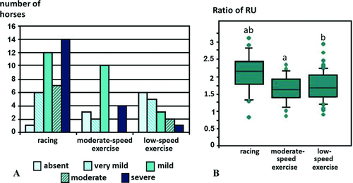
The final score for increased opacity in the third carpal bone was associated with breed (P = 0.046), work discipline (P = 0.003), and age (P = 0.031) (Fig. 3). Thoroughbreds (15/49; 30.6%) and horses < 6 years old (14/45; 31.1%) had the highest frequency of occurrence of severe increased opacity. Discipline was not associated with either medullary vascular channels (P = 0.088) or marginal lucencies (P = 0.308). Large medullary lucencies were associated with discipline (P < 0.001); they were only seen in racehorses. The ratio of radiopharmaceutical uptake was not associated with breed (P = 0.231) or age (P = 0.248), but was associated with discipline (P = 0.015) (Fig. 3). There was no association between increased radiopharmaceutical uptake grades and discipline, breed, or age.

Radiographic evidence of osteoarthritis of the middle carpal joint was associated with DRIRU (P = 0.006) and C3IRU (P = 0.026) grades in dorsal images and the final score of increased opacity in the third carpal bone (P = 0.003), but not with the ratio of radiopharmaceutical uptake (P = 0.164). There was an association between the ratio of radiopharmaceutical uptake and the final score of increased opacity in the third carpal bone (P = 0.013; rs[Spearman's rank correlation coefficient] = 0.46). There was an association between the final score of increased opacity in the third carpal bone and DRIRU grade in dorsal images (P = 0.003; rs = 0.56) and C3IRU grade in dorsal images (P = 0.002; rs = 0.59). C3IRU grade in dorsal images was associated with the TS (P = 0.002; rs = 0.58) and the TAA (P = 0.001; rs = 0.60). Six of nine (66.7%) horses with moderate or intense DRIRU had moderate or severe increased opacity in the third carpal bone; 6/9 (66.7%) horses with moderate or severe increased opacity in the third carpal bone had moderate or intense DRIRU.
Horses with medullary vascular channels in the third carpal bone had a lower ratio of radiopharmaceutical uptake (P = 0.026), lower DRIRU (P = 0.012), and C3IRU (P = 0.007) grades in dorsal images and lower TAA (P = 0.020) than those without medullary vascular channels. The horses with large medullary lucencies had a higher final score of increased opacity in the third carpal bone (P < 0.001) than those without large medullary lucencies. Three horses with large medullary lucencies had scintigraphic examination; all had a ratio of radiopharmaceutical uptake > 2, two had moderate C3IRU in dorsal images and one did not have subjective increased radiopharmaceutical uptake. Horses with marginal lucencies had higher C3IRU (P = 0.021) and DRIRU (P = 0.008) grades in dorsal images and a higher final score of increased opacity in the third carpal bone (P < 0.001) than horses without marginal lucencies.
The lameness group was associated with the TS (P < 0.001), the TAA (P < 0.001), and the final score (P < 0.001) of increased opacity in the third carpal bone (Table 4). The lameness group was associated with marginal lucencies (P = 0.003) and large medullary lucencies (P < 0.001), but not with medullary vascular channels (P = 0.184) (Table 4). The lameness group was associated with the ratio of radiopharmaceutical uptake (P = 0.037), DRIRU (P = 0.015), and C3IRU (P = 0.034) grades in lateral images and DRIRU (P < 0.001) and C3IRU (P = 0.005) grades in dorsal images (Table 4). Eight of 13 (61.5%) horses with moderate or intense DRIRU and 28/31 (90.3%) horses with moderate or severe increased opacity in the third carpal bone had lameness related to the middle carpal joint. Eight of 20 (40.0%) and 28/47 (59.6%) horses with lameness related to the middle carpal joint had moderate or intense DRIRU or moderate or severe increased opacity in the third carpal bone, respectively. Seventeen of 47 (36.2%) horses with lameness related to the middle carpal joint had no significant detectable radiographic abnormalities.
Fifteen horses in group MC had a DPr-DDiO image of the distal row of carpal bones acquired bilaterally. The final score for increased opacity in the third carpal bone was higher in the lamest limb than in the contralateral limb of seven horses, lower in the lamest limb than in the contralateral limb of one horse, and the same in both limbs of seven horses; six of which were bilaterally lame. There was no difference in final score between the lamest limb and contralateral limb (P = 0.331).
Twenty horses in group MC had scintigraphic examination. The DRIRU grade in dorsal images was the same in both limbs of 10 horses, higher in the lamest limb than in the contralateral limb of seven horses, and lower in the lamest limb than in the contralateral limb of three horses. Thirteen of 20 horses had a higher ratio of radiopharmaceutical uptake in the lamest limb than in the contralateral limb. There was no difference between the lamest limb and contralateral limb for either DRIRU grade in dorsal images (P = 0.208) or the ratio of radiopharmaceutical uptake (P = 0.279).
Discussion
In accordance with our hypothesis, increased opacity in the third carpal bone was found in horses of all disciplines, with racehorses having the highest frequency. The ratio of radiopharmaceutical uptake was associated with the work discipline. There was an association between the grade of increased opacity and ratio of radiopharmaceutical uptake and increased radiopharmaceutical uptake grades in the third carpal bone. Increased opacity in the third carpal bone, increased radiopharmaceutical uptake grades, and ratio of radiopharmaceutical uptake were associated with lameness related to the middle carpal joint.
The finding of a significant association between lameness related to the middle carpal joint and ratio of radiopharmaceutical uptake and increased radiopharmaceutical uptake grades highlights the clinical significance of focal increased radiopharmaceutical uptake detected subjectively. Dorsal images were more sensitive for detection of C3IRU and DRIRU than lateral images. In lateral images, interpretation is confounded by the superimposition of the fourth carpal bone over the third carpal bone, which may mask subtle increased radiopharmaceutical uptake in the third carpal bone. The objective evaluation was performed in lateral images because the distance between the camera and the carpi is smaller in lateral images than in dorsal images, resulting in superior image resolution and more accurate positioning of the RsOI. The DRROI encompassed the entire distal row of carpal bones that may have caused a dilution effect of focal increased radiopharmaceutical uptake in the third carpal bone. Thus, subjective evaluation may be more sensitive for focal increased radiopharmaceutical uptake,11 which may explain the lack of association between the C3IRU in lateral images and the ratio of radiopharmaceutical uptake.
An association between the presence of increased radiopharmaceutical uptake and increased opacity in the third carpal bone has been reported.11 In the present study, there was a correlation between the grade of increased opacity in the third carpal bone and increased radiopharmaceutical uptake grades and ratio of radiopharmaceutical uptake. Despite the association between increased opacity and increased radiopharmaceutical uptake, increased radiopharmaceutical uptake could be seen without detectable radiographic abnormalities and increased opacity was not always associated with increased radiopharmaceutical uptake. Increased radiopharmaceutical uptake primarily reflects increased osteoblastic activity;16 therefore, the presence of increased radiopharmaceutical uptake depends upon the stage of the disease. It is possible that the presence of increased radiopharmaceutical uptake in the absence of increased opacity in the third carpal bone reflects early disease prior to lesions becoming evident radiographically. Extensive mineralization or primary bone necrosis can be present in the distal phalanx without increased radiopharmaceutical uptake.17 Therefore, the presence of moderate or severe increased opacity in the third carpal bone without increased radiopharmaceutical uptake may reflect old, advanced osseous disease of the third carpal bone. In the present study, two horses had moderate or severe increased opacity in the third carpal bone without increased radiopharmaceutical uptake, both of which had lameness related to the middle carpal joint. Intense increased radiopharmaceutical uptake was observed in three horses; all had lameness related to the middle carpal joint and increased opacity in the third carpal bone.
Radiographic signs of osteoarthritis of the middle carpal joint were associated with the grade of increased opacity in the third carpal bone and the increased radiopharmaceutical uptake grades. Cartilage damage has been found overlying sclerotic bone in the third carpal bone.18 Increased bone mineral density and increased thickness of the calcified cartilage of the third carpal bone increases stiffness, resulting in reduction in the shock-absorbing capacity, thereby increasing the risk of shear-induced damage to the overlying articular cartilage.18-20
Subchondral lucencies in the third carpal bone have been described as the result of osteochondral collapse or acute fracture of the proximal surface of the third carpal bone and may be a prelude to slab fractures.21, 22 Therefore, in the present study, fractures and subchondral lucencies were grouped and referred to as large medullary lucencies. All horses with large medullary lucencies had increased opacity in the third carpal bone and had lameness related to the middle carpal joint. Increased density and stiffness of the subchondral bone of the third carpal bone could predispose to failure of the subchondral bone and result in fractures and subchondral lucencies.4, 23 Marginal lucencies have been described as vascular channels 24, 25 and as irregular lucent areas not associated with damage of the dorsal rim of the third carpal bone.21 However, in the current study their margins were generally more irregular than those of medullary vascular channels and they sometimes seemed to merge and form larger lucencies. Moreover, marginal lucencies were overrepresented in horses with increased radiopharmaceutical uptake, increased opacity in the third carpal bone, and lameness related to the middle carpal joint. Increased radiopharmaceutical uptake and TAA of increased opacity in the third carpal bone were associated with a reduced frequency of medullary vascular channels. Appositional new bone formation in the trabecular bone of the third carpal bone may cut off vascular channels and lead to ischemic necrosis in a sclerotic third carpal bone,25, 26 possibly explaining the negative association between the presence of medullary vascular channels and the ratio of radiopharmaceutical uptake, increased radiopharmaceutical uptake grades, and TAA of increased opacity in the third carpal bone.
Increased opacity in the third carpal bone was most common in racehorses, as previously documented,10, 12, 19 and may be a consequence of the high loads sustained particularly by the radial facet of the third carpal bone during high-speed exercise.27 There is little data on disorders of the third carpal bone in sports horses.6, 7 This may be due either to a low frequency of occurrence of this injury in sports horses or a failure to identify the condition. Herein, we identified increased opacity and increased radiopharmaceutical uptake in the third carpal bone in horses performing moderate- or low-speed exercise. Increased opacity in the third carpal bone has been described as a spectrum between normal adaptation to training and a pathologic condition.4, 10-13 Increased opacity in the third carpal bone has been reported in racehorses before they enter high-speed exercise training 11, 12 indicating that speed was not the only factor determining the development of increased opacity in the third carpal bone. Other biomechanical factors may also contribute. The effect of high-speed exercise on increased radiopharmaceutical uptake in the third carpal bone remains controversial. Exercise on a high-speed treadmill did not result in an increase in increased radiopharmaceutical uptake in the third carpal bone.28, 29 However an increase in increased radiopharmaceutical uptake in the third carpal bone was observed in a group of Standardbred racehorses submitted to race training.11 In the present study, racehorses had a higher ratio of radiopharmaceutical uptake than horses performing moderate- or low-speed exercise, but no difference was observed in increased radiopharmaceutical uptake grades.
No significant differences in grade of increased opacity, increased radiopharmaceutical uptake grade, and ratio of radiopharmaceutical uptake were observed between the lamest limb and contralateral limb. Although lameness may be more severe in one limb or manifest unilaterally, in some horses abnormal bone modeling may occur bilaterally, supporting other studies that reported increased opacity in the third carpal bone in the nonlame limb of unilaterally lame horses.11 In the present study, there was an association between lameness related to the middle carpal joint and increased opacity in the third carpal bone; 90.3% of horses with moderate or severe increased opacity in the third carpal bone had lameness related to the middle carpal joint. Lameness related to the middle carpal joint occurred in 30/76 (39.5%) and in 6/7 (85.7%) of racehorses in training with moderate or severe increased opacity in the third carpal bone.11, 12 Moderate or severe increased opacity is not always associated with lameness and does not always prevent successful training. However when associated with lameness or poor performance, moderate or severe increased opacity in the third carpal bone is of likely clinical significance. Moderate or severe increased opacity in the third carpal bone was observed in 59.6% horses with lameness related to the middle carpal joint and 36.2% horses with lameness related to the middle carpal joint had no significant radiographic abnormalities. Osseous pathology of the carpus, including abnormal mineralization of the third carpal bone, has been documented in lame horses using magnetic resonance imaging in the absence of detectable radiographic abnormalities.30 It is also possible that other causes of lameness not detected radiologically such as tearing of the intercarpal ligaments, synovitis, a cartilage defect, or other osseous pathology occurred in some horses.10, 30, 31
In summary, our findings indicate that high-speed exercise may not be the only determining factor in the development of osseous disease of the third carpal bone. Although increased radiopharmaceutical uptake and increased opacity in the third carpal bone were associated, they were not always observed concurrently. Both increased opacity and increased radiopharmaceutical uptake were overrepresented in horses with lameness related to the middle carpal joint, especially if moderate or severe. Large medullary lucencies and marginal lucencies in the third carpal bone were associated with lameness related to the middle carpal joint.
ACKNOWLEDGMENTS
We thank the referring veterinary surgeons, without whom this study would not have been possible, Marcus Head from Rossdales Equine Hospital for providing access to radiographs and case records, and Tim Watson for statistical advice.


