FRACTURES OF THE DISTAL PHALANX AND ASSOCIATED SOFT TISSUE AND OSSEOUS ABNORMALITIES IN 22 HORSES WITH OSSIFIED SCLEROTIC UNGUAL CARTILAGES DIAGNOSED WITH MAGNETIC RESONANCE IMAGING
Presented at the American Association of Equine Practitioners, 56th Annual Convention, 2010, Baltimore, Maryland.
Abstract
Ungual cartilage ossification in the forelimb is a common finding in horses. Subtle abnormalities associated with the ungual cartilages can be difficult to identify on radiographs. Magnetic resonance (MR) imaging findings of 22 horses (23 forelimbs) with a fracture of the distal phalanx and ossified ungual cartilage were characterized and graded. All horses had a forelimb fracture. Eleven involved a left forelimb (seven medial; four lateral), and 12 involved a right forelimb (five medial; seven lateral). All fractures were nonarticular, simple in configuration, and nondisplaced. The fractures were oriented in an axial proximal to abaxial distal and palmar to dorsal direction, and extended from the base of the ossified ungual cartilage into the distal phalanx. The fracture involved the fossa of the collateral ligament on the distal phalanx in 17 of 23 limbs. The palmar process and ossified ungual cartilage was abnormally mineralized in all horses. Ligaments and soft tissues adjacent to the ossified ungual cartilages were affected in all horses. The routine site of fracture in this study at the base of the ossified ungual cartilage extending into the distal phalanx suggests a biomechanical cause or focal stress point from cycling. The ligamentous structures associated with the ungual cartilages were often affected, showed altered signal intensity as well as enlargement and were thought to be contributing to the lameness. In conclusion, ossified ungual cartilages may lead to fracture of the palmar process of the distal phalanx and injury of the ungual cartilage ligaments.
Introduction
Ossification of the ungual cartilages in the forelimb is a common finding in Finnhorses, draft breeds, and Brazilian Jumper horses.1–3 The cartilages start as hyaline and progress with age to fibrocartilage and/or ossify.4 Ossification of the cartilages follows distinct patterns,5 with the most common being ossification extending proximally from the palmar process followed by ossification via a separate center of ossification.3 The cause of the ossification is unknown but may be associated with concussion, poor conformation, or poor farriery.6 Evidence in Finnhorses points to a hereditary link for ossification of the ungual cartilages.7
The clinical significance of ungual cartilage ossification is often unknown,8 and may be considered an incidental finding. However, a fracture of an ossified ungual cartilage can be a significant source of lameness.9 Furthermore, signs associated with ossification include shortening of the stride or slight lameness.5,10 This is often exacerbated when the horse is trotted in the direction of the affected foot.10 Ossified ungual cartilages cannot function in the expanding and contracting normal physiologic function.11 This may interfere with several ligaments that attach to the ungual cartilages, causing adaptive changes,12 and may also increase the strain on the ossified cartilages.
At least five ligaments are intimately associated with the ungual cartilages.12,13 These include the: (1) chondrocoronal ligament that connects the dorsal proximal aspect of the ungual cartilages to the dorsomedial/lateral margin of the middle phalanx; (2) chondrotendonous ligament that connects the dorsal aspect of the cartilages to the extensor process of the distal phalanx; (3) chondrocompedal ligament that attaches the palmar proximal aspect of the cartilages to the distal aspect of the proximal phalanx; (4) chondrosesamoidean ligament attaches the axial aspect of the ungual cartilages to the abaxial margins of the distal sesamoid bone; and (5) chondroungular ligament that connects the abaxial distal margin of the ungual cartilages to the palmar process of the distal phalanx. Additionally, there are paratendonous fibers of the deep digital flexor tendon that attach to the axial surface of the cartilages and fibers associated with the collateral ligaments of the distal interphalangeal joint that connect to the dorsal margin of the ungual cartilage.12,13
Abnormalities or fracture associated with the ungual cartilages can be difficult to identify on radiographs. While radiography is the traditional imaging technique to evaluate the foot, is not sensitive to soft tissue abnormalities. Marked ossification of the ungual cartilages has the potential to be clinically significant.2 Nuclear scinitigraphy used in combination with radiographic evaluation may provide clinically relevant information.8,14 Increased radiopharmaceutical uptake in the palmar process of the distal phalanx is also associated with magnetic resonance (MR) imaging abnormalities.15
Our aim was to describe MR imaging findings of soft tissue structures associated with ossified ungual cartilages and associated fracture of the distal phalanx as well as osseous changes associated with the fracture.
Materials and Methods
Clinical and diagnostic imaging records from nine veterinary hospitals between January 2004 and January 2009 were reviewed. Horses in which ossification of the ungual cartilage was 25% or greater with sclerosis (low signal intensity on T2, PD, and T1-weighted sequences) and fracture of the distal phalanx ipsilateral to the ossified ungual cartilage in the lame forelimb were selected and included in the study. Patient selection included fractures that extended from the axial aspect of an ossified ungual cartilage dorsally and abaxially to the palmar aspect of the solar margin of the distal phalanx were included. Sclerosis was characterized by low signal intensity on T1-weighted and proton density (PD) images.16 Horses that had a transversely oriented linear hyperintensity through an ossified ungual cartilage between separate centers of ossification or fracture were excluded. Twenty-two horses were identified. Breed distribution included 10 Warmbloods, five Quarter Horses, two Thoroughbred/Thoroughbred crosses, four Fresian/Fresian crosses, and one Standardbred. One horse had bilateral fractures of the palmar aspect of the distal phalanx. The lameness grade in all horses ranged between 2 and 3 on a scale of 0–5 as outlined by the American Association of Equine Practitioners and the lameness significantly improved (60% or greater) with bilateral perineural anesthesia of the palmar digital nerve.
One patient was imaged using a 1.5 T magnet,* and one was imaged using a 0.3 T system† and the remaining 20 with a 0.27 T system imaged.‡ Transverse, dorsal, and sagittal plane images were obtained using PD, T1-weighted fast spin echo (T1), T2-weighted fast spin echo (T2), T1-weighted three-dimensional gradient echo (3D), and short τ inversion recovery (STIR) sequences. Studies acquired with the 0.27 T system also included the T2* gradient echo (GRE) sequence (Table 1). Three horses had recheck MR imaging examinations at 14, 4, and 8 months (one examination each) to assess fracture healing. Two horses had initial MR imaging examinations and were reexamined following activity approximately 3 weeks later because of acute lameness exacerbation.
| Sequence | TR (ms) | TE (ms) | Flip Angle | FOV (cm) | Matrix Size | Slice Thickness (mm) |
|---|---|---|---|---|---|---|
| 0.27 T | ||||||
| 3D GRE T1 | 23 | 7 | 60 | 17 × 17 | 256 × 256 | 2.5 |
| 3D GRE T2* | 40 | 20 | 40 | 17 × 17 | 256 × 256 | 2.5 |
| STIR | 1800 | 90 | 90 | 19.2 × 19.2 | 256 × 256 | 5 |
| FSE T2 | 1800 | 90 | 90 | 17 × 17 | 256 × 256 | 5 |
| 0.3 T | ||||||
| PD | 1550 | 26 | 90 | 23 × 23 | 288 × 208 | 4 |
| STIR | 3040 | 24 | 90 | 22 × 22 | 192 × 184 | 4 |
| T2 FSE | 3590 | 80 | 90 | 20 × 20 | 192 × 192 | 3 |
| 1.5 T | ||||||
| T1 GRE VIBE FS | 9.3 | 3.5 | 12 | 17.5 × 17.5 | 320 × 240 | 2 |
| T2-STIR | 6720 | 74 | 90 | 15 × 15 | 320 × 240 | 3 |
| PD | 3790 | 30 | 90 | 15 × 15 | 320 × 240 | 3 |
| T2 FSE | 3790 | 119 | 90 | 15 × 15 | 320 × 240 | 3 |
- TR, time of repetition; TE, time of echo; NEX, number of excitations; FOV, field of view; GRE, gradient echo; STIR, short τ inversion recovery; FSE, fast spin echo; PD, proton density; FS, fat saturation.
Ligaments associated with the ungual cartilages, the collateral ligaments of the distal interphalangeal joint, and osseous changes associated with the distal phalanx were scored by consensus by a board-certified radiologist (N.W.) and a radiology resident (K.S). Fractures were characterized as articular or nonarticular. The degree of signal alteration on any sequence and thickening of the chondrosesamoidean ligament, chondrocoronal ligament, collateral sesamoidean ligament, fibrous attachments between the collateral ligament of the distal interphalangeal joint and the ungual cartilages, and the collateral ligaments of the distal interphalageal joint were documented as none, slight, mild, moderate, and severe (0–4). The area affected of the specific ligament and osseous structure (1–25% slight, 26–50% mild, 51–75% moderate, and 76–100% severe), the degree of signal increase, and margin of the structure were taken into account when assigning a grade (Table 2). Signal alteration in the ligaments was used in the scoring and was increased with PD, T2-weighted sequences, and STIR or decreased with PD, and T2-weighted sequences consistent with inflammation/degenerative changes and fibrosis/scarring, respectively. Sclerosis of the ossified ungual cartilage and distal phalanx, hyperintensity surrounding the fracture site on STIR images, and degree of ossification of the ungual cartilages were characterized as none, slight, mild, moderate, and severe (0–4), based on percentage of affected area (1–25% slight, 26–50% mild, 51–75% moderate, and 76–100% severe). The junction of the ungual cartilage and distal phalanx was defined at the level of the fossa of the collateral ligament of the distal interphalangeal joint on a gross anatomic specimen.4 Low to intermediate signal on PD, T1-weighted sequences above of the level of the fossa was considered mineralization of the ungual cartilage. The affected forelimb and laterality of the fracture were documented.
| Grade 0 | Uniform signal intensity. Smooth margin. Medial to lateral symmetry in size and shape. Area affected 0% |
| Grade 1 | Slight signal increase, decrease, or mottled signal. Asymmetry and/or slight margin irregularity. Slight increase in size. Loss of periligamentous facscial planes. Area of single ligament affected: 1–25% |
| Grade 2 | Mild increase, decrease, or mottled signal intensity and/or distortion of shape and size increase. Loss of periligamentous fascial planes. Area of single ligament affected 26–50% |
| Grade 3 | Moderate increase, decrease, or mottled signal intensity. Shape change. Enlargement. Loss of periligamentous fascial planes. Area of single ligament affected: 51–75% |
| Grade 4 | Severe increase, decrease, or mottled signal intensity. Shape change. Enlargement. Loss of periligamentous fascial planes. Area of single ligament affected: 76–100% |
Results
All patients had a forelimb fracture(s). Eleven fractures involved the left forelimb (seven medial; four lateral) and 12 involved the right forelimb (five medial; seven lateral) (Table 3). All of the 23 fractures were nonarticular, were simple in configuration and nondisplaced. The fractures were located in the palmar aspect of the distal phalanx and were oriented in an axial proximal to abaxial distal and palmar to dorsal orientation extending from the base of an ossified cartilage (Fig. 1). The fracture appeared as a linear area of increased signal intensity on T1-weighted FSE, T2-weighted GRE and PD sequences. Sclerosis affected the palmar process and ossified ungual cartilage ipsilateral to the fracture in all limbs (Fig. 2). Ossification of the contralateral ungual cartilage was comparable to the affected side; and sclerosis was not present in the contrallateral ossified ungual cartilage with the exception of the horse with bilateral fractures were present. Ossification grades of the ungual cartilage ipsilateral to the fracture included 3 limbs with mild ossification, 11 limbs with moderate ossification and nine limbs with severe ossification. Increased signal intensity on STIR sequences was associated with the fracture of the distal phalanx in 21 of 23 forelimbs.
| MRI Abnormality | Affected Limbs (n=23) | Grading | |||||
|---|---|---|---|---|---|---|---|
| None | Slight | Mild | Moderate | Severe | |||
| Chondrosesmoidean ligament* | 20 | 2 | 1 | 6 | 13 | 0 | |
| Chondrocoronal ligament* | 19 | 2 | 1 | 12 | 5 | 1 | |
| Fibrous attachment of collateral ligament* | 18 | 4 | 4 | 6 | 7 | 1 | |
| Collateral ligament of distal interphalangeal joint* | 20 | 3 | 1 | 7 | 10 | 1 | |
| Collateral sesamoidean ligament | 23 | 0 | 1 | 9 | 13 | 0 | |
| Distal phalanx abnormal mineralization | 23 | 0 | 1 | 2 | 11 | 9 | |
| Collateral cartilage ossification | 23 | 0 | 0 | 3 | 12 | 8 | |
| Hyperintensity in distal phalanx in STIR images | 22 | 1 | 3 | 9 | 8 | 2 | |
| Collateral ligament fossa involvement | 17 | ||||||
| Laterality of fracture | Medial=13 | Lateral=10 | |||||
| Affected forelimb | Left=11 | Right=12 | |||||
- * Ligaments not assessed in one patient due to motion . Recheck examinations not included . MRI, magnetic resonance imaging; STIR, short τ inversion recovery.
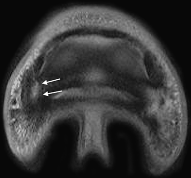
Transverse proton density image at the level of the distal phalanx of case 21. Medial is to the left. A hyperintense fracture line extends from the axial aspect of the medial palmar process, dorsally and abaxially through the palmar margin of the fossa of the collateral ligament of the distal phalanx (arrows). Moderate decreased signal intensity, consistent with sclerosis, is present in the medial palmar process of the distal phalanx.
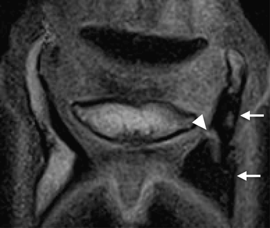
Dorsal T1-weighted gradient echo (GRE) image through the ossified ungual cartilages and the navicular bone. Medial is to the right. The ungual cartilages are ossified extensively. There is sclerosis, characterized by hypointense signal of the medial ossified cartilage and medial palmar process of the distal phalanx (arrows). Sclerosis is not present in the lateral aspect of the distal phalanx. A linear hyperintense fracture line (arrowhead) in the distal phalanx extends from the axial margin of the ungual cartilage distally and dorsally into the distal phalanx.
In the two horses receiving repeat MR imaging after lameness exacerbation, the distal phalanx fracture extended from the axial aspect of an ossified cartilage through the fossa of the collateral ligament of the distal interphalangeal joint to the solar margin of the distal phalanx.
Seventeen of the 23 fractures extended through the palmar margin of the fossa of the collateral ligament insertion on the distal phalanx (Fig. 1). Twenty of 23 had increased signal intensity and enlargement of the collateral ligament of the distal interphalangeal joint on the ipsilateral side of the fracture (Fig. 3). Twenty of the 23 affected limbs had abnormal mottled to decreased signal on T1- and T2-weighted sequences and thickening associated with the chondrosesamoidean ligament associated with the fracture site (Fig. 4). The chondrosesamoidean, chondrocoronal, collateral ligament of the distal interphalangeal joint and fibrous attachments between the collateral ligaments and the ungual cartilage could not be evaluated in one horse because of patient motion. The chondrocoronal ligament ipsilateral to the fracture was affected in 19 of 23 forelimbs characterized by either increased signal on PD, T2, and STIR sequence or decreased signal on PD, and T2 images and thickening (Fig. 5). In one horse the chondrocoronal ligament could not be evaluated. Eighteen of 23 forelimbs had abnormalities associated with the fibrous attachment between the collateral ligament and the ungual cartilage associated with the fracture. This appeared as loss of anatomic definition and thickening of this tissue. The fascial planes adjacent to the collateral sesamoidean ligament were indistinct secondary to abnormalities of the soft tissue structures in this region (Fig. 5). The collateral sesamoidean ligament had altered signal intensity and thickening in all affected forelimbs. The abnormalities in the collateral sesamoidean ligament were located along the abaxial margin of the middle phalanx, ipsilateral to the fracture.
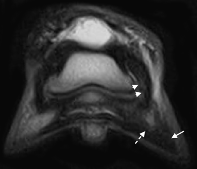
Dorsal T2-weighted fast spin echo image. Medial is to the left. The lateral collateral ligament of the distal interphalangeal joint has moderate hyperintensity that is most conspicuous in the distal axial aspect of the ligament (arrowhead). The ligament is diffusely thickened. The lateral palmar process of the distal phalanx has decreased signal intensity indicative of sclerosis (arrow). The area of increased signal intensity in the region of sclerosis had concurrent increased signal intensity on short τ inversion recovery (STIR) images indicating an active fracture (dashed arrow). The fracture line is immediately palmar to this image.
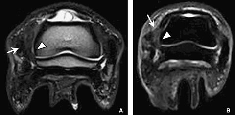
Dorsal oblique T2-weighted fast spin echo (A) and transverse short τ inversion recovery (STIR) (B) images through the navicular bone and middle phalanx. Medial is to the right. There is increased signal intensity and moderate thickening of the lateral chondrosesamoidean ligament (arrow head). The lateral ungual cartilage and adjacent fascia are thickened with loss of normal anatomic definition (white arrow). In image B, the fibrous attachment between the lateral collateral ligament of the distal interphalangeal joint and ungual cartilage is thickened (white arrow).
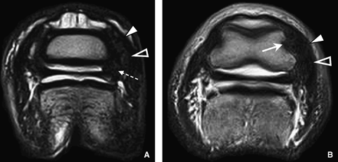
Transverse T2-weighted fast spin echo images at the level of the middle phalanx illustrating soft tissue abnormalities that can occur in conjunction with fracture of distal phalanx and ossified ungual cartilage. Medial is to the right. The medial chondrocoronal ligament (A) is severely thickened with a mottled signal pattern (white arrowhead on A). There is loss of the normal fascial planes (open arrowheads) adjacent to the collateral sesamoidean ligament, which is also diffusely thickened along the medial aspect of the middle phalanx. The medial collateral ligament and the fibrous attachment between the collateral ligament and the ungual cartilage (white open arrow on B) are moderately enlarged and primarily hypointense with a mottled signal pattern suggesting concurrent degenerative injury and scarring. The lateral soft tissue structures are normal.
Of the three patients had follow-up MR imaging to monitor fracture healing, the fracture was characterized by incorporation of the fracture line into the parent bone (Fig. 6). Sclerosis of the palmar process and ossified ungual cartilage was still apparent. The soft tissue structures were unchanged. Complete healing was noted at 14 months in one horse but clinical improvement was present at 8 months.

Sagittal (A, B) and dorsal (C, D) T1-weighted gradient echo sagittal through the palmar processes and ungual cartilages of the distal phalanx. Medial and dorsal are to the left. Parts A and B are from a different horse than parts C and D. (A) Initially, there was a hypointense signal characteristic of sclerosis in the medial aspect of the distal phalanx extending into the ossified ungual cartilage. A hyperintense linear fracture that extends distally and dorsally is present on the abaxial margin of the distal phalanx (white arrow heads). (B) Four months follow. Note the incorporation of the fracture line into parent bone and the fracture is less distinct. The sclerosis of the distal phalanx is relatively unchanged (white arrow). (C) On initial presentation, there is hypointense signal characteristic of sclerosis in the medial aspect of the distal phalanx extending into the ossified ungual cartilage. There is a proximal axial to distal abaxial oriented fracture of the lateral palmar aspect of the distal phalanx. (D) Fourteen months later there is complete osseous incorporation of the fracture line. Moderate abnormal hypointense signal consistent with sclerosis persists in the lateral palmar process of the distal phalanx.
Discussion
Horses in this study had lameness that was localized to the foot, ossification of the ungual cartilage, sclerosis of the ossified cartilage, and palmar process of the distal phalanx with a fracture involving the palmar aspect of the distal phalanx. The fracture was ipsilateral to the sclerotic ossified ungual cartilage. The fracture in conjunction with soft tissue abnormalities was the probable source of lameness.
The distal phalanx fracture appeared as a linear hyperintensity on PD, T1-, and T2-weighted sequences. Hyperintensity in STIR images extending beyond the fracture line into the surrounding bone suggested an active injury. Differential diagnoses for the hyperintensity in the STIR images was edema, hemorrhage, and/or necrosis.17
Osteosclerosis has been linked to chronic cycling possibly leading to increase chance of injury.18 Bone sclerosis can appear as a signal void on MR imaging because of low- PD and extremely short T2 relaxation times.16 There is greater mineralization in the medial palmar process of the distal phalanx in working horses and there may be a predilection for injury at these sites.15 In the equine carpus, sclerosis of radial fossa of the third carpal bone is considered the earliest sign of degeneration.19 Sclerosis can also be classified as fatigue damage, leading to weakened bone and decreased ability to withstand stressors.18 Sclerosis may lead to fracture.18,19 Fatigue injury in bone can often lead to insufficiency fractures or stress fractures and in humans these often accompanied by bone sclerosis.16 Although sclerosis is generally thought to be degenerative, the sclerosis may also be associated with healing.20 The horses wherein sclerosis was identified first followed by fracture later suggests that sclerosis of an ossified ungual cartilage and the associated palmar process of the distal phalanx predisposes to fracture.15
In the current study, there was sclerosis of the palmar process of the distal phalanx and concurrent fracture in all horses. There is a predilection for fracture of the medial palmar process,21 suggesting a contribution from load bearing. We found no predilection for medial vs. lateral location of the fracture. This may reflect a different etiology in our horses. However, there was a predilection for the fracture to occur at the base of an ossified ungual cartilage as noted by others.9 Palmar process may also may also occur in limbs that have no ossification of the ungual cartilage.21
The fact that all fractures in this study angled distally and dorsally from the base of an ossified ungual cartilage through the distal phalanx towards the solar margin suggests a biomechanical cause or focal stress point from cycling. Specific oblique radiographic views and nuclear scintigraphy are recommended for diagnosing lesions involving the palmar process of the distal phalanx or ossified ungual cartilage fractures.9,14,15,21 In this study, radiographs of the affected foot were not always available and the specific radiographic views to optimize evaluation of the palmar process were acquired. Although oblique views may have allowed the fracture to be seen in some horses in this study, there may not be a strong correlation between sclerosis in the palmar process of the distal phalanx in MR imaging and radiographic changes.15 Furthermore, occult fractures of the palmar process may not be seen with radiography,22 necessitating cross-sectional imaging such as computed tomography or MR imaging to eliminate superimposition.
As with previous studies involving the equine foot,23,24 simultaneous injuries associated with ligamentous structures of the foot were present. This relationship makes MR imaging an important diagnostic tool for evaluation of the foot. Ligamentous injury, specifically injury to the collateral ligament of the distal interphalangeal joint, may predispose the joint to instability and possibly lead to fracture of the palmar process of the distal phalanx.21 In present series, the ligamentous structures associated with the ungual cartilages and the collateral ligament of the distal interphalangeal joint were often affected and were characterized altered signal intensity and thickening. As with others,24,25 the frequency of the collateral ligament involvement suggests a relationship between ossified ungual cartilage and injuries to both the distal phalanx and collateral ligaments.
Abnormalities of ungual cartilage may be a source of lameness, but there is little evidence to support this.2,25 We found multifocal abnormalities in the ungual cartilage ligaments in addition to the fractures. An association between ligament scarring, characterized by low signal intensity on PD, and T2-weighted images, and chronic pain has been made in human knee collateral ligaments.26 Often, changes seen in affected ligaments associated with ossified ungual cartilages were consistent with scarring characterized by enlargement and low signal intensity on all sequences. However, histopathologic confirmation of scarring was lacking in this case series, which is limitation of the study. With the frequency that ungual cartilage ligaments were affected in the current study, the ligament scarring could also be contributing to the lameness in addition to the distal phalanx fracture.
It was not possible for us to determine whether the initial abnormality was the ligamentous injury associated with an ossified ungual cartilage or the sclerosis of an ossified ungual cartilage and palmar process. Ligamentous strain may be important in the ossification process of the ungual cartilages.27 Additionally, osseous remodeling and deposition can occur at the site of ligamentous attachment secondary to increased strain.28 As the ungual cartilages are integral in dissipation of energy,29 it is alternatively possible, that as the cartilages ossify, the surrounding ligaments structures assume more of the energy leading to injury. Sclerosis of an ossified ungual cartilage of the foot and palmar process of the distal phalanx has not been associated with ligament injuries, but ossified cartilages have been associated with distal interphalangeal collateral desmopathy.15,24 Sclerosis in the distal phalanx may result from concussive forces, load bearing, farriery, or conformation.15 We found the ungual cartilage ligaments affected commonly when there is ossified sclerotic ungual cartilage. Perhaps, the ligamentous structures of the foot and ossified ungual cartilage are responsible for the clinical signs noted in the current study.
Healing of the palmar process fracture was evaluated in three horses and was characterized by osseous incorporation of the fracture and reduced conspicuity of the fracture line. Initially, the fracture appeared as a linear area of increased signal intensity on T1-weighted FSE, T2-weighted GE and PD images. It is possible that the initial appearance of the fracture line represents fibrous union or immature fibrosis. However, complete healing noted in one horse was characterized by isointensity to surrounding bone and loss of fracture margins. In humans, the majority of stress fractures heal completely within 6–8 weeks.16 Fractures of the equine distal phalanx may take longer to heal. Bony union of nonarticular fractures of the palmar process, on average, require 8 months to heal.30 As the foot accepts the entire load of the horse, the biomechanical loading is persistent and there is continued micromotion at the fracture site. In addition, the distal phalanx has a vestigial periosteum. This may account for increased healing time of the distal phalanx.
Little is known about return to athletic performance with injuries involving the ligamentous structures associated with ossified ungual cartilages accompanying a palmar process fracture. The prognosis for most palmar process fractures is considered good with most horses returning to soundness.30 Radiographic studies did not address soft tissue injuries; however, it is possible that the fractures did affect the soft tissues, given the intimate nature of the osseous and soft tissue structures of the foot and time to return to function may be in part be influence by ligamentous injury. The return rate to function with collateral ligament desmosis of the distal interphalangeal joint is 60% with a mean time of 54 weeks.31 The average healing time for ligamentous and osseous injury of the palmar process is similar requiring approximately 8 months to a year for resolution.30,31 In horses with extensively ossified ungual cartilages there is a strong association with injury to collateral ligament and distal phalanx,25 and this may influence healing and return to function. Healing and return to function is injury and severity dependent and should be considered for prognosis. More information on this topic is needed.
In conclusion, ipsilateral desmopathy often accompany a palmar process fracture occurring in a sclerotic ossified ungual cartilage. Sclerosis of an ossified ungual cartilage may lead to fracture of the affected palmar aspect of the distal phalanx.
Footnotes
ACKNOWLEDGMENTS
Sincere appreciation to the following veterinarians for contributing cases to this paper: Scott Taylor, Rick Howard (Arizona Equine Medical and Surgical Centre), Paul McClellan (San Dieguito Equine Group), Richard Markell (Ranch and Coast Equine Practice), Sarah Gold, Brendan Furlong (Equine MRI of New Jersey), Kelly Tisher, Lois Toll (Littleton Equine Medical Center), Elizabeth Davidson, Midge Leitch, Dean Richardson (New Bolton Center), Nathaniel White II (Marion duPont Scott Equine Medical Center), Joe Davis (Piedmont Equine Practice), Carter Judy (Alamo Pintado Equine Hospital), Stacy Kent (Equigen, LLC), Doug Langer (Wisconsin Equine Clinic and Hospital) John Peloso (Equine Medical Center of Ocala), Erin Denny-Jones (Florida Equine Veterinary Service), Peter Blauner, and Jessica Leammle.


