LETTER TO THE EDITOR
Editor:
In a recent article Freer and Scrivani described findings of magnetic susceptibility artifacts of the vertebral column.1 In addition to describing the findings and the source of the artifacts, the authors suggest a spin echo (SE) sequence with short echo time (TE) as the optimal selection for imaging areas affected by this artifact. We have only recently acquired a magnetic resonance imaging scanner* and have encountered numerous patients with susceptibility artifacts. By varying parameters on both patients and cadaver trials, we found that the diagnostic value of the images can be improved. These trials were based on review articles and techniques for artifact reduction.2–4 It is not the purpose of this letter to go into the details of physics or the relationship of various factors. However, as it was not covered in the author's discussion, I think it may be useful to summarize operator-selectable options that may reduce susceptibility artifacts, sometimes sufficiently to salvage an otherwise non-diagnostic study.
After choosing a short TE SE or fast spin echo (FSE) sequence and ensuring that there has been patient-specific shimming of the field, additional measures that can be taken include the following: (1) decreased voxel size: increased matrix, and/or decreased slice thickness, and/or decreased field-of-view; (2) increased receiver bandwidth; and (3) change in the frequency encoding direction. This includes considering changing slice direction.
The first two have the negative effect of reducing the signal to noise ratio. Increasing the number of excitations can be done concurrently to maintain image quality. The resulting increase in time required for acquisition is substantial but can remain manageable by acquiring only the most important slices and use of an FSE technique instead of the typical standard SE for T1-weighted sequences. The FSE sequence will often decrease the artifact due to the multiple 180° refocusing pulses. The increased acquisition time might lead to problems with motion artifacts, but this has not seemed to be an issue with spine, head, or joint imaging, and options such as respiratory gating can be employed to mitigate these effects if necessary.
The third step may result in undesirable phase-encoded flow artifact propagation into the area of interest, and will alter the chemical shift artifact direction. There are other operator-selectable parameters for flow artifact reduction, if necessary, and a bandwidth increase will help reduce chemical shift artifacts.
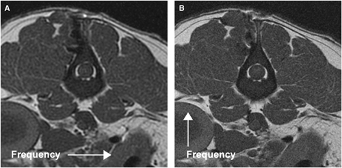
Figure 1 is a cadaver image. The lumbar epaxial muscles were dissected, a metal surgical suction tip was positioned over the wound and tapped with a high-speed burr, the tissues apposed with external pressure padding and T1-weighted images acquired. Figure 1A is an SE image obtained with typical parameters used on patients and Fig. 1B is the same slice acquired with FSE, increased matrix, increased bandwidth and a reversed frequency encoding gradient direction.

T1-weighted magnetic resonance image of lumbar spine with susceptibility artifact. Both images acquired with repetition time, 400 ms; short echo time, 14 ms; slice thickness, 3 mm; and field-of-view, 16 cm. (A) SE: matrix, 256 × 256; bandwidth, 15.6 kHz; number of excitations (NEX), 2; and frequency encoding direction right-left. (B) FSE: matrix, 512 × 512; bandwidth, 31.2 kHz; NEX, 4; and frequency encoding direction ventral–dorsal.


