ULTRASONOGRAPHIC EVALUATION OF NORMAL CANINE ILIOPSOAS MUSCLE
This paper was presented in abstract form at the 2005 ACVR Annual Scientific Meeting, Chicago, IL.
Abstract
The aim of this study was to describe the normal ultrasonographic appearance of the iliopsoas muscle and related landmarks. Hind limbs of five dog cadavers with no history of lameness were evaluated. The origin and mid-body of the psoas major and the common insertion of the iliacus and psoas major on the lesser trochanter of the femur were identified and evaluated. New methylene blue was injected under ultrasonographic guidance at the three sites. Dissection was performed to confirm placement of the dye. The L3 origin, mid-body, and insertion of the muscle were identified in all dogs and were consistent in appearance and compatible with the general appearance of muscle and tendons. The L2 origin was consistently difficult to image. The same ultrasound technique was subsequently applied to four healthy dogs, and consistent images of the iliopsoas muscle and associated landmarks were obtained. In this study, the major structures of the iliopsoas could be identified and in all dogs had a similar appearance. Ultrasound is an important tool for the diagnosis of musculotendinous injury and may be useful for identification of ilipsoas injury as a cause of lameness in the dog.
Introduction
The diagnosis of canine lameness arising from muscle injury is challenging. The exact prevalence of canine muscle or tendon injury is unknown, and this syndrome may be underdiagnosed.1 One cause of pelvic limb lameness in dogs is injury to the iliopsoas muscle group. Injury to the iliopsoas due to known trauma is recognized,2–4 but iliopsoas injury is also implicated in more subtle lameness in sporting dogs.5 Clinically, such patients have lameness accompanied by pain upon extension and internal rotation of the hip joint, and during direct palpation of the iliopsoas muscle.2–4,6 Typically, there are no radiographic abnormalities and the diagnosis is usually presumptive.
The iliopsoas muscle group is composed of two muscles: the psoas major and the iliacus. The group lies ventral to the quadratus lumborum muscle and dorsal to the psoas minor muscle. The psoas major muscle originates on the transverse processes of L2 and L3 and extends caudally, where it attaches at its mid-body along the ventral and lateral surfaces of the L4 through L7 vertebral bodies. As it passes over the ilium, it receives the iliacus muscle from the ventral surface of the ilium. The two muscles form the iliopsoas muscle and then insert as a common tendon on the lesser trochanter of the femur. The iliopsoas muscle acts to draw the pelvic limb forward by flexing the coxofemoral joint. When the femur is fixed in position, it also acts to flex the vertebral column or fix it in position. It is innervated by branches of the lumbar nerves and femoral nerve.7
The appearance of an injured iliopsoas muscle in the dog in sonographic, computed tompgraphy (CT), and magnetic resonance (MR) images has previously been described.2–4,8 The general ultrasonographic location of the iliopsoas muscle has also been reported.9 However, neither a standard, simple method of imaging the iliopsoas muscle group sonographically, nor the normal ultrasonographic appearance, has been described. The purpose of this study is to describe the normal ultrasonographic anatomy of the iliopsoas muscle and related landmarks to develop a simple, standard, and comprehensive method for scanning this muscle group.
Materials and Methods
Ultrasonographic examination of the iliopsoas musculature was performed on five medium and large-breed canine cadavers. The dogs died or were euthanized because of disease unrelated to the musculoskeletal system. The medical record of each dog was reviewed and no clinical history of lameness was found. The patients were 2–11 years old and weight ranged from 17 to 41 kg. The ultrasonographic examination was performed within 24 h of death. Bilateral scans were performed on each cadaver using an 8–5 mHz curvilinear and a 12–5 mHz linear transducer.*
The dogs were in dorsal recumbency, the medial thigh and lateral abdomen were clipped and cleaned, and coupling gel was applied to the skin. The hip joint was moderately extended for the duration of the scan (Fig. 1). The entire length of the iliopsoas muscle group was scanned and longitudinal and transverse images were obtained of the origin, mid-body, and insertion. The ultrasonographic appearance of the muscle and related landmarks were described at each of these three sites. New methylene blue agent† was then injected with a 21-G × 1.5-in. needle via ultrasound guidance into the tendon insertion and intramuscularly at the origin and mid-body (Fig. 2). The scan was then repeated on the opposite side of the patient. Total scan time for each patient was 20–30 min. Postmortem examination was performed on all five dogs within 12 h of sonography. The iliopsoas muscles were exposed in situ by dissection of the overlying structures to evaluate dye placement at the muscle origin, mid-body, and insertion. Digital photographs were made to document the dissection and confirm dye placement.
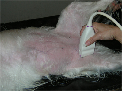
The patient is placed in dorsal recumbency, the medial thigh and lateral abdomen are clipped and cleaned, and ultrasound-coupling gel is applied to the skin. The hip joint is moderately extended and a 12–5 mHz linear transducer is used.
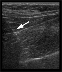
A longitudinal ultrasonographic image documenting needle (arrow) placement via ultrasound guidance into the mid-body of the psoas major muscle, before new methylene blue dye injection. Cranial is to the left. The dye placement was subsequently confirmed grossly.
Four clinically normal active medium and large-breed dogs subsequently underwent identical ultrasonographic examination of the iliopsoas musculature using the described technique. No sedation was used. Longitudinal and transverse images were again obtained of the muscle origin, mid-body, and insertion. The ultrasonographic anatomy of the musculature of these patients was compared with the ultrasonographic anatomy of the postmortem patients.
Results
The origin from L3 was visualized by first identifying the sacrum in longitudinal fashion and then moving the probe cranially, counting down from L7 to L3. Once the L3 vertebral body was identified in longitudinal orientation, the probe was shifted laterally along the transverse process, a clearly demarcated hyperechoic structure with far-field shadowing. The origin of the psoas major muscle was easily visualized arising caudally from the transverse process as hypoechoic muscle interspersed with obliquely oriented hyperechoic, linear fibers (Fig. 3). The origin of the psoas major muscle on the transverse process of L3 was easily identified in all attempts. However, the origin from L2 was consistently difficult to image, due to increased abdominal depth in the cranial abdomen at this level.
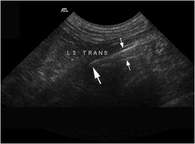
Longitudinal ultrasonographic image of the origin of the psoas major muscle. The psoas major muscle originates from the transverse process of L3 (large arrow) and is visualized extending caudally as hypoechoic muscle interspersed with obliquely oriented hyperechoic, linear fibers (small arrows delineate the dorsal and ventral margins).
To identify the mid-body of the psoas major muscle in a longitudinal orientation, the vertebral bodies of L4–L7 were first identified in long axis. The large mid-body muscle belly could be visualized ventral to the vertebral bodies, and was similar in appearance to the muscle at the origin: hypoechoic muscle interspersed with hyperechoic, linear structures. The linear fibers were oriented in a fashion parallel to the long axis of the muscle (Fig. 4A). To identify the mid-body in transverse orientation, the vertebral bodies of L4–L7 and their associated transverse processes were identified in short axis. The mid-body could be visualized ventral to and between each vertebral body and its transverse process (Fig. 4B). In a transverse orientation, both left and right psoas major muscle groups were symmetric. The origin of the iliacus muscle was difficult to visualize as a separate muscle from the psoas major. It was visualized in only one limb of one of the cadavers, and was not identified in any of the live, healthy patients.
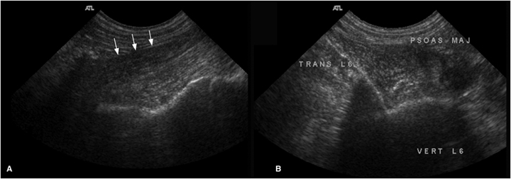
Ultrasonographic images of the mid-body of the psoas major muscle. (A) Longitudinal orientation. The muscle is identified ventral to the vertebral bodies as hypoechoic muscle interspersed with hyperechoic, linear structures. The ventral margin of the muscle is marked (arrows). (B) Transverse orientation. The muscle is identified ventral to and between each vertebral body and its transverse process.
For assessment of the insertion, the ultrasound transducer was initially placed in a transverse plane on the medial thigh at the level of the mid-diaphysis of the femur. The medial aspect of the femur was then followed proximally toward the coxofemoral joint (Fig. 5A) until the lesser trochanter of the femur was identified. The lesser trochanter was easily visualized in short and long axis fashion as a small, focal protrusion from the medial surface of the femur several centimeters (dependent upon patient size) distal to the coxofemoral joint. The iliopsoas muscle tendon could easily be visualized inserting on the lesser trochanter (Fig. 5B). Occasionally a slight rotation of the cranial aspect of the probe toward midline was required in order to obtain a more longitudinal view of the tendon (Fig. 6A). The final structure evaluated at the insertion site was the myotendinous junction, which was also easily visualized in all dogs. With the tendon in a longitudinal orientation, the probe was moved slightly cranially, while fanning from side to side, until a clear demarcation could be seen between the tendon and the adjacent muscle (Fig. 6B).
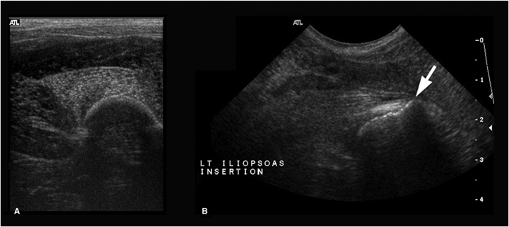
Transverse ultrasonographic images at the region of insertion of the iliopsoas. (A) The medial aspect of the femur, visible as a crescent-shaped hyperechogenicity with distal acoustic shadowing. (B) The lesser trochanter (arrow) is visible as a focal protrusion from the medial surface of the femur several centimeters distal to the coxofemoral joint. The iliopsoas tendon of insertion is visible as a linear hyperechoic structure attached to the trochanter.
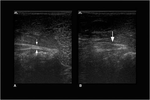
Longitudinal ultrasonographic images of the tendon of insertion (A) and myotendinous junction (B). (A) A slight rotation of the probe toward midline allows a more longitudinal view of the tendon of insertion, visible as a linear hyperechoic structure (arrows delineate the margins). (B) The myotendinous junction (arrow) is visible as a clear demarcation between the tendon (right side) and the adjacent muscle (left side).
The insertion of the iliopsoas muscle on the lesser trochanter of the femur was easily identified in all dogs. Its appearance was typical of other muscular tendons: short, linear, and hyperechoic. The muscle at the myotendinous junction was fan shaped, with a linear muscle fiber pattern characterized by hypoechoic muscle interspersed with hyperechoic, linear structures oriented obliquely.
Postmortem examination revealed accurate placement of new methylene blue dye in four out of five cadavers. In one cadaver in which the dye placement failed, the failure occurred only at the site of origin. Grossly, the dye was present in the cranial mid-body at the level of L4 instead of at the origin on L3. All significant anatomic structures were easily visualized grossly.
Once the ultrasonographic examination of the cadavers was completed, the findings were applied to ultrasonographic examination of the four healthy dogs. These examinations revealed similar anatomic findings to the cadavers, and all significant structures at the origin, mid-body, and insertion were identified in all four dogs.
Discussion
The origin and mid-body of the psoas major muscle and the common myotendinous junction and tendon of insertion of the iliacus and psoas major were reliably identified and had consistent ultrasonographic imaging characteristics. Other important related landmarks, including the transverse processes of the mid to caudal lumbar vertebrae, the ventral vertebral bodies, the lesser trochanter, and the surface of the femur, were also consistently identified. The identification of the muscle and related landmarks was subsequently confirmed using dye injections and postmortem inspection. The one instance of improper dye placement was most likely due to operator inexperience as dye was placed one vertebral body caudal to the origin site on L3 and this error occurred in the first cadaver entered into the study.
Ultrasonographic evaluation of muscle and tendon is commonly performed in equine and human sports medicine. Although anamnesis in canine patients may be somewhat less complete or accurate than in human patients, some specific physical examination findings may lead a clinician to suspect iliopsoas injury. Because the iliopsoas muscle group serves to flex the hip and is medially located, pain associated with injury to these structures is reportedly identified with simultaneous extension and internal rotation of the hip joint, as well as during direct palpation of the iliopsoas myotendinous junction and insertion. Confirmation of a soft tissue injury, by conventional means, is limited to palpation or radiographic abnormalities such as avulsion fractures of the lesser trochanter in the absence of ultrasound.
There are several ultrasonographic changes we could expect with traumatic injury to the iliopsoas muscle. These include muscle enlargement with interior hypoechoic regions which have been reported to represent edema, inflammation, and hemorrhage in the acute stage of injury.2,3,10–13 In chronic canine muscle injuries, hyperechoic areas are reported and interpreted to be consistent with fibrosis or ectopic mineralization.2,3,11–13 In addition to traumatic muscle injuries and muscle strains, ultrasound may be useful in diagnosing other iliopsoas muscle pathology that has been reported in canines, such as abscesses or neoplastic processes.14,15
CT and MR imaging have also been used effectively in diagnosing canine iliopsoas muscle injuries.4,8 These modalities also allow visualization of nearby muscles and nerves, which could be useful in defining the full extent of muscular, vascular, and nerve pathology associated with a disease process.4,8,14,15 CT and MR imaging were able to detect iliopsoas injury in humans in a significantly larger proportion of affected patients than ultrasound.16 However, similar comparative data are not available for animals. The different anatomic conformation of dogs vs. humans may allow ultrasound to be more accurate than in humans.
During the examination of the healthy animals, sedation was not used. However, it should be emphasized that dogs' iliopsoas muscle injuries can be painful, and the point pressure of the ultrasound transducer can further exacerbate this problem. Hence, sedation may be required for certain patients. Additionally, to diagnose iliopsoas injury, veterinarians must be aware of the condition and trained to perform a directed physical examination. Ultrasound should be used to support the physical examination findings. In humans, there are useful techniques for evaluating the area of origin and insertion of the iliopsoas muscle such as functional testing, direct palpation, and passive stretching of the muscle. These techniques are not very technically demanding but they do require training and adequate anatomic knowledge.17
In conclusion, ultrasound examination of the iliopsoas muscle is simple and quick to perform, and important structures are consistently visualized. Ultrasound may be a useful tool in evaluating iliopsoas muscle injury. Hence, it is important to develop knowledge of the normal anatomy of the iliopsoas muscle and related landmarks, and to determine a simple, efficient technique for comprehensively scanning the muscle. Further studies are indicated to evaluate cases of presumed iliopsoas muscle injury using the knowledge of anatomy and technique determined in this study, and to compare the accuracy of various imaging modalities in diagnosing injuries.


