EFFECT OF CONTRAST MEDIA FORMULATION ON COMPUTED TOMOGRAPHY ANGIOGRAPHIC CONTRAST ENHANCEMENT
Abstract
The characteristics of contrast media formulation (mgI/ml, osmolarity, and viscosity) are generally not considered important in computed tomography (CT) angiography in animals. The purpose of this study was to assess the contrast effect in CT angiography as a function of contrast media formulation, with a constant iodine dose. The contrast effects of three contrast media with different iodine concentrations were compared by administering identical iodine dosages (mgI/kg). The contrast effects of the three contrast media differed, and the area under the time–attenuation curve of iohexol 350 mgI/ml, which had the highest iodine concentration, was the lowest. It was hypothesized that the contrast effect of a contrast medium decreases with higher iodine concentration because of the high amount of residual iodine present in the circulatory system from the injection site to the portion immediately before the great vessels. In addition, the influence of osmotic dilution on contrast media with high osmolarity was also considered. In conclusion, the contrast effect varies with different contrast media formulations, even when the same iodine dosage is administered.
Introduction
Veterinarians typically consider the following parameters when using contrast medium for contrast-enhanced computed tomography (CT): fluid volume per kilogram body weight (ml/kg), iodine dosage (mgI/kg), injection speed (ml/s), and iodine delivery rate (mgI/s). In general, the contrast medium formulation (mgI/ml, osmolarity, and viscosity) is not considered.
Contrast media with high iodine concentration are expected to provide high contrast because X-ray absorption is proportional to iodine concentration.1 However, the theoretical contrast effect cannot be guaranteed in patients because of the transit time of the contrast medium and its subsequent dilution.2–4 Therefore, reproducible results cannot be realized with the same administration method without taking contrast media formulation into consideration.
The purpose of this study was to determine the effect of contrast medium formulation on the contrast effect in CT angiography.
Materials and Methods
Iohexol 350 mgI/ml (IOH350),* Iohexol 180 mgI/ml (IOH180),† and Iodixanol 320 mgI/ml (IDX320)‡ were used in this study (Table 1). All contrast media were stored at 37°C until just before examination.
| Properties | IOH350 | IOH180 | IDX320 |
|---|---|---|---|
| Iodine concentration (mgI/ml) | 350 | 180 | 320 |
| Osmolarity (mmol/kg) | 844 | 408 | 290 |
| Viscosity (37°C, mPa s) | 10.6 | 2.0 | 11.4 |
| Molecular weight | 821.14 | 821.14 | 1550.19 |
Four healthy beagle dogs, aged 2–4 years with a mean body weight of 10.2 kg were used. Studies were carried out in a crossover method with the same four dogs used three different times. All experiments were approved by the animal experimental guidelines of the Obihiro University of Agriculture and Veterinary Medicine.
Anesthesia was induced with intravenous propofol§ through a 20.0 gauge catheter in the left cephalic vein. Anesthesia was maintained with a continuous infusion of 20.0 mg/kg/h propofol. All dogs were intubated and in ventral recumbency on the CT table. Pulse, blood pressure, and respiration were monitored. In addition, before the injection of each contrast medium, pulse and blood pressure were recorded for each dog because these parameters affect the rate of contrast medium delivery.
All CT images were obtained using a multidetector-row CT.¶ Contrast media were injected using an auto-injector∥ through a 20.0 gauge catheter in the right cephalic vein. Image acquisition was performed during a single breath-hold.
Comparison of Contrast Effect using Abdominal Aorta Time–Attenuation Curves (TAC)
This examination was performed using an identical iodine dosage of 600 mgI/kg and an injection rate of 1.3 ml/s. The scan location was at the level of the middle of the left kidney. Dynamic CT image acquisition was initiated at the time of contrast medium injection for 70 s at 5 s intervals (120 kVp, 200 mA, 5 mm slice, and 1.0 s/rotation). To measure enhancement, regions of interest were drawn over the abdominal aorta by using an image processing workstation,# and TAC were generated. The TAC, peak CT value, area under the curve (AUC), and time-to-peak (TTP) values of each contrast medium were compared.
Visual Assessment of 3D Angiographic Images
This examination was performed using an iodine delivery rate of 400 mgI/s obtained by setting an identical iodine dosage (600 mgI/kg) and total injection time (15 s). CT images were obtained from the diaphragm to the distal end of the femur immediately after contrast medium injection (135 kV, 150 mA, 0.75 s/rotation, 2.0 mm slice thickness, and 1.0 mm reconstruction pitch). All image comparisons were performed using 3D images generated with an identical opacity curve (the same color map). The 3D images were then converted to MPEG files, which could be observed from all directions on a personal computer by dragging the mouse.
Using the MPEG files, four grades of the angiographic quality of each contrast medium were assessed by four veterinarians (M.K., K.Y., J.S., and Y.M.) who were not engaged in the 3D image generation and who were unaware of the contrast medium used in image formation. The assessment criteria were as follows: (1) observation of the bifurcation of the abdominal aorta, (2) observation of the femoral artery to the distal end of the midsection of the femur, (3) observation of the bifurcation of the caudal vena cava, and (4) observation of the femoral vein up to the distal end of the midsection of the femur. Each criterion was assigned 1 point, and the mean value of the four observers was calculated.
A post hoc test was used and P values <0.05 were considered to be significant. Kendall's coefficient of concordance was used to measure the level of agreement between the reviewers in the 3D visual assessment.
Results
In all experiments, no significant differences were observed in pulse or blood pressure. Furthermore, there were no characteristic changes in pulse, blood pressure, and respiration during and after the injection of contrast medium.
Comparison of Contrast Effects with the Abdominal Aorta TACs
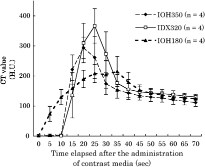
The TACs of the abdominal aorta are shown in Fig. 1. The peak contrast effect was largest in the IDX320 group, followed by the IOH350 and IOH180 groups (Table 2). The peak value in the IDX320 group was significantly higher than in the IOH180 group, but no significant difference was observed between the peak values in the IDX320 and IOH350 groups.

Time–attenuation curve of the abdominal aorta. Contrast effects were different regardless of the same iodine dosage (mgI/kg) and injection speed (ml/s).
| Contrast Medium | Mean Peak CTValue (HU) | SD |
|---|---|---|
| IDX320 | 365.87* | 57.28 |
| IOH350 | 292.74 | 35.72 |
| IOH180 | 214.23* | 50.02 |
- * P<0.05.
- CT, computed tomography; HU, Hounsfield units; SD, standard deviation.
The AUC was the highest in the IDX320 group, followed by those in the IOH180 and IOH350 groups (Table 3). The AUCs of the IDX320 and IOH180 groups were significantly higher than that of the IOH350 group; however, there was no significant difference between the AUCs of the IDX320 and IOH180 groups.
- †,‡ P<0.05.
- AUC, area under the curve; TDC, time–density curve; SD, standard deviation.
The TTP was the highest in the IOH180 group, followed by those in the IDX320 and IOH350 groups (Table 4). There were significant differences between the TTP values of the IOH180 and IOH350 groups as well as between those of the IOH180 and IDX320 groups.
- §,∥ P<0.05.
- TDC, time–density curve; TTP, time to peak; SD, standard deviation.
In addition, the times required for contrast medium detection, which indicated the transit time of the contrast medium to the abdominal aorta, were 5, 15, and 16.3±2.5 s after IOH180, IOH350, and IDX320 administration, respectively. The time for contrast medium detection in the IOH180 group was significantly less than in the IOH350 and IDX320 groups.
Visual Assessment of 3D Angiographic Images
Figure 2 shows the 3D angiographic images of a dog that were used for visual assessment. The mean value of the contrast effects were 2.1±6.1, 2.1±4.6, and 1.6±4.6 for the IDX320, IOH180, and IOH350 groups, respectively. Intraobserver agreement between the four veterinarians was characterized by a significant positive association (Kendall's W=0.9652; P<0.0001).

Three-dimensional angiographic images from the same dog used for visual assessment: (A) IDX320, (B) IOH180, and (C) IOH350. The contrast effect value of IOH350 was lower than those of IDX320 and IOH180.
Discussion
To compare the contrast effects using the abdominal aorta TACs, three contrast media formulations with different iodine concentrations were administered using an identical iodine dosage (600 mgI/kg) and identical injection speed (1.3 ml/s). Despite administering the same iodine dose, the same TAC was not obtained, and significant differences were noted in the peak contrast effect and AUC.
The reason for the low peak value of IOH180 was considered to be a result of the low iodine-delivery rate due to the high fluid volume, which requires a longer duration for injection.5 The AUC should remain constant, provided the iodine dose remains constant because the AUC reflects the administered iodine dose. However, in this study, the AUC of the IOH350 group was lower than those of the IOH180 and IDX320 groups. In contrast, no significant difference was observed between the IDX320 and IOH180 groups. In addition, based on visual assessment of the 3D angiographic images, the contrast effect of IOH350 was the lowest. The iodine dose and total injection time were identical in the administration protocol used for the 3D images. Therefore, in theory, the contrast effect of IOH350 should have been same as that of the other two contrast media as the iodine delivery rate (mgI/s) was identical.6 Therefore, it can be assumed that the effective iodine dose of IOH350 that reached the scan region was the lowest among the three contrast media. When a contrast medium is administered with an auto-injector, the residual contrast medium between the cephalic vein and the cranial vena cava following the injection is not flushed into the cardiovascular system at the same speed as the initial volume of contrast medium. As a result, in contrast media with high iodine concentrations, the contrast effect is less than that obtained using contrast media with low iodine concentrations because of the high amount of residual iodine present in the circulatory system from the injection site to the portion immediately before the great vessels.2,7 In other words, the reason for the lower AUC and 3D visual assessment grades in the IOH350 group was considered to be the residual iodine dose up to the cranial vena cava.
In humans, the residual volume of the contrast medium from the antecubital vein to the superior vena cava is reported to be approximately 30 ml in a person weighing 60 kg.2 The mean body weight of the dogs in this study was 10.2 kg. Based on the available data,2 the residual volume of the contrast media in this study should have been 5.0 ml based on the mean body weight of the dogs. Furthermore, 5 ml IOH350 corresponds to 1750 mgI, and 5 ml IDX320 corresponds to 1600 mgI. Therefore, based on this hypothesis, 150 mgI iodine (3% of the total iodine dose) results in a difference of as much as 73.13 Hounsfield units (HU) (Table 2). However, according to a previous study that compared IOH350 and IOH300,2 when the residual volume was presumed to be 30 ml for a person weighing 60 kg, the residual iodine dose difference was 4%, and the peak CT value difference was 14.2 HU. Taking into account the higher difference in the CT values between the IOH350 and IDX320 groups than that could be expected based on the abovementioned report, another reason for the observed differences may exist other than the residual amount of contrast medium.
One proposed hypothesis for this discrepancy is the difference in the osmolarity of IOH350 and IDX320. IOH350 is a monomeric contrast medium, and its osmolarity is 844 mmol/kg. IDX320 is a dimeric contrast medium with a near-isotonic osmolarity of 290 mmol/kg. It is known that compared to low-osmolarity contrast media, decreased contrast effects are observed for high-osmolarity contrast media due to osmotically induced dilution.4,8,9 This raises the possibility that the lower contrast effect of IOH350 compared with IDX320 was due to osmotic dilution between the cephalic vein and the abdominal aorta.
In addition, contrast media were injected from peripheral veins and the CT value was measured in the abdominal aorta. However, the contrast effect of IOH350 has been reported to be higher than that of IDX320 when the contrast media were injected through the left atrium, and the CT value was measured in the carotid artery.1 This indicates that if contrast media are injected through the left atrium, the contrast effect depends on the iodine concentration and not on the osmotic dilution. This also shows that high-osmolarity contrast media are probably diluted in the lungs when they are transferred from the arterial system to the venous system as the lungs have a rich capillary bed (osmotic dilution by extravascular fluid). However, further research is required to verify this hypothesis.
On the other hand, there was no significant difference in the AUC and 3D visual assessment grades between the IDX320 and IOH180 groups. Given that the contrast effect depends on the residual contrast medium, the AUC and 3D grades for IDX320, which has a high iodine concentration, should be lower than observed for IOH180 because of the high residual volume present in the vessels between the cephalic vein and the cranial vena cava. The osmotic hypothesis can also be applied to this question. The osmolarity of IDX320 is 290 mmol/kg, i.e. nearly isotonic, as already mentioned. In contrast, the osmolarity of IOH180 is 408 mmol/kg. Possibly because of the higher osmolarity of IOH180 than IDX320, the AUC of the former decreased to the same level as that of the latter due to osmotic dilution. However, in this study, it was not possible to verify which factor (residual contrast medium or osmotic dilution) had a greater influence on contrast effects or the degree of such an influence. In the future, studies for the comparison of contrast effects by flushing the residual contrast medium with saline will be required.7,10,11
Additionally, the detection time in the IOH180 group was significantly higher than those in the IOH350 and IDX320 groups. It is assumed that the vascular transit speed of IOH180 to the region of interest was higher because of its lower viscosity (2.0 mPa s). On the other hand, it is assumed that the detection time was not influenced by the difference between the viscosities of IOH350 (10.6 mPa s) and IDX320 (11.4 mPa s).
In conclusion, it is not appropriate to expect the same contrast effect with different contrast media formulations even if the same iodine dose is used. Furthermore, using contrast media with a high iodine concentration does not necessarily guarantee high contrast effects. In addition to the administration method, the iodine concentration and physiochemical properties of the contrast media formulations being used should be considered to ensure examination reproducibility.


