Giant retinal tear after pneumatic retinopexy
Editor,
Most giant retinal tears are idiopathic (Wilkinson & Rice 1997). Males with pathological myopia are at risk. The second most common cause of giant retinal tears is blunt trauma. Surgical manipulation such as anterior vitrectomy can also be a cause (Wilkinson & Rice 1997). We report a case of giant retinal tear developed after pneumatic retinopexy and illustrate the pathogenesis of this rare and vision-threatening complication.
A 54-year-old, non-myopic Chinese man presented with unprovoked floaters in the left eye. Visual acuity (VA) was 20/20 in both eyes. Anterior segments were unremarkable. Fundal examination of the left eye showed a superior rhegmatogenous retinal detachment extending from the 10 to the 2 o’clock position, with a small break and lattice degeneration at 12 o’clock. The right fundus was unremarkable.
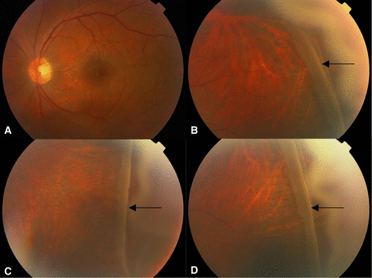
The patient underwent pneumatic retinopexy with cryopexy around the primary break and injection of 0.3 mL 100% C3F8 gas. Postoperatively, sitting upright with facedown posture was recommended. Ten days later, the superior retinal detachment was completely reattached. However, a giant retinal tear extending from 12 to 4 o’clock with pars plana detachment was noted (1, 2).

Fundus photographs of the left eye after pneumatic retinopexy (A) The posterior pole is flat. (B−D) Giant retinal tear with rolled edge extending from 12 to 4 o’clock with pars plana detachment after pneumatic retinopexy.

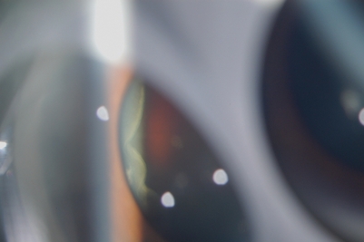
Slit-lamp photograph of the newly developed giant retinal tear.
Scleral buckling, pars plana vitrectomy, perfluorocarbon liquid, endolaser, cryopexy and 20% SF6 gas−fluid exchange were performed. The retina remained flat with VA of 20/70 at 8 months postoperatively. There was mild nuclear sclerosis pending cataract extraction.
Pneumatic retinopexy is a safe retinal reattachment procedure involving the transconjunctival injection of gas into the vitreous cavity, cryopexy or laser retinopexy, and posturing. Complications of this procedure are rare, and consist of new retinal break formation, subconjunctival gas, subretinal gas, gas entrapment at the pars plana, shift of detachment into previously attached macular and delayed subretinal fluid absorption (Hilton et al. 1990). Among these, new retinal break formation is a relatively common complication (incidence of 13%) with a favourable outcome (Hilton et al. 1990). It is believed to be the major reason for the lower initial success rate associated with pneumatic retinopexy (Hilton et al. 1990; Holz & Mieler 2003). By contrast, new giant retinal tear is a serious, vision-threatening complication and is exceedingly rare after pneumatic retinopexy.
Partly detached posterior vitreous is believed to be responsible for giant tear formation (Holz & Mieler 2003). Our patient’s young age suggests incomplete liquefaction and detachment of the posterior vitreous preoperatively. Because of its small initial volume, the intraocular gas may inadvertently migrate into the retro- hyaloid space. With subsequent expansion, it may lead to further dissection of the interface between the posterior vitreous and the internal limiting membrane of the retina (Freeman et al. 1988; Holz & Mieler 2003). This may produce excessive vitreoretinal traction leading to giant retinal tear formation, as in our case.
Giant retinal tear is a serious complication and remains a surgical challenge because of the high incidence of proliferative vitreoretinopathy and re-detachment with which it is associated (Ghosh et al. 2004). Vitrectomy combined with scleral buckling, perfluorocarbon liquid, laser, cryopexy, and gas or silicone oil is the mainstay of management (Ghosh et al. 2004). Preoperative assessment for the presence of posterior vitreous detachment, avoidance of injection of intraocular gas into retro-hyaloid space and frequent postoperative examination for new retinal breaks are important precautions. Prior to the procedure, patients should be informed that giant retinal tear is a remote but possible serious complication of pneumatic retinopexy.


