Stimulation of Pneumocystis carinii Encystment Following Necropsy
The cyst is an integral life cycle stage of Pnewnocystis carinii (P. carinii) [4]. The stimulating signal that triggers encystment is not known, but has been proposed that crowding or diminished energy supply may favor conversion to this more rigid form. A rat model of P. carinii pneumonia was utilized to determine if a change in environment will induce encystment; specifically, whether there is an increase in cyst number in infected lungs after the lungs are removed from the host animal.
MATERIALS AND METHODS
Female Sprague-Dawley rats were infected by transtracheal inoculation with approximately 7.8 × 1088 organisms. At specific time pints the rats were anesthetized and exsanguinated by cardiac puncture, and their lungs were removed. Severity of infection was determined as previously described [l]. Slides were prepared and fixed in methanol at the time of necropsy and every 24 hr of the next 96 hr. Lungs were stored at 4°C in a humidified chamber. Impression smears of infected lung tissue were blinded, then counted for trophozoite number by Giemsa stain and cyst number by GMS. Inoculum organisms were obtained from eight different rats, designated 1-1867. 1-1827, 1-2297. 1-2243, 1-2246, 1-2301, 1-2302, and 1-1444. For 1-1444, at least three rats were assessed at each time point.
Twenty-four hours after sacrifice, P. carinii organisms were isolated from some lungs by the method of Chin and Clarkson [2]. Cyst viability was assessed by the method of Maher et al. [3]. One-way ANOVA or Student's t-test was performed where appropriate to determine significant differences among or between samples using the Sigmastat software package (Jandel, Scientific).
RESULTS AND DISCUSSION
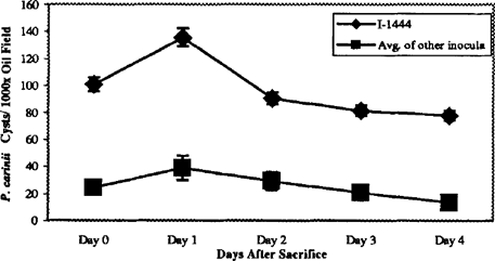
Vitality of the cysts at 24 hr as assessed by an RT-FCR viability assay showed that 80% of the cysts were viable [3]. For each animal, the number of cysts was consistently increased 24 hr after sacrifice of the animal and then decreased over the next 72 hr until the number was 40% below that originally noted in the tissue (Figure 1).

Increase in P. carinii cyst number in P. carinii-infected rat lung after sacrifice. Five different aliquots of P. carinii inoculum organisms were used to transtracheally inoculated immunosuppressed rats. After 42 days, infected lungs were removed and impressions smears were madle at the time of sacrifice and every 24 hr for the next 4 days. With every inoculum tested, there was an increase in the number of cysts in the lung tissue after 24 hr incubation at 4°C.
One set of animals, inoculated with organisms that had originally been isolated in 1995 from a particular animal (1-1444) displayed more cysts than animals inoculated with P. carinii harvested from all other animals at every time point analyzed. At 24 hr after sacrifice, when cyst number is highest, animals inoculated with 1-1444 had significantly more cysts than animals inoculated with P. carinii from other rats (Figure 2).

1-1444 P. carinii inoculum produces more cysts than other P. carinii inocula. The average cyst number per lOOOx oil immersion field from the five inocula shown in Figure 1 was compared to the cyst number present at the same time points in lungs from rats infected with 1-1444 inoculum of P. carinii. At each time pint, the number of cysts in 1-1444 infmed rats was significantly higher than that of the average infaed rat (p<0.0001). The increase in cyst number after 24 hr incubation at 4°C was also noted in 1-1444 infeued rats.
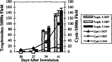
To determine whether this increase in cyst number was an indication of greater infection as a whole, additional animals were infected with 1-1444, 1-1827, or 1-1867 trophozoites. The 1-1444 animals showed more cysts at 20, 27, 34, and 41 days after transtracheal inoculation than has been previously noted in this laboratory. Trophozoite numbers in animals infected with each of the three inocula were similar at all time points, indicating that 1-1444 does not represent merely a more virulent isolate of P. carinii (Figure 3). 1-1444 did show more cysts than either of the other two inocula at all time points assessed (Figure 3). indicating that perhaps these organisms are prone to encystment.

Increased cyst number in I-1444 rats is not a function of more severe infedon. I-1444 was compared to two other lineages of P. carinii hoculum organisms for cyst and trophozoite numbers early and late in infection. At each time point studied h e cyst number in 1-1444 infected rats was higher than for other inocula. while the trophomite number was roughly equal in all inocula at all time points. The tr0phozoite:cyst ratio for 1-1444 was 60:40 at Day 41, as compared to 90:10 for 1-1867 and 1-1827.
These results imply that some P. carinii organisms are more prone to encystment than others and that encystment is stimulated by some factor present in dying tissue or lost from vital tissue. Used in conjunction with the cyst enrichment protocol described previously [3]. these data may provide impetus for exploring signals of encystment as well as a basis for studying the differences between P. carinii trophozoites and cysts in order to better understand P. carinii virulence factors and disease pathogenesis.


