In vitro Activity of Antimitotic Compounds Against the Microsporidium Encephalitozoon intestinalis
Cyclin-dependent kinase (CDK) are major players in the progression of the eukaryotic life cycle and inhibition of CDK clearly arrest cell proliferation [7]. Theses enzymes represent attractive potential targets for antiparasitic chemotherapy [5]. The effect of a collection of antimitotic compounds rich in CDK inhibitors has been tested on a mammalian cell line infected with Encephalitozoon intestinalis, a microsporidium causing intestinal and systemic infection in immunocompromized human subjects [4].
MATERIALS AND METHODS
Infection and treatment of cell cultures
Rabbit kidney cells (RK13) were cultivated in Lab-Tek® slides, in RP-MI 1640 medium supplemented with 8% heat-inactivated fetal calf serum (56°C for 30 rain), streptomycin (100 ug/ml), penicillin (100 U/ml) and L-glutamine (2 mM). Spores from E. intestinalis collected from RK13 cells monolayers as described [9] were added to cultured cells at a ratio of 1 spore/10 cells. At the time of infection, different doses of the inhibitors (0.01 to 3 μM) were added to the cultures. After 48 hrs of incubation the cultures were fixed in ethanol at -20°C for 10 min and then incubated for 1 h at 37°C with the monoclonal antibody (MAb) M1.6C1.2C11 [1]. This MAb was raised against a surface protein of sporogonic stages, i.e. sporonts, sporoblasts and spores, produced in the parasitophorous vacuoles (PVs) within 24 hrs after infection. PVs containing the parasites were counted in the Lab-Tek® slides using an epifluorescence microscope. The effect of the inhibitors on the morphology of E. intestinalis and RK13 cells was examined by electron microscopy
Determination of IC50 values on parasites and host cells
The immunofluorescence assay (IFA) data were plotted and the IC50 values determined using the Cricket Graph software. Microculture plates (Falcon, 96-well flat bottom) were prepared with RK13 cells. One μCurie of [3H] thymidine (5 Ci/mmol) and the appropriate amount of inhibitor was added to each well. Plates were incubated for 48h and [3H] thymidine incorporation was measured with a scintillation counter (BECKMAN). The IC50 values determined using the Cricket Graph software.
RESULTS AND DISCUSSION
Effect of compounds on parasites and host cells
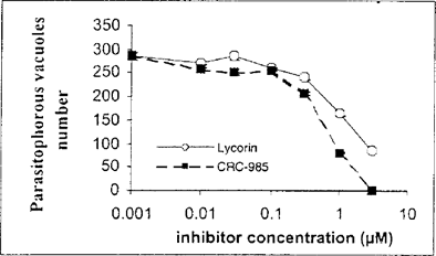
The effect of 526 inhibitors was evaluated from the number of PVs detected by IFA. We report the effect of two of these molecules: lycorine, an alkaloid isolated from the bulbs of several plants (Lycoris radiata, Narcissus pseudonarcissus, Amaryllis belladona.) and CRC-985, a staurosporine-ribose conjugate. Both compounds had a good inhibitory effect on parasite development, with IC50 values of 1.4 μM for lyconne and 0.75 μM for CRC-985 (Fig. 1).

In vitro effect of lycorin and CRC-985 on parasite development (means ± standard deviations of triplicate culture).
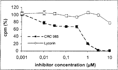
We next measured the effect of the same inhibitors on host cells in a standard [3H] thymidine incorporation assay. The IC50 values obtained from the graph (Fig. 2) for the lycorin was around 10 μM, and thus 10 fold higher than parasite IC50. For CRC-985, the IC50 value was 0,70 μM almost identical to that for the parasite (0.75 μM).

In vitro effect of Lycorine and CRC-985 on RK13 cells (means ± standard deviations of triplicate culture).
Morphological effects of antimitotic compounds
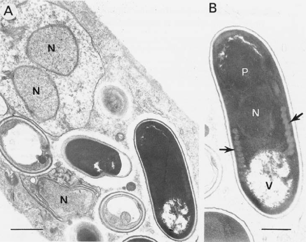
The gradual resorption of infection caused by the inhibitors in cell cultures was confirmed by transmission electron microscopy. Large PVs containing all developmental stages including numerous spores were seen in cultures treated with the lower doses. At the concentration of 0.5 μM, a reduction in the size of PV was observed, that corresponded to a decrease in the production of sporogonic stages. Curiously some of the spores produced in these vacuoles were larger (up to 4.0 μm) than those of the species usually seen (1.5–2.0 × 1.0–1.2 μm) (Fig. 3A).

Transmission electron micrographs showing alterations of parasite development in cultures treated with 0.5 μM CRC985. A. Abnormal swelling of a binucleate meront (top), presumably resulting from a disregulation of cytodieresis. The large electron dense spore measures 4.0 × 2.0 μm, instead of 2.0 × 1.0 μm as seen in untreated spores. B. Detail showing the 9 coils of the polar tube (arrows) instead of the 6 to 7 observed in the species. N, nucleus; P, polaroplast; P, posterior vacuole. Bars = 0.5 μm. Large sporogonic stages were also seen in cultures treated with 0.5 μM lycorin.
Additionally, 8 to 9 coils of the polar tube were numbered in these spores instead of the 6–7 characteristic Fig. 3B). The dividing meronts seen in these PVs became rounded instead of elongated (Fig. 3A). At a concentration of 1 μM, sporogonic stages were not (or rarely) seen in the PV, whilst meronts were still observed. Some isolated spores were apparently internalized by phagocytosis.
Thus a dose-dependent inhibition of infection occurred in cultures treated with the compounds tested in this study. Both IFA and electron microscopy suggest that the molecules altered the intracellular development of the parasites, especially sporogony. TEM examination of cultures treated with a dose of 1 μM confirmed the drop in the spore production, and also showed the persistence of meronts which were not detected by the MAb directed against a sporogonic stage protein. The correlation between the low number of generated spores and their larger size reflects the reduction of the mitotic activity of sporonts. Likewise, the electron dense nucleus seen in RK13 cells, more especially in those treated with CRC-985 suggests some alteration of their division which could occur after the prophasic condensation of chromosomes.
The decrease in the production of developmental stages and their concomitant enlargement was observed in the microsporidian Encephalitozoon cunicuili treated with albendazole, an inhibitor of tubulin polymerisation which also alters the mitotic activity of the parasite [3]. Lycorine is chemically related to Pancratistatin and 7-deoxynarciclasin, two antitumoral compounds shown to have antimicrosporidial properties (Ouarzane Amara et al., to be published elsewhere). CRC-985 is related to staurosporine, a compound which inhibits a broad range of protein kinases. Staurosporine inhibits proliferation and causes an abnormal swelling of Leishmania promastigotes, without affecting their differentiation to amastigotes, but the mechanism of action of staurosporine is in this case not elucidated [2]. In our study, the similar effects of Lycorine and CRC-985 tend to indicate that the targets of both inhibitors are regulators of the microsporidian cell cycle. A gene encoding a cdc2-related kinase has been isolated in E. cuniculi [8] and more recently in E. intestinalis (Thellier, Ouarzane-Amara, Doerig, unpublished). The recent classification of microsporidia with the fungi [10] suggests that a single CDK may control progression through the different phases of their cell cycle, as is the case in yeasts [6]. Thus the information reported here points to new potential therapeutical tools which certainly deserve further characterization, and identifies molecules that may prove promising for investigating microsporidian development.
ACKNOWLEDGMENTS
Study supported by a grant of SIDACTION and PRFMMIP Programme (French Ministry of Research).


