Hand-Assisted Laparoscopic Ovariohysterectomy in the Mare
No reprints available.
Supported in part by the Patricia Bonsall Stuart Awards for Equine Studies.
Presented at the American College of Veterinary Surgeons 13th Annual Veterinary Symposium, Washington, DC, 2003.
Abstract
Objective— To develop a minimally invasive, hand-assisted laparoscopic ovariohysterectomy (HALS-OHE) technique in the mare and to evaluate safety and any associated complications.
Study Design— Experimental study.
Animals— Eight, 11–24-year-old mares with anatomically normal urogenital tracts.
Methods— The surgical technique was developed in 2 non-survival mares and subsequently evaluated in 6 survival procedures. Food was withheld for 48 hours, then mares were anesthetized and positioned in dorsal recumbency for laparoscopic surgery. A hand access device (Omniport™) was placed followed by 4 laparoscopic portals. Transection of the ovarian pedicles and broad ligaments was achieved using a combination of a laparoscopic stapling instrument (Endo GIA II), an ultrasonically activated instrument (Harmonic Scalpel®), and endoscopic clips (Endo Clip II ML). The genital tract was exteriorized through the laparotomy, and the uterus transected and sutured in a conventional pattern. Horses were evaluated through postoperative day 14 when a necropsy was performed.
Results— Four mares recuperated well after surgery, 1 mare was euthanatized because of bilateral femur fracture during anesthetic recovery, and another developed severe pleuropneumonia. At necropsy all but 1 abdominal incision was healing routinely. One mare had abscessed along the celiotomy incision and developed visceral adhesions. Uncomplicated healing of transected mesovarial, mesometrial, and uterine remnants was observed.
Conclusions— Ovariohysterectomy in horses can be accomplished using HALS technique.
Clinical Relevance— HALS-OHE technique represents a minimally invasive and technically feasible alternative for conventional OHE. Careful patient selection and preparation may reduce the complications observed. The HALS technique may be useful in other laparoscopic surgical procedures.
Introduction
EQUINE OVARIOHYSTERECTOMY (OHE) is an uncommonly performed but technically demanding surgery that has been associated with a high degree of invasiveness and morbidity.1,2 The most common indication for OHE in mares is treatment of chronic pyometra non-responsive to medical management. Other indications include chronic intramural hematoma, removal of uterine tumors, removal of macerated fetuses, and cervical aplasia with mucometra.1,3–5 Conventional OHE in the mare is performed by caudal ventral median celiotomy.1,6,7 Reported disadvantages of this approach include the requirement for a large incision, poor intraoperative visibility and exposure, and excessive postoperative morbidity.1,2
Laparoscopic techniques have the recognized advantages of increased intraoperative visualization, smaller incisions, less extensive manipulation of the abdominal viscera, reduced postoperative pain, and reduced periods of convalescence, compared with corresponding procedures performed in horses using conventional methods.8,9 A recent advance in human laparoscopic surgery has been hand-assisted laparoscopic surgery (HALS), a technique where the surgeon's hand can be inserted into the laparoscopic field while maintaining laparoscopic observation.10,11 The hand is either introduced through a mini-laparotomy alone or through a special hand port device, which allows a complete seal between the surgeons arm and the device, thus maintaining abdominal insufflation.
Using HALS technique, complex laparoscopic procedures have been facilitated in humans by the coordinate manual manipulation and retraction of tissues and manipulations of transabdominally placed instruments.12,13 Additionally, larger organs (spleen, kidney, uterus) can be exteriorized from the abdomen through the device.13,14 HALS combines the convenience and safety of conventional open surgical techniques with the advantages of minimally invasive surgery.15
Our objectives were to evaluate potential use of a HALS technique for equine abdominal surgery through development of a minimally invasive, HALS-OHE technique for mares and to evaluate safety and complications associated with this technique in normal mares. We hypothesized that a HALS technique could facilitate OHE in the mare.
Materials and Methods
Mares studied were donated because of problems unrelated to the reproductive tract. A non-survival pilot study was performed on 2 mares to develop the HALS-OHE technique. Six mares were used to evaluate the method and any associated complications. Preoperative evaluation included physical and gynecologic (transrectal palpation, transrectal ultrasound, vaginal examination) examinations as well as complete blood count and chemistry profile.
Survival surgery involved 1 quarter horse and 5 thoroughbred mares that weighed 442–567 kg and were 11–24-years old. One mare was a maiden (1) and 2 mares (5, 6) were in estrus at the time of surgery.
Food and bedding were removed 48 hours before surgery. Mineral oil (3.7 L) was administered by nasogastric tube 48 and 24 hours before surgery. Procaine penicillin G (22,000 U/kg, intramuscularly [IV]) and flunixin meglumine (1.1 mg/kg, intravenously) were administered before surgery and every 12 hours after surgery for 60 hours. Before induction of anesthesia, feces were manually evacuated from the rectum to reduce potential interference with the laparoscopic field.
Horses were administered xylazine hydrochloride (0.5–1.2 mg/kg IV) and guaifenesin (64–86 mg/kg IV). General anesthesia was induced with ketamine hydrochloride (2.2 mg/kg IV) and maintained by gas inhalation; positive pressure ventilation was used. All mares were administered butorphanol tartrate (0.02 mg/kg IV) at the beginning of surgery and again 1 and 2 hours after induction. Monitoring included measurement of direct arterial blood pressure, arterial blood gases, end-tidal CO2 concentration, and electrocardiography. Xylazine hydrochloride (50–100 mg IV) was administered after mares were positioned in the anesthesia recovery room.
Anesthetized horses were positioned in dorsal recumbency. The ventral abdomen was clipped from the xiphoid to the perineum and laterally to the folds of the flank. The surgical field was prepared for aseptic surgery.
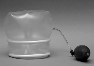
The Omniport™ (Advanced Surgical Concepts, Dublin, Ireland) hand and laparoscopic instrument access port for laparoscopic assisted surgery was used in this study. The Omniport™ (Fig 1) consists of an inflatable polyurethane cuff forming a 1-chamber pneumohelix with 2 integrated rings and is available in 2 sizes (50 or 80 mm distance between the 2 rings) to accommodate patients with a range of abdominal wall thickness.

Omniport™—a hand access port for laparoscopic-assisted surgery .
Surgical Technique
A 11-cm skin incision was made over the cranial aspect of the longitudinal intermammary groove. The incision continued through the subcutaneous tissue, the medial suspensory laminae, and the linea alba, bisecting the mammary gland. The hand access port was lubricated with 1% carboxymethylcellulose (Aqualon®, Hercules, Wilmington, DE), both rings were compressed into an oval configuration, and the device was obliquely inserted into the abdomen. The outer ring was gently exteriorized from the abdomen whereas the other ring was positioned within the abdomen against the peritoneum. Care was taken to ensure that abdominal viscera were not positioned between the device and the peritoneum.
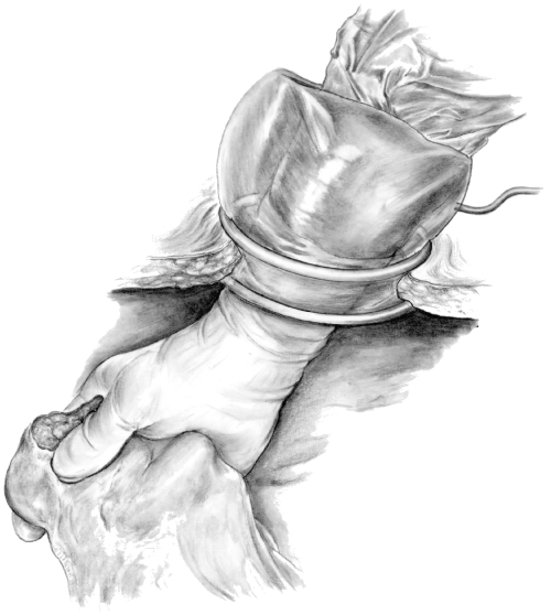
The left ovary was approached first in each mare with the primary surgeon positioned on the left side of the horse. The 1st assistant, positioned on the right side of the horse, inserted the right hand through the hand access port into the abdomen and the device was inflated using the rubber bulb inflation pump to affect a seal between the forearm and the incision (Fig 2). Carboxymethylcellulose was used to ease hand insertion. A 1.5-cm incision was made through the skin and linea alba over the umbilicus. Using the hand placed into the abdomen, the 1st assistant elevated the ventral abdominal wall and provided counter pressure while an 11-mm laparoscopic sleeve with sharp pyramidal trocar was passed into the abdomen. The sharp trocar was removed and replaced by a laparoscope (Hopkins® Telescope, 10 mm × 57 cm, 30° angle; Karl Storz Endoscopy, Goleta, CA). A 2nd assistant, also positioned on the right side of the horse, controlled the laparoscope.

The assistant surgeon's hand placed through the hand access device into abdomen .
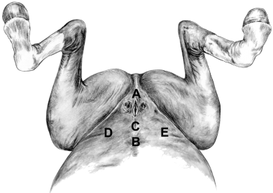
Capnoperitoneum was established to 10–15 mm Hg and maintained using an automatic insufflator (Electronic Laparoflator 26012, Karl Storz Endoscopy), the surgical table was tilted approximately 13° in a head-down position, and mares were rotated slightly to the right of dorsal recumbency. An instrument portal was similarly made on the ventral midline slightly cranial to the midpoint between the umbilicus and the cranial aspect of the hand access port laparotomy incision (Fig 3). Laparoscopic claw forceps was passed through this portal and used by the 1st assistant to manipulate the ovary and mesometrium. The left ovary was placed manually under cranial traction and a 3rd (left ovarian) portal was made approximately 5 cm cranio-lateral to the ventral projection of the ovary. A 1.5-cm incision through the skin and external rectus sheath was made, and a 12-mm laparoscopic sleeve with sharp pyramidal trocar (Endopath® 512, Surgical Trocar 12 mm × 100 mm; Ethicon Endo-Surgery, Cincinnati, OH) was passed through the abdominal wall. The left ovary was immobilized manually and infiltrated with 15 mL 2% mepivacaine delivered through a laparoscopic injection needle (Karl Storz Endoscopy) passed through a reduction sleeve placed in the left ovarian portal.

Location of the caudal median celiotomy for placement of the hand access port (A), the laparoscope portal (B), the instrument portal (C), the left ovarian portal (D), and the right ovarian portal (E) .
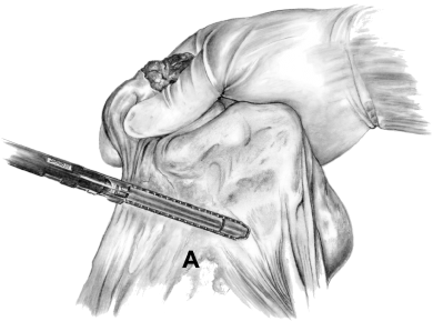
A laparoscopic stapling instrument (Endo GIA II with 3.5 mm × 60 mm cartridge; United States Surgical Corp., Norwalk, CT) placed through the left ovarian portal was used to transect the mesovarium that had been manually placed perpendicularly into the instrument jaws (Fig 4). Subsequently, Laparoscopic Coagulating Shears™ (Harmonic Scalpel®, 15 mm straight blade; 10 mm × 34 mm; Ethicon Endo-Surgery) were inserted through the same portal and used for transection of the mesometrium. Most of the mesometrium was transected using the blunt blade at power setting 3 as described.16 When large vessels were encountered, the flat blade surface was applied proximal and distal on the vessel, followed by transection between these areas. When hemostasis after use of the Harmonic Scalpel® was incomplete, 9 mm titanium endoscopic clips (Endo Clip II ML; United States Surgical Corp.) were applied to provide hemostasis. On some occasions, large vessels were first skeletonized with the Harmonic Scalpel® and endoscopic clips applied proximal and distal before transection. Mesometrial transection was continued to the level of the caudal uterine body.

Manually assisted placement of the laparoscopic stapling instrument on the mesovarium (A). The assistant surgeon's right hand is holding the left ovary, and gently stretching the mesovarium to permit perpendicular placement of the instrument jaws .
The mare was then rotated to a slightly left oblique position and the positions of the surgical team reversed. A 4th (right ovarian) portal was made craniolateral to the right ovary and the right mesovarium and mesometrium was transected as described above.
The hand access port was deflated and removed, and the operating table was returned to a horizontal position. The uterus and ovaries were exteriorized through the caudal median celiotomy. Remaining caudal uterine arterial branches along the body of the uterus were ligated with 2-0 polyglactin 910 and transected. Two large Carmalt forceps were applied transversely across the uterine body at the most caudal accessible extent and the uterus was transected between them. The uterine stump was closed using 1 polydioxanone in a Parker–Kerr pattern. The linea alba was closed with 2 polyglactin 910 in a simple continuous pattern, the mammary gland and subcutaneous tissue were apposed with 2-0 polyglactin 910 in a simple continuous pattern. Skin incisions were closed using 2-0 nylon in a simple continuous pattern for the celiotomy incision and 2 or 3 simple interrupted sutures for the laparoscopic portal incisions.
Operative and recovery time, hemorrhage, and methods of hemostasis as well as complications during and after surgery were recorded. Operative time was defined as the time from the first skin incision to completion of skin closure. Recovery time was the time between disconnecting the horse from the anesthetic circuit until the horse was standing.
Horses were stall confined after surgery except for short periods of exercise walking in hand, twice daily beginning the day after surgery. Feed was gradually reintroduced, starting with minimal amounts offered hourly from the day of surgery. Physical examinations were performed hourly during the first 8 hours after surgery, every 6 hours during the next 24 hours, twice daily through day 7 and once daily for the remainder of the study. Variables included general attitude, pulse and respiratory rate, body temperature, appetite, borborygmi, defecation, urination, and subjective evaluation of incisional healing. The packed cell volume (PCV) and the plasma total protein concentration were determined daily for 3 days after surgery.
Fourteen days after surgery, mares were euthanatized and abdominal cavities examined. The remnants of the broad ligaments and reproductive tract were evaluated for inflammation, adhesions, hematoma formation, and character of healing. Samples from the amputated genital tract transection line were obtained and fixed in 10% neutral buffered formalin. Tissue specimens were subsequently embedded in paraffin, sectioned at 5 μm, and stained with hematoxylin and eosin. Histologic evaluation was performed to determine the level of transection (uterus or cervix).
Results
Intraoperative
Insertion of the Omniport™ was quick and easy. The 50 mm ring distance device was used in 2 mares and the 80 mm ring distance device in 4 mares. The hand port facilitated maintenance of abdominal insufflation while the first assistants hand and forearm was positioned in the abdominal cavity. Small volumes of gas escaped during arm insertion and removal through the portal. Manipulation of the genital tract and intestine located in the caudal abdomen was readily performed under laparoscopic observation. Lifting the ovary and the uterus and gently stretching the mesovarium and mesometrium, respectively, facilitated accurate placement of the stapling instrument and Harmonic Scalpel®. During transection of the mesometrium, digital palpation also permitted identification of larger vessels that required occlusion before division. Generally, there was good exposure and visibility of the ovaries, uterus, and mesometrium with occasional interference by large intestine. When this occurred, the viscera could be repelled with the intra-abdominal hand and the genital tract lifted for optimal instrument placement. Paresthesia and fatigue of the intra-abdominal hand occurred periodically and was most often associated with overinflation of the hand port and sustained digital traction during the isolation and positioning of blood vessels for occlusion, respectively.
Ovarian artery hemostasis and mesovarial transection was complete in all mares. Hemostasis of large vessels within the mesometrium using the Harmonic Scalpel® was inadequate in 3 mares (1, 4, 5); hemorrhage was controlled by subsequent application of endoscopic clips. In some instances occlusion of large vessels was performed with endoscopic clips before transection with the Harmonic Scalpel®. Generally, branches of the caudal uterine artery were occluded and transected using the Harmonic Scalpel® during laparoscopy. In 3 mares (3–5), right caudal uterine arterial branches were ligated and transected after the uterus was exteriorized. Transection and closure of the uterine stump presented no difficulties, though the exteriorized uterus was under moderate tension. Surgical time ranged from 150 to 210 minutes and decreased as the experience of the surgical team with the procedure increased.
No substantial intraoperative anesthetic complications were observed. Mare 1 had bradycardia (heart rate <20 b.p.m.) and occasional 2° atrioventricular block, and mare 2 had a few ventricular premature contractions. All mares had episodes of hypotension (mean arterial pressure <70 mm Hg), which responded to treatment with dobutamine (1–5 μg/kg/min). Anesthetic recovery times ranged from 40 to 100 minutes in 5 horses. One mare (3) was unable to rise and was subsequently euthanatized.
Postoperative
Pulse and respiration rates, and body temperatures returned to or remained within normal limits during the first 24 hours after surgery in 4 mares (1, 2, 5, 6). Five hours after surgery, mare 2 had signs of abdominal discomfort (heart rate 68 b.p.m recumbency) that resolved after administration of 100 mg xylazine hydrochloride. Mare 1 had pyrexia (102.4°F) 4 hours after surgery that resolved after a scheduled administration of flunixin meglumine. Mare 6 developed intermittent pyrexia (<102.0°F) from the 3rd to 5th postsurgical day that resolved after administration of 0.5–1.1 mg/kg flunixin meglumine once daily for 3 days. The 4 mares had good appetites and were fed ad libidum within 24 hours after surgery. Borborygmi were slightly decreased on the day of surgery, but returned to normal during the subsequent 24 hours. Defecation and urination was noted in all 4 mares within the first 24 hours after surgery. Mare 4 did not return to normal after surgery but appeared depressed and had a decreased appetite throughout the entire postoperative evaluation period. Because of intermittent tachycardia, tachypnea, and pyrexia, the mare was administered flunixin meglumine and procaine penicillin from the 5th postsurgical day onwards. Based on clinical and ultrasonographic evaluations, pleuropneumonia was suspected and confirmed on necropsy on the 12th day.
All but 1 surgical wound had uncomplicated healing. Mare 6 developed purulent discharge from the celiotomy incision on the 13th day. In mares 5 and 6 transient hematoma formation was encountered at 1 ovarian laparoscopic portal site. Three mares (2, 4, 6) developed mild-to-moderate ventral edema. Infrequently, small volumes of serosanguineus vaginal discharge was observed in 4 mares (1, 2, 5, 6) and persisted for 5 days in 1 mare.
Except for 2 mares, PCV and the plasma total protein concentration remained within physiologic ranges postoperatively. A decline in PCV was observed in mare 1 (from 39% to 26%) and mare 4 (from 38% to 20%).
Necropsy Findings
Surgical incisions were healing normally in all but mare 6. In this mare, subcutaneous suppuration of the caudal median celiotomy incision, adhesions between the descending colon, its mesocolon, and the abdominal incision, as well as, multiple small fibrous tags (10–20 mm length) on the serosa of the cecum and ascending colon were observed. Adhesion formation to the surgical site was not observed in any other mare. In mare 1, a 50 mm × 50 mm area of peritoneum ventrolateral to remnant of the left broad ligament was covered by a thin layer of fibrous tissue. The mare euthanatized in the recovery stall had bilateral femoral fractures. Mare 4 on the 12th day had severe locally extensive, acute to subacute pleuropneumonia and a moderate subacute generalized peritonitis. Bacterial cultures obtained from multiple organs yielded Pasteurella multocida, Klebsiella pneumonia, Brevundimonas sp. and α-hemolytic Streptococcus sp.
The transection line of the mesovarium, mesometrium, and uterine remnant were healing routinely in all 5 mares recovered from anesthesia. A smooth, 1 mm layer of tan-pink colored fibrous tissue covered the staples along the ovarian pedicle and the suture line on the uterine remnant. Small (5–10 mm diameter), firm, brown nodules along the mesometrial transection line were observed in the vicinity of larger blood vessels. Caudal to the more highly vascular area, the mesometrial boundary appeared as a thin line without discoloration or thickening. Mare 5 had 2 organized hematomas approximately 60 mm diameter within 1 mesometrium and a small seroma in the contralateral mesometrium. Histologic evaluation of the amputated genital tracts indicated that transection was completed within uterine tissue in all horses.
Discussion
We have described a minimally invasive HALS technique for OHE in mares with normal urogenital tracts. Although the indications for HALS-OHE in mares may be limited, the technical advantages of HALS technique may be applicable to other equine abdominal procedures currently performed using either conventional or laparoscopic techniques.
The hand access port allowed manual access to the abdominal cavity under laparoscopic observation while capnoperitoneum was effectively maintained. The use of the intra-abdominal hand facilitated protection of viscera during laparoscopic portal placement, stabilization of ovaries during injection of anesthetic, and the ligation and division of the mesovarium and broad ligament. Additionally, viscera were manually retracted to improve observation, and prompt hemostasis was achieved by digital pressure when hemorrhage occurred.
Advantages described for HALS in humans, include reduced surgical time compared with routine laparoscopic methods and the requirement for less technical expertise, resulting in procedures with reduced degree of difficulty.10,17 The capability for tactile examination eliminates one of the major disadvantages reported for laparoscopic procedures.10,12,14 Lastly, the retrieval of a large tissue specimen (e.g. kidney, tumor, uterus) can be readily performed through the hand access port.10,13,14,17
The reported disadvantages of HALS compared with standard laparoscopy in humans includes the potential reduction of internal and external surgical work space in small patients, the time required for device placement, difficulties in maintaining abdominal insufflation, reduced cosmesis, increased morbidity compared with standard laparoscopic incisions, and the expense of the device.10,14,17,18 In contrast with humans, the large size of the equine abdomen provided ample surgical workspace and no interference between the hand and laparoscopic instrument manipulation; but, also resulted in the disadvantage of requiring 3 scrubbed surgeons, similar to a previous report of conventional OHE in horses.1 We observed that placement of the hand access port required little time and was technically simple. Though intended for single use, it is possible that the cost of the Omniport™ device ($350) may be recovered over several cases as devices were gas sterilized and used for multiple horses without loss of function in our study. It was not determined how many times the hand access devices could be reused without loss of function.
Techniques have been recently described for HALS-OHE and hand-assisted left-sided nephrectomy in standing horses.19,20 In these reports a hand port device was not used. Loss of abdominal insufflation was reported but it did not prevent completion of the procedures. However, it is important to note that adequate laparoscopic observation depends to a greater extent upon maintenance of adequate abdominal insufflation for horses in dorsal recumbency compared with standing laparoscopic procedures.
Optimal use of the hand access port is influenced by several factors including port location, incisional length, and hand port size. In our study, the hand port celiotomy incision was positioned just cranial to the pubic brim to maximize genital tract exteriorization after transection of the mesovarium and mesometrium. Selection of the Omniport™ size (80 mm versus 50 mm ring distance) was made by estimation of body wall thickness once the caudal midline laparotomy was completed. For human patients the 50 mm version is recommended for body walls <40 mm thick and the 80 mm ring distance for body walls between 40 and 70 mm.21 Insufficient incisional length for placement of the hand portal device has been reported to result in obstruction of circulation in the surgeons hand, resulting in paresthesia.15,18 Occasional paresthesia observed in this study was alleviated by reducing the cuff inflation pressure of the hand portal device. In our study, an incisional length of approximately 11 cm was adequate to comfortably accommodate the forearm of the 1st assistant and was adequate for removal and resection of normal urogenital tracts. Clinical conditions such as pyometra may require the length of the incision to be increased to permit safe exteriorization. We believe the resulting incision would be considerably <40 cm length reported for conventional OHE in horses.1
In our study the surgical table was inclined approximately 13° from horizontal (the maximum possible). This is considerably <30° incline previously reported for ventral laparoscopic ovariectomy;22 however, exposure of the ovaries and uterus during the surgery was judged to be adequate in our study. The need for urinary bladder catheterization as previously reported was not apparent.22 In addition to inclination of the surgical table, tilting the mares slightly to the side opposite the side being operated displaced the abdominal viscera and was judged to be important for laparoscopic observation.
A variety of methods have been reported for achieving hemostasis of the ovarian and uterine arteries during laparoscopic and non-laparoscopic ovariectomy and OHE in horses.1,2,5,16,19,22–25 In our study, hemostasis of the ovarian pedicle was consistently achieved by single application of a laparoscopic stapling instrument (60 mm × 3.5 mm staples). Although reported previously that the use of the Harmonic Scalpel® produced adequate hemostasis of the ovarian pedicle,16 the surgical time saved by using a laparoscopic stapler was thought to be advantageous in our study. Hemostasis of the vessels within the mesometrium was not always achieved using the Harmonic Scalpel®. The Harmonic Scalpel® has a reported 85% success rate of sealing arteries up to 3.5 mm in diameter, pressurized to 300 mm Hg, at a power setting of 3.26
The size of the vasculature in the mesometrium has not been determined, and it is likely increased in multiparous mares and those with chronic uterine disease. It is possible that the larger vessels within the mesometrium exceeded the capacity of the Harmonic Scalpel® to seal consistently. Because of branching of the middle uterine artery at approximately 5 cm from the uterus,27 transection of the mesometrium closer to the uterus necessitates the division of more but smaller vessels. In our study endoscopic vascular clips consistently provided complete hemostasis, though the larger vessels required skeletonization of the vessel with the Harmonic Scalpel® before clip application for secure closure. Digital manipulation allowed positioning of the vessel for accurate clip application. A time-saving but more expensive alternative would be the second application of the laparoscopic stapling instrument to incorporate the middle uterine artery and its branches bilaterally. Ligation of the caudal uterine artery at the level of the caudal uterus and cervix was performed as recommended for conventional OHE28; however, consistent with a previous report, this vessel was not encountered in all mares.1
For clinical OHE in horses it has been recommended to remove as much uterine tissue as possible.1 In our study, amputation of the genital tract was performed as caudally as possible. Histologic examination revealed that transection was completed within uterine tissue in all horses. Previous reports describe the transection within uterine, cervical, or vaginal tissue.1,3,6 In our study secure uterine stump closure was achieved using a Parker-Kerr pattern. It is possible that in clinical conditions like pyometra, the genital tract would be considerably enlarged and amputation caudal to the cervix could be achieved using the described technique.
Surgical time ranged from 150 to 210 minutes and was considered excessive. Surgical time decreased as experience of the surgical team with the procedure increased. Additional reduction in surgical time could be achieved by the use of alternative methods of hemostasis. Application of the laparoscopic stapling instruments for mesometrial transection or the use of a vessel-sealing device (LigaSure™ Atlas Laparoscopic Sealer/Divider Instrument, Valleylab, Boulder, CO) for vessel coagulation are possibilities. It is anticipated that reduction in surgical time would reduce the occurrence of the first assistant hand fatigue and may contribute to a reduced rate and severity of surgical complications.
Anesthetic recovery times in our study ranged from 40 to 100 minutes, which is comparable with a previous report on horses undergoing prolonged surgery in dorsal recumbency.29 The oldest mare (24 years) fractured both femurs during anesthetic recovery. Based on her clinical appearance during anesthetic recovery a postanesthetic myopathy or neuropathy may have occurred but was not confirmed. Surgical removal of granulosa cell tumors have been previously associated with a high incidence of paresis, and localized and generalized myositis, compared with horses undergoing other elective procedures.30
Four of 5 recovered mares were considered to have recuperated satisfactorily based on early return to normal vital signs. The signs of discomfort in mare 2 were considered mild and responded immediately to xylazine hydrochloride. Previous reports of mares undergoing conventional OHE indicated mild-to-severe postoperative pain in almost all cases.1,3,4 The vaginal discharge we observed was limited to small amounts of serosanguineus fluid and was considered normal, consistent with a report on OHE in bitches.32 The inciting cause for the pleuropneumonia and peritonitis observed in mare 4 was not identified; the mare was aged (22 years). Despite normal presurgical appearance, exacerbation of a pre-existing subclinical condition is possible, as is the development of pleuropneumonia subsequent to general anesthesia and surgery. Pleuropneumonia is a recognized postanesthetic complication in human and veterinary medicine.32,33 Additionally, the potential for increased risk for development of bacteremia and endotoxemia after pneumoperitoneum has been described in humans.34
In a recent report on HALS-OHE for removal of ovarian tumors in horses, incisional drainage or dehiscence occurred postoperatively in 6 of 10 mares.19 Wound healing complications were observed in one of the 5 horses recovered from anesthesia in our study. It is possible that the hand access device functions as mechanical protection to the wound edges during surgery. It should also be reiterated that the mares used in this study had clinically normal urogenital tracts and may be less prone to development of incisional complications than mares with uterine disease.
The overall complication rate in our study was high considering that normal mares were used; a higher complication rate might be expected in mares with clinical indications for OHE. Previous reports on mares with clinically indicated OHE described a variety of complications occurring in almost all mares. Complications reported ranged from minor problems like decreased borborygmi and decreased fecal output to severe complications, like septic peritonitis, uterine stump necrosis, hemorrhage, and death.1,3–5 In a report of conventional OHE in normal mares, the only reported complication was intraoperative or postoperative hemorrhage in 6 of 20 mares.2
A HALS technique for OHE in the dorsally recumbent mare was successfully developed. Manual access to the laparoscopic field facilitated completion of an otherwise minimally invasive procedure. We demonstrated that HALS-OHE is technically feasible, and in selected cases may provide an alternative to conventional OHE. Two of the 6 mares had serious complications leading to their death. Whether these complications were attributable directly to the HALS-OHE technique was not determined. We feel that modifications in the technique that reduce surgical time would be beneficial and that careful case selection and preparation is important for a successful outcome. Only mares with uteri amenable to exteriorization through a relatively small incision should be selected for this technique. We anticipate that the HALS technique we used could be readily modified for development of other minimally invasive procedures of the equine abdomen or to compliment established laparoscopic procedures.
Acknowledgments
The authors acknowledge Weck®, Research Triangle Park, NC for providing the Omniport™ devices used in this study. We also would like to thank Jerry Baber and Terry Lawrence for producing the illustrations in this report.


