An In Vitro Biomechanical Investigation of an Equine Interlocking Nail
Supported by the Center for Equine Health with funds provided by the Oak Tree Racing Association, the State of California pair-mutual wagering fund, and contributions by private donors; and Smith and Nephew Richards, Memphis, TN.
No reprints available.
Abstract
Objective— To determine the mechanical properties of Equine Interlocking Nail (EIN; JD Wheat Veterinary Orthopedic Research Laboratory, University of California, Davis) stabilized osteotomized tibiae and compare these variables with estimated in vivo loads.
Study Design— In vitro biomechanical investigation.
Animals— Twelve adult equine cadaveric tibiae.
Sample Population— EIN-stabilized tibiae were tested monotonically under compression, 3- and 4-point bending, and torsion. Mechanical properties were compared with estimated in vivo loads.
Results— EIN-tibial composite mean compressive yield load (11 kN) and bending moment (216 Nm) were greater than loads expected postoperatively in vivo; however, the mean torsional yield load (156 Nm) was less than that expected in vivo.
Conclusions— EIN-stabilized tibiae had compressive and bending strengths greater than those expected to maintain stability during walking in adult horses. Torsional yield strength did not appear sufficient to provide stability during walking in vivo.
Clinical Relevance— The EIN is not a feasible method of fracture repair for adult equine tibial fractures at this time, because its mechanical properties appear inadequate to withstand the postoperative torsional loads encountered during walking. Because this method of fracture repair may offer biological advantages, further modification of an interlocking nail for adult horses appears warranted.
Dult Horses with diaphyseal tibial fractures have a poor prognosis for survival,1,2 reportedly as low as 9%.3 Reduced tibial fractures must be stabilized to enable immediate loading.4 External coaptation is a poor treatment option, because the stifle joint cannot be immobilized.5,6 Dynamic compression plates (DCP) and specialty plates, including the dynamic condylar screw (DCS) and dynamic hip screw (DHS) plates (Synthes, Paoli, PA), are the strongest implants available for use in equine fracture repair7; however, clinical outcome has been disappointing.1
Despite noteworthy in vitro monotonic mechanical strength and stiffness of DCP repaired osteotomized equine tibiae,8 similarly stabilized clinical fractures have failed.9 In our experience, failure has occurred either immediately after surgery or 3 to 4 weeks later, suggesting that the mechanical properties of DCP-repaired tibiae are inadequate. Fixation techniques for equine fractures must be designed or improved to enhance the mechanical properties of stabilized equine tibial fractures.10
Optimization of bone healing will also enhance the likelihood of success by decreasing the time that fixation devices must bear load. In general, DCP application requires extensive exposure with partial disruption of portions of the periosteal and extraosseous blood supply.11 Consequently, the likelihood of infection and delayed healing are enhanced.9 Delayed bone healing prolongs implant loading,12 increasing the likelihood of fatigue failure of implants.9
Improving the success of adult equine tibial fracture repair requires a fixation technique that provides mechanical stability throughout healing and optimizes bone healing to minimize the implant-loading period. Enhanced bone healing occurred in human tibial fractures when intramedullary interlocking nail fixation was used.13–15 Improvements were attributed to nail application in a semiclosed surgical procedure that minimized disruption of the periosteal and extraosseous blood supplies, reducing the opportunity for infection and optimizing healing.15
With these considerations, our goal has been to develop an intramedullary interlocking nail technique for adult equine tibial fracture repair. Interlocking nails are presently manufactured for human,15 adolescent equine (College of Veterinary Medicine, Texas A&M University, College Station, TX), bovine, and small animal fractures (Innovative Animal Products, Gauthier Medical Inc, Rochester, MN).
Two human interlocking nails, the Universal Femoral Nail (UFN, Synthes, Paoli, PA) and the Alta Nail (Howmedica Division, Pfizer Hospital Products Group, Rutherford, NJ), are geometrically suitable but mechanically inadequate for use in adult equine tibiae (McDuffee LA, unpublished data, September 1993).16 The current study focuses on in vitro, monotonic, biomechanical evaluation of constructs repaired with an intramedullary interlocking nail (Equine Interlocking Nail [EIN]) developed for the adult equine tibia at the School of Veterinary Medicine, University of California, Davis. EIN results were compared with reported in vivo loads.17
Materials and methods
The Equine Interlocking Nail
In vitro mechanical properties of the Universal Femoral Nail,16 pilot studies conducted on the Alta Nail, and on several prototype equine interlocking nails (McDuffee LA, unpublished data, September 1994), and knowledge of the basic mechanical properties of cylinders18 were used to enhance the design of a nail and interlocking screws within the geometric constraints of the adult equine tibia. A 16-mm-diameter nail was the largest rigid nail that could be inserted with minimal medullary canal reaming, and thus optimize endosteal blood supply to fracture fragments. A hollow nail was chosen to allow use of a guide rod during insertion. A thick wall (4 mm) enhanced area and polar moments of inertia for resistance to bending and torsion. A 7.5-mm screw core diameter was chosen to maximize resistance to screw bending while keeping the insertion hole less than 20% to 30% of bone diameter to minimize the likelihood of bone failure around the screw hole.19 The nail had two screw holes located near both its proximal and distal ends to facilitate application to a large variety of diaphyseal fracture configurations. The EIN, a 16-mm-diameter, hollow, 316 L stainless-steel, intramedullary, interlocking nail with a 4 mm wall thickness, and four, 7.5-mm core diameter, self-tapping, interlocking screws with a 0.5-mm positive-profile thread (Fig 1), were manufactured (J.D. Wheat Veterinary Orthopedic Research Laboratory, University of California, Davis) and evaluated in this study.

The Equine Interlocking Nail (EIN), a closed section, hollow, 316 L stainless-steel nail, 16-mm-diameter and wall thickness ṁ 4 mm interlocking screws, are 8-mm-diameter self-tapping screws.
Study Design
Twelve adult equine tibiae (six pairs) were obtained and randomly assigned to four mechanical testing modes: axial compression (n = 3), craniocaudal 3-point bending (3), craniocaudal 4-point bending (3), and torsion (3), with the exception that no more than one tibia from a horse was assigned to a test mode. After mid-diaphyseal osteotomy, proximal and distal bone fragments were anatomically reduced and stabilized with one EIN. The mechanical properties of repaired tibial constructs were evaluated for each test group.
Specimen Collection and Preparation
Six pairs of tibiae were collected from horses (2–25 years; estimated weights > 364 kg) that had been killed for reasons unrelated to hindlimb lameness. Most horses were admitted as colic patients; none of the horses had a known history of chronic disease or metabolic disorders. Soft tissues were removed, and bones were wrapped in saline-soaked towels and stored at −20°C. Tibiae were thawed to room temperature (21–27°C) before preparation for testing.
One mid-diaphyseal, transverse 1.5-mm-thick computed tomography image (250 mA, 120 kVp, PWC = 2; GE 8800, General Electric, Milwaukee, WI) of each intact tibia was obtained for determination of the craniocaudal external radius (ro) and internal radius (ri) of each tibia. An oblique midshaft osteotomy coursing proximolateral to distomedial was created with a bandsaw. Each osteotomized tibia was stabilized with one EIN and four interlocking screws. Nail length (32 or 34 cm) was selected according to the length of each tibia. Interlocking screws were 9 cm in length.
The EIN was applied according to techniques used for reamed human interlocking nails (The Universal Nailing System Technique Guide, Synthes, Paoli, PA) with some modifications. The insertion site was located midway between the proximal aspect of the groove for the middle patellar ligament and the intercondylar eminence as previously reported.16 The osteotomy was reduced, and an insertion hole was created using 4.5-mm-, 9-mm-, and 12-mm-diameter drill bits in succession. The medullary canal was reamed using successively larger diameter reamers in 0.5-mm-diameter increments beginning with 12.5 mm, to yield an 18-mm-diameter cavity. The nail was inserted into the medullary cavity with the concave bend in the nail facing craniad using a custom-designed guide. The guide included a slap hammer for nail insertion and allowed proximal and distal screw insertion without need for fluoroscopy. Drill bits of 3.2-mm-, 5.5-mm-, and 7.5-mm-diameter were used sequentially to yield screw holes of 7.5-mm diameter. Two self-tapping interlocking screws were applied proximal and distal to the osteotomy in a medial-to-lateral direction.
The proximal and distal ends of each tibia were embedded in methyl methacrylate (Coe Tray Plastic, GC America, Chicago, IL) pedestals (15.25-cm × 15.25-cm × 5.00-cm depth) with 2 cm of potting material between the bone and the bottom of the pedestal. The pedestal incorporated a 3-cm length of bone and was at least 0.5 cm away from any interlocking screw. For tibiae in the compression group, a styrofoam plug was placed between the proximal aspect of the nail and the top of the potting jig to prevent methyl methacrylate from covering the nail and impeding extrusion during testing.
Compression
Axial compression was directed along the longitudinal axis of the midshaft of the bone.16 Each tibia was loaded (Model 809 Axial-Torsional Testing System, MTS Systems Corporation, Minneapolis, MN) in compression at a constant actuator displacement rate of 18 mm/s to 20-mm displacement. The test loading rate was determined using displacement to failure data from pilot studies, and a 0.5-second time to failure, which represents failure at half the stance time at a walk.20,21 Load and displacement data were collected at 500 Hz.
Three-Point Bending
Each tibial construct was subjected to craniocaudal 3-point bending similar to a simply supported beam.22 The cranial aspect of the methyl methacrylate pedestals contacted the outer supports of the bending fixture with an intersupport span of 35.6 cm. The central load support contacted the caudal aspect of the specimen (offset 90° from the center of the osteotomy, without contacting any part of the implant), at a loading rate of 10 kN/s to an actuator displacement of 40 mm. A craniocaudal bending loading configuration was chosen because the caudal and craniolateral surfaces of the tibia are reported to be the compression and tension surfaces, respectively.23,24 Load and displacement data were collected at 500 Hz.
Four-Point Bending
Each tibial construct was subjected to craniocaudal 4-point bending similar to a simply supported beam as described for 3-point bending. The intersupport span was 35.6 cm in length. Two inner loading points were placed 11.43 cm from the outer support points. The inner loading points were at least 2 cm from any interlocking screw and at least 1 cm from the osteotomy. The ultimate actuator displacement was 35 mm. Loading rate and data collection were identical to that for 3-point bending.
Torsion
The proximal and distal ends of each tibial construct were embedded in methyl methacrylate pedestals within torsional test fixtures attached to the load frame, with the longitudinal axis of the midshaft of the tibia aligned along the axis of rotation. Each tibia was externally rotated as is reported to occur in vivo,24 so that the cranial aspect of the tibial crest initially moved in a medial direction relative to the midshaft, at 18°/s until a rotation of 40° was attained. The loading rate was determined using displacement-to-failure data from pilot studies and was calculated to result in failure of an intact tibia in approximately 0.5 seconds. The specimens were derotated to a torsional load of zero before removal from the materials testing system. Torque and angular displacement data were collected at 500 Hz.
Post-Test Radiographs
After materials testing, potting material was removed, and craniocaudal and lateromedial radiographs (Comptar 3 CGR Medical Corporation, GE Medical Systems, Sacramento, CA) of each construct were taken to record the status of the nail and interlocking screws.
Failure Configurations
The bone and implant failure configurations were diagrammed at the completion of each test. Nails and screws were removed from the bone when necessary to determine residual deformation of the implants.
Data Analysis
Load-displacement (compression), bending moment-displacement (3-point and 4-point bending), or torque-angle (torsion) curves were constructed for each test and used for determination of mechanical properties. The yield point was determined using the offset method25 with a 2% displacement (compression and bending) or 5% rotation (torsion) offset from the linear regression line. Three- and 4-point bending moments were calculated using the equations of static equilibrium for a simply supported beam.22
The in vivo torsional load (T) for the equine tibia at a walk was derived from the torsional formula of a cylinder25 using estimates for: shear strain (γ) = 900 μ strain,24 elastic modulus (E) = 15 GPa,26 and Poisson's ratio (ν) = 0.36.27
Statistical Analysis
Mechanical properties for tibial constructs in each group were reported using descriptive statistics. The mean yield properties of the EIN-stabilized tibiae were compared with estimates for in vivo loads previously reported,16,17 and to an estimate of the torsional load calculated as described above. Statistical analyses were not performed because only single values were reported for in vivo estimates.
Results
Data from one 4-point bending specimen were lost because of a test malfunction, leaving data from a only two specimens in the 4-point bending group. Tibiae in all groups ranged from 36 to 39 cm in length. Four-point bending tibiae were 36 and 38 cm long.
Failure Configurations
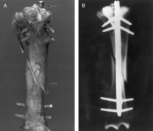
Compression constructs had overriding of the osteotomy fragments, extrusion of the nail at the insertion site, bending of the proximal and distal screws, comminution of bone at the osteotomy, and bone failure around the screws (Fig 2).

(A) Photograph of the cranial aspect of an EIN-stabilized osteotomized tibia tested to failure in axial compression. The nail extruded from the insertion site (large arrow). Note the bone fragment from the distal aspect of the proximal fragment at the osteotomy (small arrow) and comminution of the bone around the proximal screw holes (arrowhead). (B) Craniocaudal radiograph of an EIN-stabilized osteotomized tibia tested in axial compression. The proximal and distal screws are bent; however, the nail has not undergone permanent deformation.
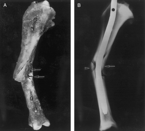
Three- and four-point bending constructs had residual nail deformation (Fig 3). There was no deformation of the screws or bone failure associated with the screws. Two 3-point bending and all 4-point bending tibiae had bone comminution at the osteotomy.

(A) Photograph of the medial aspect of an EIN-stabilized osteotomized tibia representative of the failure configuration under 3-point and 4-point bending. The nail is bent near the level of the central load point(s). The screws did not have residual deformation. Bone of both the proximal and distal fragments was comminuted adjacent to the osteotomy (arrows). (B) Mediolateral radiograph of an EIN-stabilized osteotomized tibia representative of the failure configuration under 3-point and 4-point bending. The nail is bent (arrow) near the level of the central load point(s). Bone is comminuted at the osteotomy site (arrowhead).
Torsion constructs had residual nail rotation, screw deformation, opening of the osteotomy gap, and bone failure at the osteotomy. Permanent deformation of the nail and screws was minor (Fig 4). The screws were bent in a craniocaudal direction. Two constructs had bone failure associated with distal screw holes.

(A) Photograph of the medial aspect of an EIN-stabilized osteotomized tibia tested in torsion. The proximal and distal screws are in different planes (arrows), indicating permanent rotational deformation of the implant. The screws were minimally bent (insert). Note the comminution of the caudomedial cortex at the osteotomy site involving the distal aspect of the proximal portion of the tibia (arrowhead). (B) Craniocaudal radiograph of an EIN-stabilized osteotomized tibia tested in torsion. Although the both the nail and screws had residual deformation, the implant has not failed catastrophically. The bone is comminuted at the osteotomy site (arrow), and the osteotomy gap has widened (arrowhead).
Mechanical Testing Variables
The mean (±SD) craniocaudal radius [(ro+ ri)/2] and polar moment of inertia (J) for the mid-diaphysis were 1.4 ± 0.06 cm and 9.0 ± 3.0 × 10⁻⁷ m4, respectively. The estimated torsional load, for an adult equine tibia was 350 Nm. Considerable plastic deformation occurred at the completion of all tests with mean yield loads ranging from 51% to 88% of failure loads and 49% to 60% of ultimate failure loads (Table 1). There was residual strength after failure in the constructs for all loading conditions.
| Variable/Group | Compression (n = 3) | 3-Point Bending (n = 3) | 4-Point Bending (n = 2) | Torsion (n = 3) |
|---|---|---|---|---|
| Yield load (kN, Nm)* | 11.4 ± 0.84 | NA | NA | 156.0 ± 48.8 |
| Failure load (kN, Nm) | 19.6 ± 1.3 | NA | NA | 191.0 ± 34.7 |
| Ultimate failure load (kN, Nm) | 22.3 ± 3.5 | NA | NA | 261.3 ± 41.4 |
| Yield bending moment (Nm) | NA | 186.5 ± 2.82 | 216.1 ± 1.0 | NA |
| Failure bending moment (Nm) | NA | 362.29 ± 24.0 | 410.2 ± 129.0 | NA |
| Ultimate failure bending moment (Nm) | NA | 371.54 ± 11.4 | 469.0 ± 123.1 | NA |
| Yield displacement (mm, °)† | 2.2 ± 0.7 | 5.83 ± 0.4 | 3.9 ± 0.0 | 11.4 ± 3.4 |
| Failure displacement (mm, °) | 6.8 ± 5.0 | 26.7 ± 8.3 | 19.3 ± 8.9 | 17.7 ± 9.1 |
| Ultimate failure displacement (mm, °) | 14.8 ± 4.5 | 31.9 ± 0.7 | 21.6 ± 12.2 | 34.9 ± 2.4 |
| Stiffness (kNm/mm, Nm/mm, Nm/°)† | 5.4 ± 3.0 | 41.5 ± 2.83 | 50.2 ± 7.6 | 13.4 ± 1.8 |
| Yield energy (kN ṁ mm, kNmm ṁ mm, Nm ṁ°)† | 9.2 ± 2.7 | 251.1 ± 109.8 | 458.9 ± 328.3 | 850.3 ± 422.1 |
| Failure energy (kN ṁ mm, kNmm ṁ mm, Nm ṁ°) | 67.7 ± 69.5 | 4,557.4 ± 1,507.3 | 4,038.9 ± 2,324.0 | 1,723.3 ± 1,212.9 |
| Ultimate failure energy (kN ṁ mm, kNmm ṁ mm, Nm ṁ°) | 211.7 ± 46.1 | 5,790.1 ± 816.2 | 5,271.4 ± 4,067.0 | 4,707.3 ± 110.8 |
- Abbreviation: NA, not applicable.
- *Units are given for compression and torsion, respectively.
- †Units are given for compression, 3-point bending and 4-point bending, and torsion, respectively
Discussion
The compressive and bending strengths of the EIN were greater than the strengths required to maintain stability during postoperative standing, and walking, in the adult horse.17 The torsional yield strength was less than that calculated for torsional stability in vivo, using combined in vitro and in vivo data.8
The long oblique diaphyseal osteotomy is representative of clinical tibial fracture configurations.28 Stability was evaluated with uniaxial compressive, bending, and torsional tests to simulate the different forces that act on the tibiae in vivo.17,24 Loading rates approximated loading at a walk, because mechanical properties of viscoelastic structures are load-rate–dependent.29,30 Monotonic tests simulated a single step.
Ideally, in vitro structural properties of implant-stabilized tibiae must exceed expected in vivo loads. Although in vivo strains occurring on the adult equine tibia have been measured,23,24 in vivo forces experienced by the adult equine tibiae have not been directly measured. Single estimates of compressive loads and bending moments have been calculated from in vivo strain gauge data with beam-bending theory.17 These estimates were used for comparison with the forces withstood by EIN-stabilized osteotomized tibiae.
The EIN-stabilized tibiae had adequate strength (Table 1) to withstand in vivo compressive loads during walking (0.5 kN) and trotting (3 kN)17; bending moments during standing (98 Nm) and walking (44 Nm)17; and bending moments (106 Nm) but not compressive loads (12 kN) during recovery from anesthesia.17 We chose to be conservative for our comparisons with in vivo data because loads equivalent to yield, the point at which permanent deformation is initiated,31 would likely result in instability and failure with repetitive loading. Because fatigue can also contribute to bone and implant failure,32 monotonic structural properties do not provide a complete assessment of the mechanical behavior critical for the entire healing period. Cyclic testing would provide information on the fatigue life of the nail-bone composite under repetitive loading conditions.
Previously, in vivo torsional load for the adult equine tibia at a walk (211 Nm) was estimated8 using in vivo strain gauge data24 and in vitro strain-torque curves. In an attempt to increase our confidence in this estimate, an alternate calculation, similar to that for compression and bending,17 using in vivo rosette strain gauge data24 and the torsional formula for a cylinder33 was performed, which yielded 350-Nm torque. Mathematical calculations of forces include assumptions about the structure and its material, which include: material homogeneity and isotropy, and symmetrical geometry. Bone is heterogeneous, anisotropic, and unsymmetrical. Mathematical calculations of forces occurring in long bones17,33,34 and other biological structures may be valid, but limitations must be recognized. Focusing on the mid-diaphysis where bone is fairly symmettrical may increase tolerance for these assumptions.34 The torsional or shear strain reported for the tibiae in vivo24 is a cumulative shear strain produced by bending and torsional loading configurations.18 Our calculations using this cumulative strain likely overestimate the in vivo torsional load to which our in vitro data are compared. Both estimated torsional values exceeded the in vitro torsional yield loads for EIN-stabilized tibiae indicating that torsional moments encountered by the adult equine tibia may not be resisted with this method of repair. However, considering limitations inherent in the calculation and variability between estimates of in vivo torsional loads, it is difficult to make an accurate conclusion about in vivo torsional performance of the EIN.
An EIN-stabilized tibia is likely to withstand immediate postoperative compressive and bending loads in adult horses during standing and walking, and during recovery from anesthesia in bending. Because the compressive yield load approximated the compressive load expected during conventional recovery from anesthesia, alternative recovery methods using slings or pools, which are expected to decrease forces on the limbs, may need to be considered.38 The mean torsional strength of EIN-stabilized tibiae was not sufficient to withstand estimated torsional loads during walking, indicating that the nail may fail under torsional loads in vivo.
Further enhancements in nail design to improve torsional strength and stiffness appear warranted before the EIN can be considered a feasible method of repair for adult equine tibial fractures. Considering the adequacy of compressive and bending mechanical properties of the EIN, reported biological advantages of interlocking nails for fracture repair, and successful healing in osteotomized foal femora repaired with a femoral intramedullary interlocking nail,10 further structural modification for use in adult equine fracture repair appears worthy of pursuit.
Acknowledgment
Special thanks to Steve Gage and Burt Vannucci for technical assistance, and Neil Willits, PhD, for statistical advice.


