Vanishing bile duct syndrome in a patient with advanced AIDS
Abstract
A 39-year-old HIV-infected woman developed signs and symptoms of obstructive jaundice and cholestasis. Serological tests were positive for cytomegalovirus (CMV) infection. There was no evidence of AIDS cholangiopathy in ultrasonography or magnetic resonance cholangiopancreatography (MRCP). A liver biopsy revealed marked ductopenia and the patient was diagnosed with vanishing bile duct syndrome, thought to be secondary to CMV infection as a result of profound immunosuppression. To the best of our knowledge, this is the first reported case of vanishing bile duct syndrome diagnosed in a patient with HIV/AIDS.
Case report
A 39-year-old woman presented with a complaint of fatigue, anorexia, nausea and vomiting, of duration 3 days. She also reported a 1-week history of scleral icterus, pruritis and abdominal pain. The nausea and vomiting were described as nonbilious and nonbloody, occurring three times a day, while the abdominal pain was localized to the right upper quadrant without radiation or association with meals. Past medical history was significant for HIV infection and hepatitis C virus (HCV) infection diagnosed in 1997. She was not taking any medication, including highly active antiretroviral therapy (HAART) or treatment for HCV infection, because of a history of polysubstance abuse of cocaine, alcohol and intravenous drugs. Her last documented CD4 cell count and quantitative HIV-1 RNA viral load 7 months prior to admission were 22 cells/μL (3%) and 48 026 HIV RNA copies/mL, respectively. Her weight was 43 kg and all initial vital signs were normal.
Physical examination was significant for cachexia and generalized icterus. The right upper quadrant was tender to palpation without rebound tenderness or guarding. There were no masses palpated and no evidence of ascites. Murphy's sign was negative. The liver span was measured as 5 cm. No cutaneous manifestations of chronic liver disease were noted. Initial chemistry values were significant for the following: albumin=2.9 g/dL, total bilirubin=15.4 mg/dL, direct bilirubin=11.0 mg/dL, alkaline phosphatase=2200 IU/L, aspartate transaminase=114 IU/L and alanine transaminase=29 IU/L. The partial thromboplastin time and international normalized ratio were 39 s and 1.4, respectively. Urine analysis revealed proteinuria and bilirubinuria while the CD4 cell count and HIV-1 RNA viral load were measured as 7 cells/μL (2%) and 721 000 copies/mL, respectively. HCV RNA measured by quantitative PCR was >5 000 000 IU/mL, while the a-fetoprotein level was <5 ng/mL. Other haematological values were not significant and ethanol and urine drug screens were negative.
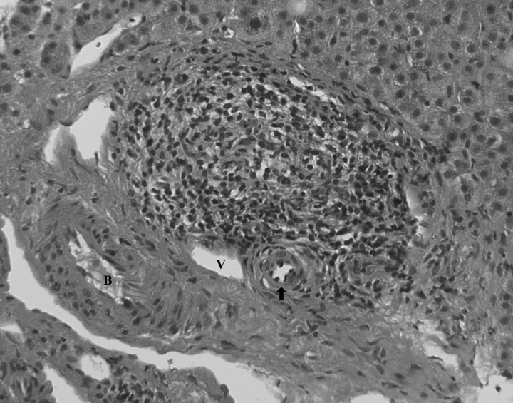
The patient's abdominal complaints did not change during the course of her admission and an abdominal ultrasound revealed no evidence of cirrhosis, focal liver lesions, intrahepatic duct dilatation or hepatic vein thrombosis. The common bile duct was measured and found to be normal. Fungal, mycobacterium, aerobic and anaerobic blood cultures as well as tests for Cryptosporidium parvum, Microsporidium and Toxoplasma were all negative. Other negative serological tests included those for hepatitis A and B, cryoglobulins, and antimitochondrial (AMA) and antinuclear antibodies (ANA). A cytomegalovirus (CMV) PCR and qualitative immunoglobulin G (IgG) antibody test were positive. A magnetic resonance cholangiopancreatography (MRCP) was performed to test for intrahepatic cholestasis and HIV cholangiopathy. There was no cholangiographic evidence of sclerosing cholangitis, discrete liver masses or extra- or intrahepatic biliary duct dilatation. A needle biopsy of the liver was performed which revealed marked ductopenia; sections of the liver showed 11 portal tracts, two of which contained bile ducts (Fig. 1). Accompanying granulomatous inflammation was also noted, and a diagnosis of vanishing bile duct syndrome was made.

Liver biopsy revealing granulomatous inflammation accompanying a portal triad consisting of a bile duct (B) and branches of a portal vein (V) and hepatic artery (arrow).
As the patient was not considered to be a transplant candidate, she declined further therapy. Her bilirubin level peaked at 36.7 mg/dL while her liver function continued to decline. One month following her initial presentation, she died from complications of liver failure.
Discussion
Vanishing bile duct syndrome (VBDS) is an infrequent cause of progressive cholestasis as a result of progressive loss of small and medium-sized intrahepatic bile ducts [1]. The condition, also termed ‘idiopathic adulthood ductopenia’, was first described in 1988 as the absence of interlobular bile ducts in at least 50% of small portal tracts, and is not associated with an underlying cause [2].
While the specific mechanism for VBDS is unknown, it has been postulated that VBDS may be immune-mediated. It has been suggested that immunological mechanisms such as the release of toxic cytokines in conditions such as primary biliary cirrhosis (PBC), primary sclerosing cholangitis (PSC) and transplant rejection play important roles in biliary destruction [3,4]. Studies have reported abnormal HLA molecules expressed on the biliary epithelium in cases of graft-versus-host disease, PBC and various other viral infections, all of which have been associated with VBDS [5–8]. CMV infection has been associated with biliary ductal destruction and is the most common viral cause of VBDS [8]. Another possible aetiology may be drug related, as more than 30 pharmocotherapies have been reported as aetiological factors in VBDS, the most frequent of which include antibiotics and psychiatric medications [9].
While the most common causes of cholestasis in patients with HIV/AIDS include AIDS cholangiopathy and sclerosing cholangitis, these were not considered to be aetiologies in this case. While AIDS cholangiopathy is a syndrome of biliary duct obstruction caused by infection-related strictures, no strictures were noted from the ultrasound or MRCP. In addition, with regard to diagnosing AIDS cholangiopathy, ultrasonography has a sensitivity and specificity of 75–97% and nearly 100%, respectively [10]. The ultrasound performed revealed no focal hepatic lesions or intrahepatic duct dilatation and normal biliary duct measurements. The diagnoses of AIDS cholangiopathy and sclerosing cholangitis are made via MRCP or endoscopic retrograde cholangio pancreatography. There was no cholangiographic proof of intra- or extrahepatic biliary ductal dilatation, strictures or irregular narrowing of the ducts, features characteristic of both diseases. Sclerosing cholangitis classically involves histopathological evidence of concentric periductal fibrosis, while periportal inflammation and necrosis, ductal proliferation and fibrosis are seen in PBC. None of these findings was noted in this presentation. Given the marked ductopenia noted from the liver biopsy and unremarkable MRCP, we felt that the most likely aetiology of the patient's cholestasis was VBDS secondary to CMV infection from profound immunosuppression. However, other aetiological possibilities should be considered. The patient suffered from chronic HCV infection, and this infection has been shown rarely to cause ductopenia and it is possible that it may have been responsible for the biopsy findings [11]. It is also possible that the patient suffered from idiopathic adulthood ductopenia unrelated to her HIV infection, in which case it may be impossible to distinguish the ductopenia from these two causes [12].
In addition to cases of liver rejection following transplantation, PBC and PSC, VBDS has also been reported in cases of Hodgkin's disease, hystiocytosis X, and adult non-Hodgkin's lymphoma [1,3,4,13,14]. To the best of our knowledge, the above presentation is the first reported case of VBDS in a patient with HIV or AIDS and should be considered as a potential aetiology for chronic intrahepatic cholestasis in such patients. Nearly all cases of VBDS require liver transplantation, as the syndrome leads to irreversible liver damage.


