The first spontaneous spawning of European hake Merluccius merluccius L.: characteristics of eggs and early larval stages
Abstract
For the first time, a spontaneous spawning of hake was recorded in Spain in April 2009. The spawn was obtained from broodstock kept in captivity for two years at the facilities of the Spanish Institute of Oceanography in Vigo (NW Spain). Eggs were transparent, spherical and had an average diameter of 1.067 ± 0.024 mm; yolk occupied the majority of egg volume. The oil droplet had a diameter of 0.27 ± 0.03 mm. The incubation period of the eggs lasted for 4 days at 14°C and the duration from hatching to the total absorption of the yolk sac was between 5–7 days after hatching, at the same temperature. Newly hatched larvae had an average total length of 3.20 ± 0.13 mm and began feeding 6 days after hatching; a daily growth rate of 0.158 mm day-1 was observed from hatching to yolk sac consumption. This paper describes the daily evolution of biometric and morphological characteristics of the different stages of embryos and larvae of European hake up to the age of 19 days.
Interest has recently increased in the cultivation of hake on an industrial scale. The attempt to grow hake in captivity has been reported in Chile using Merluccius australis (Hutton, 1872) albeit with limited success (Bustos & Landaeta 2005). Regarding the European hake, larval rearing experiments were carried out using stripped eggs from wild specimens in Norway, reporting the growth of juveniles up to 250 days (Bjelland & Skiftesvik 2006). In Spain, Iglesias, Lago, Sánchez and Cal (2010) described the conditions of capture, transport and conditioning, as well as the diets used and the first data on growth of adult hakes in captivity.
In previous studies (Coombs & Mitchell 1982; Marrale, Alvarez & Motos 1996), the embryonic development and yolk sac larvae from wild-caught population have been described. In our work, a spontaneous spawning obtained from a hake broodstock kept in captivity for 2 years is reported for the first time in Europe. The morphological features and biometric changes of the early development stages (eggs and larvae) of hake as well as the first attempt of larval rearing of this species in Spain are described.
Broodstock source and conditioning
Hake broodstock were obtained from fish caught in the Ría de Vigo (NW Spain) during the years 2007 and 2008 following the methodology described by Iglesias et al. (2010). In the laboratory, 50 male and female hakes ranging from 34 to 47 cm in length were kept together in an 8 m3 tank with an open water system at a flow of 500 L h−1. Water temperature and salinity ranged between 13 and 14°C and from 32 to 34 g L−1 respectively. Fish were maintained on a natural photoperiod and low light intensity and they were not sampled to avoid disturbing their behaviour.
Hake were fed with live sand eel (Ammodytes sp.) for the first weeks and frozen sardine (Sardina pilchardus) later on. After 2 months, a semi-moist feed consisting of fish flour (35%), fish (30%), squid (17%), mussel (18%) and vitamin premix (6 mg kg−1), was supplied every 2 days ad libitum.
Spawning and incubation
The first spontaneous spawn was registered in the morning of 2 April 2009, between 09.00 and 11.00 hours. A volume of 60 mL of floating eggs (n = 52 000) was collected from the outflow of the broodstock tank. The number of eggs per mL was estimated by counting the eggs in five replicates of 1 mL. Eggs were transparent, spherical and had a diameter of 1.067 ± 0.024 mm. They had a single golden oil droplet of 0.24 mm in diameter. The floating eggs were introduced into a 150-L incubator connected to an open water circuit (2.5 L min−1) maintained at 14°C. Hake eggs have a hydrofuge nature and tend to float exposing part of their surface above the water level (Coombs & Mitchell 1982). To avoid this, the water inlet (2.5 L min−1) flowed through a horizontal pipe of 4 cm in diameter, perforated with holes of 1 mm, which caused the water to trickle out obliquely. This flow moved the eggs gently down from the surface of the water and caused a slight rotation around the central outlet. Bustos, Landaeta, Bay-Schmith, Lewis and Moraga (2007), used a similar system placed vertically inside the incubation tank.
Embryonic development
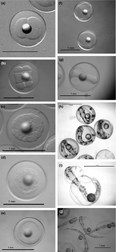
Eggs harvested with two mitotic divisions were estimated to be fertilized 2 h previously, according to Marrale et al. (1996) (Fig. 1a). In a period of 2–5 h, the first mitotic divisions were observed (Fig. 1b and c). Morula appeared 5 h after fertilization and exhibited a finely segmented yolk (Fig. 1d). Twenty-four hours later (at 14°C), the blastula was formed with the blastomeres observed at the animal pole (Fig. 1e). Two hours later, the yolk was covered by blastomeres and finally the gastrula stage was attained (Fig. 1f).

After 48 h, the embryo was observed and somites became evident, the head was formed and optic calyx differentiated (Fig. 1g). The onset of heart beat and notochord formation were also observed. Soon after (50 h), pigmentation of the embryo was noted. At 72 h, the larval tail occupied 75% of the length of egg circumference (Fig. 1h); chromatophores in the head and tail had proliferated and otoliths were clearly distinguished. Four hours later, the tail was longer than the total egg perimeter, and the retina formation was initiated. At 80 h post-fertilization, muscle contractions were observed, and hatching began 3.5 days after fertilization (Fig. 1i). At 90 h, 50% of the eggs had hatched and 6 h later the remaining 50% did so (Fig. 1j). Larvae hatched at a mean total length of 3.20 ± 0.13 mm.
Our data on eggs diameter and TL of hatched larvae agree with Russell (1976), who reported a range of 0.94–1.03 mm for egg diameter and a newly hatched larval total length of about 3.0 mm. Moreover, Bjelland and Skiftesvik (2006) reported an egg diameter at fertilization of 1.07 ± 0.01 mm and a larval total length of 3.0 mm. Bustos and Landaeta (2005) described M. australis egg diameters between 0.9 and 1.1 mm and a standard length of 2.8 mm for newly hatched larvae.
The incubation period of 4 days at 14°C is in accordance with Bjelland and Skiftesvik (2006), who reported a value of 4 days at 14.5°C.
Early larval stages
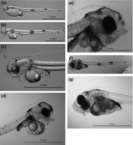
Newly hatched larvae exhibited a free, passive swimming behaviour and three typical dark pigmented vertical bands on the post-anal area (Fig. 2a). The dorsal area of the head and the oil droplet were also pigmented, showing dendritic chromatophores. The mouth is not functional at this stage. One day after hatching (1 DAH), larvae reared at 14°C were very similar to newly hatched larvae (Fig. 2b), adopting a vertical position in the water column and remaining quiet most of time. At 2 DAH, larvae swam freely in the water column, yolk sac length decreased considerably (Table 1) and eyes showed high differentiation with initial stages of pigmentation. A short digestive tract was observed. At 3 DAH, the caudal fin began to differentiate and the first sketch of lower jaw appeared. Eyes darkened and the dorsal part of the digestive tract was spotted with melanophores (Fig. 2c).

| Age | N | TL (mm) | SL (mm) | DW (mg) | OD Ø (mm) | YS L (mm) |
|---|---|---|---|---|---|---|
| 0 | 19 | 3.20 ± 0.13 | 3.05 ± 0.13 | 0.064 ± 0.01 | 0.27 ± 0.003 | 1.05 ± 0.08 |
| 1 | 10 | 3.52 ± 0.23 | 3.39 ± 0.23 | 0.074 ± 0.01 | 0,25 ± 0.010 | 1.03 ± 0.06 |
| 2 | 10 | 3.78 ± 0.25 | 3.63 ± 0.25 | 0.082 ± 0.01 | 0.24 ± 0.007 | 0.79 ± 0.07 |
| 3 | 10 | 4.08 ± 0.12 | 3.93 ± 0.12 | 0.103 ± 0.02 | 0.22 ± 0.012 | 0.60 ± 0.06 |
| 4 | 10 | 4.11 ± 0.11 | 3.98 ± 0.12 | 0.104 ± 0.02 | 0.19 ± 0.013 | 0.54 ± 0.06 |
| 5 | 10 | 4.14 ± 0.11 | 4.01 ± 0.09 | — | 0.17 ± 0.009 | 0.44 ± 0.06 |
| 6 | 20 | 4.15 ± 0.08 | 4.00 ± 0.09 | 0.107 ± 0.02 | 0.14 ± 0.010 | 0.33 ± 0.03 |
- Age in days; N, sample size; TL, total length; SL, standard length; DW, dry weight; OD, oil drop; YS L, yolk sac length.
In larvae at 4 DAH, the eyes were completely darkened and the notochord was also pigmented. Mouth opening first occurred at 5 DAH (Fig. 2d), and at 6 DAH, the mouth differentiated into upper and lower jaws and the larvae began to feed exogenously (Fig. 2e). At 7 DAH, larvae already had a functional gastrointestinal tract, the pectoral fins and swim bladder were well differentiated, and absorption of oil droplet continued (Fig. 2f). Larvae were now capable of attacking prey with quick and precise movements.
The biometric characteristics of hake larvae from hatching to total yolk sac absorption are shown in Table 1. Larvae showed a gradual increase in size from 3.20 ± 0.13 mm (day 0) to 4.15 ± 0.08 mm (6 DAH). During the same period, the yolk sac gradually decreased until total absorption (7 DAH), while eye diameter did not vary substantially. Larvae dry weight varied from 0.06 to 0.11 mg from hatching to yolk sac absorption. Total yolk sac absorption between 5 and 7 DAH observed in the present study agrees with values reported by Bjelland and Skiftesvik (2006), as well as the age at start-feeding (6 DAH). Bustos and Landaeta (2005) indicated 9 days for the complete yolk sac consumption in M. australis larvae cultured at 11.5°C and a larval growth of 0.153 mm day−1 similar to our data (0.158 mm day−1).
Larval rearing
Six DAH larvae were transferred to a 500-L rearing tank connected to a stagnant green water system, where one-third of the tanks volume was replaced every 2 days. Temperature, salinity, oxygen concentration and prey density were recorded daily. Initial larval density was 10 L−1 and they were fed the rotifer at a concentration of 2 rotifers mL−1, previously enriched with the microalgae Isochrysis galbana which was maintained at a concentration of 150 000 cells mL−1 in the rearing tank. Larvae were reared at 14°C until day 19 (Fig. 2g), when they attained a total length of 4.81 ± 0.41 mm (4.58 ± 0.53 mm standard length) and 0.146 ± 0.60 mg (dry weight). Bjelland (2001) reported a standard length of 4.5 mm for 24-day-old larvae reared at 12.5°C.
Growth from hatching until yolk sac absorption was 0.158 mm day−1. The growth equation was: length = 3.2194 age0.1443.
At day 19, an unexpected increase in water temperature up to 16.5°C caused the death of all larvae. This fact could indicate a low tolerance for rearing hake at temperatures over 15°C, but this assumption requires further confirmation.
In conclusion, after demonstrating that it is possible to obtain spontaneous spawning in captivity, and having described the morphological characteristics of eggs and larvae of European hake, it is time to start the next phase of research: the larval culture.


