Individual combinations of danger signals synergistically increase FVIII product immunogenicity
Abstract
Introduction
The most severe side effect in haemophilia A treatment is the development of antifactor VIII antibodies, also called inhibitors. Why inhibitors develop in a proportion of treated patients while others are unaffected still remains unanswered. The presence of immunological danger signals, associated with events such as infection or surgery, has been proposed to play a role. Previous studies demonstrated that the presence of the bacterial molecule lipopolysaccharide (LPS) can synergistically increase the activation of human DC and subsequent T cell activation by FVIII.
Aim and Methods
In the present study, we investigated whether a combination of two danger signals can further increase immune cell activation by FVIII. For this, human in vitro differentiated DC that were treated with combinations of danger signals were co-cultured with autologous primary T cells, and T cell proliferation was analysed.
Results
Interestingly, by combining LPS with a second danger signal, lower LPS concentrations were sufficient to synergistically increase DC and subsequent T cell activation by FVIII. Of note, a combination of LPS and the double-stranded RNA, polyinosinic-polycytidylic acid (poly(I:C)), was most potent in increasing FVIII immunogenicity, followed by LPS + R848 (resiquimod). However, a combination of LPS and the bacterial lipopeptide Pam3CysSK4 did not induce increased immune cell activation by FVIII.
Conclusion
Thus, individual combinations of danger signals can increase FVIII product immunogenicity. This should be considered in the treatment routine of haemophilia A patients.
1 INTRODUCTION
Haemophilia A (HA) is a hereditary bleeding disorder caused by deficiency of the coagulation protein factor VIII (FVIII).1 To control bleedings, HA patients are treated with intravenous infusion of FVIII products. Though the treatment is successful in most cases, 25%-30% of patients with severe HA develop antidrug antibodies against the infused FVIII, also called inhibitors.2 Why inhibitors develop in a proportion of treated patients while others are unaffected still remains unanswered.3 The most important risk factor for inhibitor development is the severity of FVIII deficiency.4 However, not all patients with large deletions develop inhibitors, and inhibitors can also be observed in HA patients with only minor changes in FVIII. Mechanisms leading to these discrepancies are not yet understood.3 Notably, frequent and high-dose treatments with FVIII products during periods of surgery or severe bleeds were associated with more frequent inhibitor development.5 Consequently, it has been hypothesized that immunological danger signals associated with events such as infections or periods of intensive bleedings could promote inhibitor development.5
Immunological danger signals include conserved pathogen-associated molecular patterns (PAMPs) that are recognized by certain pattern recognition receptors (PRRs). PRRs include transmembrane toll-like receptors (TLRs) or cytoplasmic proteins such as NOD-like receptors (NLRs) or RIG-I-like helicases (RLHs).6 Ligation of PRRs triggers dendritic cell (DC) maturation and cytokine production, thus effectively bridging innate and adaptive immunity.7 Ten human TLRs (TLR1-10) have been identified so far.6 Individual TLRs recognize distinct structural components of pathogens, e.g. TLR4 recognizes lipopolysaccharide (LPS), a membrane component of Gram-negative bacteria, a heterodimer consisting of TLR1 and TLR2 recognizes bacterial lipopeptides like Pam3CysSK4, TLR3 recognizes double-stranded RNA and R848 (also known as resiquimod) is recognized by TLR7/8.7 Until recently, the impact of immune cell activation by FVIII in the context of concurrent TLR triggering was unknown. We demonstrated that stimulation of human DC with FVIII products in combination with LPS induces stronger DC activation than each of the substances alone.8 We further demonstrated that addition of autologous T cells to these synergistically activated DC resulted in explicitly stronger proliferative responses of CD4+ T cells.9 Thus, the presence of only one danger signal effectively increases the innate and adaptive immune response towards FVIII.
However, under real-life conditions, HA patients usually get confronted with a variety of different danger signals instead of only one. What effects a combination of danger signals might have on the immunogenicity of infused FVIII products remains to be elucidated.
In our current study, we investigated the impact of a combined activation of TLR4 (LPS) and TLR3 (poly(I:C)), TLR4 (LPS) and TLR1/2 (Pam3CysSK4) or TLR4 (LPS) and TLR7/8 (R848) on the activation of human DC and T cells by a FVIII product. To assess DC activation, the expression of co-stimulatory molecules CD83 and CD86 was determined. For investigation of T cell activation, proliferation of autologous T cells upon addition to co-treated DC was determined.
2 MATERIALS AND METHODS
2.1 Monocyte and T cell purification
Peripheral blood mononuclear cells (PBMC) were isolated from commercially available buffy coats (Blutspendedienst) of healthy donors by density gradient centrifugation with Ficoll (Biochrom). The age of donors ranged from 18 to 65 years; male and female donors were included. Monocytes were purified from freshly isolated PBMC by positive selection using CD14 MicroBeads (Miltenyi Biotech) according to manufacturer's instructions. T cells were isolated from thawed PBMC by negative selection using Pan T cell Isolation Kit (Miltenyi Biotech).
2.2 DC assay
For DC generation, freshly isolated CD14+ monocytes were cultured in 96-well flat bottom tissue culture plates (2 × 10⁵ cells in 200 µL; Sarstedt) using X-Vivo 15 medium (Lonza) in the presence of GM-CSF and IL-4 (both at 1000 U/mL; CellGenix) for 5 days. Resulting immature DC were treated with the following: (a) 1 U/mL plasma-derived FVIII concentrate + LPS (0.01 or 0.001 µg/mL as indicated; Salmonella abortus equi; Sigma), (b) LPS + poly(I:C) (HMW; 0.5-1 µg/mL; Biomol), (c) FVIII + LPS + poly(I:C), (d) LPS + R848 (resiquimod; InvivoGen), (e) FVIII + LPS + R848, (f) LPS + Pam3CysSK4 (EMC microcollections), (g) FVIII + LPS + Pam3CysSK4 for 24 hours at 37°C. Untreated cells and cells treated with each of the substances alone (FVIII, LPS, poly(I:C), or R848) served as controls. Next day after stimulation, DC were harvested by up and down pipetting, subsequent addition of cold PBS w/o Ca2+ and Mg2+ with 1 mmol/L EDTA, and up and down pipetting again. Harvested cells were processed for flow cytometric analysis. Beside the commercially available LPS (Salmonella abortus equi, Sigma, which can contain up to 60% contaminating RNA), DC were also treated with the highly purified (HP) LPS (kind gift from Ulrich Zähringer, Research Center Borstel).
2.3 DC:T cell co-culture assay
For DC generation, CD14+ monocytes were cultured in 24-well tissue culture plates (106 cells/mL; Sarstedt) using X-Vivo 15 medium in the presence of GM-CSF and IL-4 as described above. Resulting DC were harvested for DC and T cell co-culture by repeated up and down pipetting, subsequent addition of cold PBS w/o Ca2+ and Mg2+ with 1 mmol/L EDTA, and up and down pipetting again. Harvested DC were resuspended in X-Vivo 15 medium to 5 × 10⁵/mL and serially diluted in 1:2 steps resulting in 2.5 × 10⁴, 1.25 × 10⁴, 0.6 × 10⁴ and 0.3 × 10⁴ DC/well in 100 µL in 96-well round bottom tissue culture plates. For every indicated treatment, serially diluted DC were used. DC for DC:T cell co-culture assay were treated as described for DC assay. 24 hours after DC treatment, isolated autologous pan T cells were stained with PKH26 according to manufacturer's instructions (Sigma) and resuspended in X-Vivo 15 medium (Lonza) to 106/mL followed by addition of 100 µL of this T cell suspension to the wells with pretreated DC. After 9 days of co-culture, cells were harvested and processed for flow cytometric analysis. For data analysis, the highest proliferation rate in the FVIII + LPS + second danger signal (poly(I:C) or R848) sample was identified and subsequently, for all control samples, proliferation rates from the same DC dilution step were used.
2.4 Flow cytometric analysis
For flow cytometric analysis, harvested DC from the DC assay were stained for 15 min at 4°C using the following monoclonal antibodies diluted to their optimal concentration: anti-CD83-FITC (clone HB1e) or anti-CD83-APC (clone HB15e, both from BD Pharmingen), and anti-CD86-PE (clone IT2.2, Biolegend). Harvested cells from DC:T cell co-culture assay were resuspended in buffer, and T cell proliferation was analysed as the decrease in PKH26 fluorescence intensity.
All cells were analysed using LSR II with BD FACS DIVA version 6.1.3 (BD Biosciences) and FlowJo version 7.6.5 software.
2.5 Statistical analysis
All statistical analyses were performed with GraphPad Prism, version 7.01. Statistical analyses of all data were performed using one-way ANOVA for paired data and paired t tests with Bonferroni correction for multiple pairwise comparisons. The means of following preselected pairs were compared: (a) FVIII + LPS vs FVIII + LPS + poly(I:C) and LPS + poly(I:C) vs FVIII + LPS + poly(I:C) or (b) FVIII + LPS vs FVIII + LPS + R848 and LPS + R848 vs FVIII + LPS + R848. For statistically significant results, following convention was used: *, P < .05; **, P < .01; ***, P < .001; ****, P < .0001.
3 RESULTS
3.1 Addition of a second danger signal increases LPS-dependent activation of human DC by FVIII
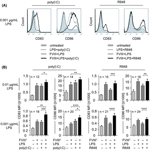
To investigate the effect of a second danger signal on the LPS-dependent activation of human DC by FVIII, we first titrated LPS and the second danger signal, namely poly(I:C) or R848 on DC in the absence and the presence of FVIII (data not shown). Interestingly, we observed that as soon as LPS is combined with a second danger signal, 10- to 100-fold lower concentrations of LPS are sufficient to achieve a strong activation of human DC by co-incubation with FVIII (Figure 1). A combination of only 0.001 µg/mL LPS with 0.5-1 µg/mL poly(I:C) or with 0.1 µg/mL R848 + FVIII leads to an increased expression of CD83 and CD86 on DC (Figure 1A). For statistical analysis of data obtained from different donors, we compared the expression of CD83 and CD86 on FVIII + LPS + poly(I:C)-treated DC with FVIII + LPS-treated DC and with those treated with LPS + poly(I:C) without FVIII (Figure 1B). The same combinations were compared for R848-treated DC (Figure 1B). Surprisingly, the synergistic effect on DC activation induced by the addition of two danger signals and FVIII is more pronounced when the lower LPS concentration of 0.001 µg/mL is used (Figure 1B). This holds particularly true for poly(I:C). Of note, the combinations of FVIII and poly(I:C) or FVIII and R848 induced no or only minor DC activation (Figure S1A). Thus, the strongest activation of human DC is achieved by a combination of two danger signals in low concentrations plus FVIII. However, not every combination of two danger signals and FVIII resulted in increased DC activation. No increased activation was observed for DC treated with LPS + Pam3CysSK4 + FVIII (data not shown).

3.2 Synergistic activation of DC by two danger signals plus FVIII translates into a stronger T cell response
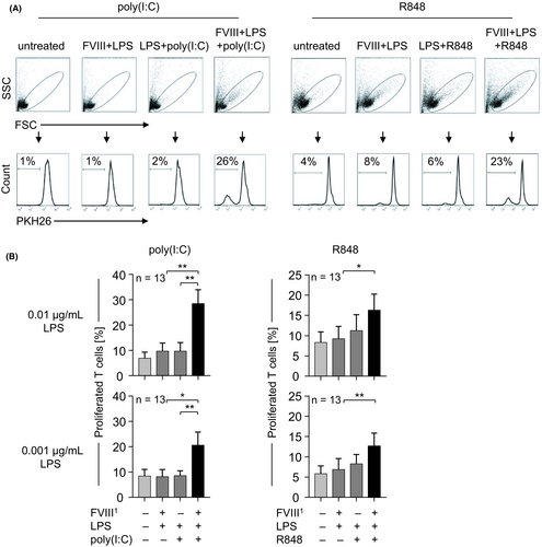
Proper activation of DC is prerequisite for T cell activation and thus initiation of an adaptive immune response. We next assessed whether the synergistic activation of DC by FVIII plus two danger signals translates into a stronger adaptive immune response, namely T cell proliferation. For that we used our previously established DC:T cell co-culture assay,9 in which autologous T cells are added to DC that were pretreated as described above and T cell proliferation is assessed upon co-culture (Figure 2). Low proliferation rates were observed for T cells added to untreated, FVIII + LPS-treated, and LPS + second danger signal-treated (poly(I:C) or R848) DC (Figure 2A and B). However, as soon as FVIII was combined with LPS and a second danger signal (poly(I:C) or R848) a strongly increased T cell proliferation was observed. This increased T cell proliferation was observed for both LPS concentrations, 0.01 and 0.001 µg/mL (Figure 2B). Thus, synergistic activation of human DC by low concentrations of two danger signals and FVIII translates into a stronger T cell response.

3.3 LPS, and not potential preparation impurities, is responsible for human DC and subsequent T cell activation by FVIII
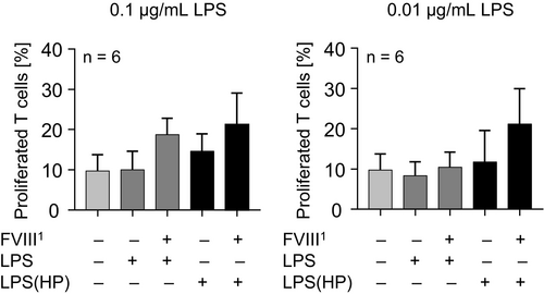
Here, and previously, we demonstrated that LPS can increase the activation of human DC and T cells by FVIII concentrates.8, 9 In these studies, we used a commercially available LPS preparation that, according to the manufacturer, can contain up to 60% contaminating RNA and up to 1% protein. These contaminating molecules may also target certain PRRs and thus induce cell activation, so that the effect of LPS on FVIII immunogenicity might not be clearly discerned. To investigate the role of LPS on FVIII concentrate immunogenicity, we performed a DC:T cell co-culture assay with the commercially available and a highly purified LPS (HP) (Figure 3). Notably, T cell proliferation rates observed for FVIII + LPS and FVIII + LPS(HP) samples did not differ significantly. In contrast, we observed a tendency that LPS(HP) induced even higher T cell proliferation rates than the conventional LPS did. Thus, we conclude that LPS itself, and not any impurities within the conventional LPS preparation is responsible for the observed increased activation of immune cells by FVIII.

4 DISCUSSION
We previously demonstrated that the concomitant presence of just one PRR ligand plus FVIII can substantially increase the immune response to FVIII. Of note, combined activation of different PRRs can result in further enhancement of innate and adaptive immune responses.10 Here we investigated whether targeting of two or more PRRs in the presence of FVIII would further enhance immune cell activation. Throughout our study, we investigated expression of CD83 and CD86 on DC since we previously showed that upregulation of these two markers correlates with the capability of DC to mediate proliferation of autologous T cells.8, 9 In turn, proliferation of autologous CD4+ T cells within the DC:T cell co-culture pinpoints towards the immunogenicity of antigen plus danger signal, since Miller et al 2018 provided first evidences for accumulation of FVIII-specific T cells upon LPS plus FVIII stimulation.9 We found that the most potent combination investigated was LPS + poly(I:C) + FVIII, inducing the strongest upregulation of the maturation marker CD83 and the co-stimulatory molecule CD86 on DC, followed by the combination of LPS + R848 + FVIII. This synergism was even more pronounced in the DC:T cell co-culture assay resulting in significantly increased T cell proliferation rates as soon as all three substances were present. Interestingly, targeting an extracellular TLR (TLR4) together with an intracellular or endosomal PRR in combination with FVIII resulted in the strongest immune cell activation (Figures 1 and 2). This is in line with previous publications reporting that combinations of ligands for cell surface TLRs with agonists for intracellularly located, nucleic acid-sensing TLRs enhance Th1-polarizing cytokine release from myeloid cells.11-13
In contrast, no increased expression of DC activation markers was observed for the combination of PRR ligands Pam3CysSK4 and LPS, targeting two extracellular TLRs (TLR1/2 and TLR4) (data not shown). This result is supported by previous findings that failed to observe synergistic action of TLR2 and TLR4 agonists 11 and, instead, rather observed cross-tolerance induction by either TLR2 or TLR4 ligands.14 Moreover, it was demonstrated that stimulation of human monocyte-derived DC with LPS + poly(I:C) or LPS + R848 resulted in the production of a different set of cytokines than with a combination of LPS + Pam3CysSK4.15 Of note, among detected cytokines upon LPS + Poly(I:C) or LPS + R848 treatment, the highest concentrations were detected for the cytokines IL-6, TNF-α and the Th1-polarizing cytokine IL-12p70.15 In line with this, it was previously demonstrated that induction of type I interferon (IFN) by LPS was detrimental for IL-12p70 synthesis and a Th1 response.11 Thus, the presence of bacterial PAMPs and type I interferon secretion, which are both observed in infection, represent important co-factors that mediate immunogenictiy of FVIIII.
Our findings indicate that enhancement of FVIII immunogenicity is mainly driven by simultaneous engagement of different classes of TLRs, thus mimicking the presence of bacterial pathogens containing cell wall-derived PAMPs and of viral pathogens containing viral nucleic acids.
Why some TLR combinations induce synergism while others do not, could result from differential integration of signalling pathways downstream of the respective PRR. Such differences could lie in differences in the quantitative induction of key adaptor molecules such as IRAK4, GSK3β and TIRAP/Mal, and in the PKB/Akt-mTOR or Erk.16-19
Of note, by the combination of two different danger signals plus FVIII, even lower concentrations of the danger signals, in particular LPS, were required to induce a synergistic DC activation and subsequent T cell activation. While we used an LPS concentration of 0.1 µg/mL in our previous studies,8, 9 10- to 100-fold lower concentrations were required as soon as LPS was combined with 0.1 µg/mL R848 or 0.5 µg/mL poly(I:C). The strongest synergism was observed only if LPS was present (Figure S1B). The combination of poly(I:C) and R848 with FVIII in absence of LPS resulted in less DC activation (Figure S1B). Bacterial endotoxins such as LPS are frequently detected in low amounts (1-15 pg/mL) in the plasma of healthy donors.20 These plasma endotoxin levels can increase in different clinical setting for example upon (sub-) clinical infections or in patients with a leaky-gut syndrome.21 We show here that under in vitro conditions a combination of 1000 pg/mL LPS plus a second danger signal and FVIII is sufficient to induce a strong DC and subsequent T cell activation. This situation might be even more pronounced in HA patients who are frequently exposed to endogenous damage-associated molecular patterns (DAMPs) from dying cells at the bleeding site in addition to those stimuli present in healthy human beings as well. Together with a viral infection and subsequent release of viral RNA and/or type I IFN release the patient could be predisposed to develop FVIII antibodies and FVIII-specific T cells. Of course, we are aware of the fact that DC and T cells of healthy donors might not entirely resemble the in vivo conditions within haemophilia A patients. However, to our knowledge, co-cultivation of monocyte-derived DC with primary autologous T cells is one of the best currently available in vitro models.
5 CONCLUSION
Taken together, our data suggest that a combination of different PRR ligands is even more potent in increasing FVIII immunogenicity than only one ligand. Combinations of ligands targeting extracellular and intracellular PRRs seem to be most effective. This could be of special interest for the treatment of HA patients suffering from a (subclinical) bacterial and/or viral infection. Also, in patients suffering from an extensive bleeding both extra- and intracellular PRRs would be triggered. To avoid this, treatment of HA patients in those situations might be avoided/postponed.
ACKNOWLEDGEMENTS
We are grateful to Stefanie Kronhart and Dorothea Kreuz for excellent technical assistance.
DISCLOSURES
The authors stated that they had no interests which might be perceived as posing a conflict or bias.
AUTHOR CONTRIBUTIONS
LM collected, analysed and interpreted the data, generated figures and wrote the manuscript; JK and CS performed experiments; KMH performed statistical analysis of the data; IB-D. interpreted the data and helped with the manuscript text; ZW designed experiments, interpreted the data and wrote the manuscript.


