Cross-sectional area reference values for peripheral nerve ultrasound in adults: a systematic review and meta-analysis—Part I: Upper extremity nerves
Pitarokoili and Krogias made equal contributions.
See editorial by S. Schreiber and A. Haghikia on page 1435
Abstract
Background and purpose
Measurement of the cross-sectional area (CSA) of peripheral nerves using ultrasound is useful in the evaluation of focal lesions like entrapment syndromes and inflammatory polyneuropathies. Here, a systematic review and meta-analysis of published CSA reference values for upper extremity nerves was performed.
Methods
Available to date nerve ultrasound studies on healthy adults were included and a meta-analysis for CSA was provided of the following nerves: median nerve at the wrist, forearm, upper arm; ulnar nerve at the Guyon's canal, forearm, elbow, upper arm; radial nerve at the upper arm. Regression and correlation analyses for age, gender, height, weight, geographic continents and publication year are reported.
Results
Seventy-four studies with 4186 healthy volunteers (mean age 42.7 years) and 18,226 examined nerve sites were included. The calculated mean pooled CSA of the median nerve at the wrist was 8.3 mm2 (95% confidence interval [95% CI] 7.9–8.7, n = 4071), at the forearm 6.4 mm2 (95% CI 5.9–6.9, n = 3021), at the upper arm 8.3 mm2 (95% CI 7.5–9.0, n = 1388), of the ulnar nerve at the Guyon's canal 4.1 mm2 (95% CI 3.6–4.6, n = 1688), at the forearm 5.2 mm2 (95% CI 4.8–5.7, n = 1983), at the elbow 5.9 mm2 (95% CI 5.4–6.5, n = 2551), at the upper arm 6.6 mm2 (95% CI 5.1–6.1, n = 1737) and of the radial nerve 5.1 mm2 (95% CI 4.0–6.2, n = 1787). Substantial heterogeneity across studies (I2 > 50%) was found only for the radial nerve. Subgroup analysis revealed a positive effect of age for the median nerve at the wrist and for height and weight for different sites of the ulnar nerve.
Conclusion
The first meta-analysis on CSA reference values for the upper extremities with no or only low heterogeneity of reported CSA values in most nerve sites is provided. Our data facilitate the goal of an international standardized evaluation protocol.
INTRODUCTION
Peripheral nerve sonography has been increasingly used in the past 10 years for measuring the cross-sectional area (CSA) of the peripheral nerves. This allows recognition of focal CSA increase in entrapment syndromes as well as in hereditary and inflammatory polyneuropathies [1, 2. The first report about normal CSA values was published 1991 from Buchberger et al. reporting on CSA values of the median nerve at the wrist [3 however, the CSA was calculated from mediolateral and anteroposterior diameter. During the following years, further authors measured the CSA by tracing nerve boundaries instead of calculating it from diameters. Due to an increasing use of nerve ultrasound in daily clinical practice several study groups published reference values for CSA for a number of anatomical sites [4-7. However, reported reference values show a great variability of measured CSA, for example ranging from 8 to 40 mm2 for the tibial nerve in the popliteal fossa [8. Some authors reported the effects of age, weight and gender on CSA [5, 9 whilst others could not confirm any association [10. Therefore, a meta-analysis on CSA reference values of peripheral nerves measured by nerve ultrasound was performed. In this first part of our analysis, the results referring to the nerves of the upper extremities are reported.
METHODS
This meta-analysis is reported according to the Preferred Reporting Items for Systematic Reviews and Meta-analyses (PRISMA) guideline [11. A systematic literature search of MEDLINE and the Scopus database was performed by two independent reviewers (AF and KP). The following search terms were used for the database search: ‘nerve AND (sonography OR ultrasound) AND (normal values OR reference values)’. All studies found by February 2020 were independently reviewed by AF and KP. The title and abstract of the studies were screened on whether the studies matched the content of this meta-analysis. Only studies measuring the CSA of peripheral nerves with B-mode sonography and healthy adult subjects were included. The following nerves were analysed: median nerve at the wrist, forearm, upper arm; ulnar nerve at the Guyon's canal, forearm, elbow, upper arm; radial nerve at the upper arm; nerve roots C5, C6, C7; vagal nerve at the carotid sheath; fibular nerve at the fibular head, popliteal fossa; tibial nerve at the popliteal fossa, malleolus; sural nerve at the leg and malleolus.
Information from the abstract and main manuscript of the identified studies was extracted on characteristics of study participants (including mean age, sex, height, weight, year and country in which the study was performed), data on methodology (ultrasound device, transducer frequency, measurement sites) and mean CSA of peripheral nerves (including SD).
This procedure was performed separately by two investigators (AF and KP). Disagreements were resolved by discussion between the two review authors and the senior author (CK). The study protocol was not registered in any database.
Statistical analyses
Pooled estimates of mean CSA in both the overall and subgroup analyses were calculated with the random-effects model (DerSimonian–Laird model). Multivariate meta-regression analyses of CSA measurements with age, height, weight and male prevalence were also performed under the random-effects model. The equivalent z test was performed for each pooled estimate, and for p < 0.05 it was considered statistically significant. Heterogeneity amongst studies was assessed with the Cochran Q and I2 statistics. For geographic subgroup analyses, a standard test for heterogeneity across subgroup results was used to investigate for potential differences between subgroups. All analyses were conducted in STATA/MP 16.0 (StataCorp) with the metan and metareg packages. Data are available on request from the authors.
RESULTS
Included studies
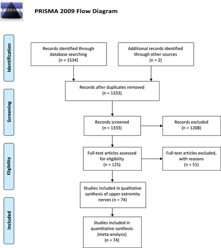
In all, 1333 potentially relevant studies were found from which 125 eligible studies were retained for full-text evaluation after screening titles and abstracts. Fifty-one studies were excluded after consensus of the two reviewers, most of them due to missing information about standard deviation or due to missing data for upper extremity nerves (Table S1). Seventy-four studies were included in the review and meta-analysis (Table S2, Figure 1). Full references of included studies (13-79) and excluded studies (80-130) are listed in Appendix S1. Twenty-three studies were conducted in Europe, nine in America, 35 in Asia, five in Australia and three studies in Africa (one study included healthy persons also from India and The Netherlands). The range of frequencies used as ultrasound probe was between 3 and 18 MHz. A total of 18,226 measures of upper extremity nerves of cumulatively 4186 healthy people were analysed. All studies included healthy adults older than 18 years. Mean age was 42.7 years. The mean proportion of female patients was 58%.
Cross-sectional area reference values and heterogeneity of reported values
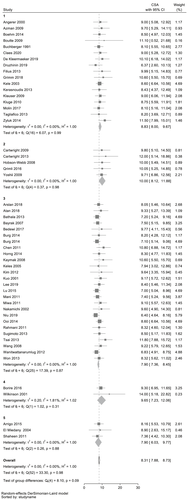
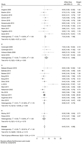
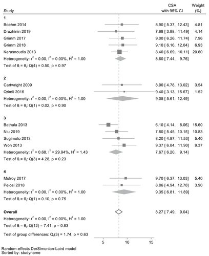
The calculated mean pooled CSA of the median nerve at the wrist was 8.3 mm2 (95% confidence interval [95% CI] 7.9–8.7 mm2, n = 4071), at the forearm 6.4 mm2 (95% CI 5.9–6.9 mm2, n = 3021) and at the upper arm 8.3 mm2 (95% CI 7.5–9.0 mm2, n = 1388).
The calculated mean pooled CSA of the ulnar nerve at the Guyon's canal was 4.1 mm2 (95% CI 3.6–4.6 mm2, n = 1688), at the forearm 5.2 mm2 (95% CI 4.8–5.7 mm2, n = 1983), at the elbow 5.9 mm2 (95% CI 5.4–6.5 mm2, n = 2551) and at the upper arm 6.6 mm2 (95% CI 5.1–6.1 mm2, n = 1737).
The calculated mean pooled CSA of the radial nerve at the upper arm at the spiral groove was 5.1 mm2 (95% CI 4.0–6.2 mm2, n = 1787).
A substantial heterogeneity across studies reporting mean CSA values of the radial nerve (I2 = 56.66%) was found and low heterogeneity across studies reporting mean CSA values of the median nerve at the forearm (I2 = 22.91%) and the ulnar nerve at the Guyon's canal (I2 = 9.95%). No heterogeneity was found across studies reporting CSA values for all other sites.
These results are shown in Table 1. Figures 2-4 show results of the meta-analysis of CSA for the median nerve. Results for the radial and ulnar nerves are given in Figures S1–S5.
| Nerve | Site | N studies | N nerves | Mean CSA (mm2) | 95% CI (mm2) | Heterogeneity (I2) (%) |
|---|---|---|---|---|---|---|
| Median nerve | Wrist | 53 | 4071 | 8.3 | 7.9–8.7 | 0.00 |
| Forearm | 29 | 3021 | 6.4 | 5.9–6.9 | 22.91 | |
| Upper arm | 13 | 1388 | 8.3 | 7.5–9.0 | 0.00 | |
| Ulnar nerve | Guyon's canal | 14 | 1688 | 4.1 | 3.6–4.6 | 9.95 |
| Forearm | 19 | 1983 | 5.2 | 4.8–5.7 | 0.00 | |
| Elbow | 23 | 2551 | 5.9 | 5.4–6.5 | 0.00 | |
| Upper arm | 18 | 1737 | 6.6 | 5.1–6.1 | 0.00 | |
| Radial nerve | Upper arm | 14 | 1787 | 5.1 | 4.0–6.2 | 56.66 |
- Abbreviation: CI, confidence interval.
Correlations of CSA with age, gender, publication year, height, weight and geographic differences Small but significant effects of age could be recorded in univariate regression analysis for the median nerve at the wrist (meta-regression coefficient 0.066, p = 0.018), which in multivariate regression analysis was not significant anymore (p = 0.229). For height a weak significant association to CSA was found in univariate regression analysis for the ulnar nerve at the elbow (coefficient 0.160, p = 0.017) and at the upper arm (coefficient 0.149, p = 0.012). Small significant effects of weight to CSA were found for the ulnar nerve at the elbow (coefficient 0.105, p = 0.030) and at the upper arm (coefficient 0.121, p = 0.005). In multivariate regression analysis, these effects were not found to be significant. The CSA of the ulnar nerve at the Guyon's canal was reported to be higher in studies conducted in America than in Europe and Asia (p = 0.04). This was also the case with regard to the CSA of the radial nerve (p < 0.001). For other nerve sites, geographic continents had no effect on CSA. A later publication year resulted in slightly lower CSA values for the ulnar nerve at the Guyon's canal (r = −0.187, p = 0.022), but not in the other nerve sites. This effect was found to be significant also in multivariate regression analysis (p = 0.046). There were no significant differences of CSA in the analysis of gender proportion.
The results of meta-regression analyses are shown in Table 2 and Figures S6–S9.
| Nerve | Site | Meta-regression analysis mean age | Meta-regression analysis height | Meta-regression analysis weight | Meta-regression analysis male proportion | Meta-regression analysis publication year | Test of group differences geographic regions, p |
|---|---|---|---|---|---|---|---|
| p (coefficient) | p (coefficient) | p (coefficient) | p (coefficient) | p (coefficient) | |||
| Median nerve | Wrist | 0.018 (0.066) | n.s. | n.s. | n.s. | n.s. | 0.09 |
| Forearm | n.s. | n.s. | n.s. | n.s. | n.s. | n.s. | |
| Upper arm | n.s. | n.s. | n.s. | n.s. | n.s. | n.s. | |
| Ulnar nerve | Guyon's canal | n.s. | n.s. | 0.07 (0.075) | n.s. | 0.022 (−0.187) | 0.04 |
| Forearm | n.s. | n.s. | 0.09 (0.065) | n.s. | n.s. | n.s. | |
| Elbow | n.s. | 0.017 (0.160) | 0.030 (0.105) | n.s. | n.s. | 0.09 | |
| Upper arm | n.s. | 0.012 (0.149) | 0.005 (0.121) | n.s. | n.s. | 0.09 | |
| Radial nerve | Upper arm | n.s. | n.a. | n.a. | 0.060 (−13.112) | n.s. | 0.00 |
DISCUSSION
For the first time, the results of a meta-analysis of ultrasound CSA nerve reference values are presented in this study.
High variability accompanied by a substantial heterogeneity of reported reference values of the upper extremity nerves was found in this meta-analysis only for the radial nerve. This is probably because the radial nerve is more difficult to examine in its spiralling course around the humerus. Therefore, technical and methodological differences in the different studies and ultrasound protocols probably arise especially for the radial nerve. For the measurement of CSA, the majority of studies used a tracing method of the inner borders of the hyperechogenic rim surrounding the hypoechogenic nerve. However, a description of the investigation site with anatomical landmarks differed in accuracy throughout the publications. Only studies in which regions were specified were included but it cannot be ruled out that anatomical differences contribute to the heterogeneity of reported CSA values of the radial nerve. An exact definition of the examination site is dependent on the purpose of the examination. Strictly defined anatomical landmarks seem most important for entrapment syndromes, for example the median nerve at the wrist, should be measured just proximal of the possible compression site of the flexor retinaculum to detect entrapment-related CSA enlargement. On the other hand, in inflammatory polyneuropathies it appears to be more important to investigate the whole nerve dynamically as the exact location of potential enlargement is different in each patient.
Overall, polyneuropathy ultrasound protocols using the anatomical regions described in this meta-analysis have been established in the last few years, as can be recognized from the large number of studies investigating these specific nerve sites. Hereby meta-analytical data are provided for these regions and therefore it is recommended that these examination sites are used, to reach an internationally standardized paraclinical evaluation. Still, each ultrasound laboratory should compare its own normal values to the results of this meta-analysis and aim to reduce deviations as much as possible.
In this meta-analysis, correlation of the CSA of the median nerve at the wrist with age suggests that age could be a risk factor for asymptomatic morphological changes at typical entrapment sites. However, most studies examined patients with a maximum mean age of 60 years. Only one group, Cartwright et al, [6 recruited elderly people with a mean age of 82 years revealing the largest CSA reference values (Figure 4). Therefore, correlation of CSA at entrapment sites with age should be confirmed in further studies with patients older than 60 years. The correlation of CSA with height and weight was only found for examinations of the ulnar nerve, not for the median and radial nerves. Why this correlation only existed for the ulnar nerve remains unclear. Overall, the correlations of CSA with age, height and weight were weak, so that age-, height- and weight-dependent standard values for the CSA of adult patients do not appear to be necessary in everyday clinical practice.
The fact that higher CSA reference values were found for the radial and ulnar nerves in American studies could result either from methodological differences in ultrasound examinations between these regions or from actual different population characteristics. Again, internationally standardized protocols are necessary to avoid methodological differences. The number of available studies per continent was small for the analysis of geographic differences, and therefore results must be interpreted with caution.
One limitation for the meta-analysis is that the associations detected at the population level might not be present at the individual patient level (ecological fallacy). Also, the lack of significant associations could be attributed to vast heterogeneity between studies rather than the lack of the presence of true associations. Beyond this, the influence of the ultrasound device or the frequency of the ultrasound probe to CSA values could not be analysed as various devices were used and frequencies were only given as a range (i.e. 8–15 MHz in the majority of publications). Moreover, in a few publications different anatomical regions were examined, that is, the deep branch of the radial nerve, which were not included in this analysis.
In this first part of our meta-analysis only results for upper extremity nerves are reported. For other nerves such as lower extremity nerves and brachial plexus which are technically more difficult to examine, an even higher heterogeneity must be expected.
In conclusion, reference values of the CSA of the upper extremity nerves are provided that are based for the first time on a meta-analysis of 18,226 examined nerve sites. The heterogeneity of reported studies was substantial only for the radial nerve probably due to methodological differences. All other analysed nerve sites showed no or low heterogeneity of reported values. Subgroup analysis revealed age as a possible risk factor for asymptomatic morphological changes with increased CSA only for the median nerve at the wrist as a typical entrapment site. Correlations of CSA with age, height and weight were weak. Our meta-analytical data facilitate the goal of an international standardized evaluation protocol.
ACKNOWLEDGEMENTS
Open access funding was enabled and organized by DEAL project of Ruhr-University Bochum.
CONFLICT OF INTERESTS
Anna Lena Fisse: No disclosures related to this work. Aristeidis H. Katsanos: No disclosures related to this work. Ralf Gold: Received consultation fees and speaker honoraria from Bayer, Schering, Biogen Idec, Merck Serono, Novartis, Sanofi-Aventis and Teva; he also acknowledges grant support from Bayer Schering, Biogen Idec, Merck Serono, Sanofi-Aventis and Teva, none related to this paper. Kalliopi Pitarokoili: Received travel grants and speakers’ honoraria from Novartis, Biogen Idec, Teva, Bayer, Celgene, CSL Behring and Grifols all unrelated to the paper. Christos Krogias: Received travel grants and honoraria from Bayer Vital and Daiichi-Sankyo.
AUTHOR CONTRIBUTIONS
Anna Lena Fisse: Designed study, collected data, analysed data, drafted paper. Aristeidis H. Katsanos: Analysed data, revised paper. Ralf Gold: Designed study, revised paper. Christos Krogias: Designed study, analysed data, revised paper. Kalliopi Pitarokoili: Designed study, collected data, revised paper.
Open Research
DATA AVAILABILITY STATEMENT
The data that support the findings of this study are available from the corresponding author upon reasonable request.


