Early View
Video Article
Free Access

Gel-immersion-assisted endoscopic injection sclerotherapy under observation with Texture and Color enhancement imaging for esophageal varices

Tsunetaka Kato

Tsunetaka Kato

Department of Endoscopy, Fukushima Medical University Hospital, Fukushima, Japan

Department of Gastroenterology, Fukushima Medical University School of Medicine, Fukushima, Japan

Search for more papers by this author
Takuto Hikichi

Corresponding Author

Takuto Hikichi

Department of Endoscopy, Fukushima Medical University Hospital, Fukushima, Japan

Corresponding: Takuto Hikichi, Department of Endoscopy, Fukushima Medical University Hospital, 1 Hikarigaoka, Fukushima-City, Fukushima 960-1295, Japan. Email: [email protected]

Search for more papers by this author
Takumi Yanagita

Takumi Yanagita

Department of Endoscopy, Fukushima Medical University Hospital, Fukushima, Japan

Department of Gastroenterology, Fukushima Medical University School of Medicine, Fukushima, Japan

Search for more papers by this author
First published: 03 April 2025

Abstract

Watch a video of this article.

BRIEF EXPLANATION

Intravascular endoscopic injection sclerotherapy (EIS), an effective treatment for esophageal varices (EVs), is technically more challenging than endoscopic variceal ligation (EVL).1 In conventional EIS, gas insufflation reduces the EV diameter, making the puncture harder and less precise. To address this issue, gel immersion EIS (GI-EIS) has recently been developed.2-4

GI-EIS offers several advantages. Retaining the gel within the esophageal lumen ensures a clear and stable endoscopic field while reducing the esophageal wall tension, preserving the variceal diameter, and facilitating sclerosing agent injection (Figure 1). However, reduced mucosal tension surrounding EVs can sometimes make them appear less prominent, posing challenges in selecting the optimal puncture site.
Details are in the caption following the image
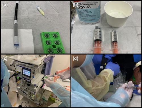
Gel immersion endoscopic injection sclerotherapy under texture and color enhancement imaging. (a) An endoscope (GIF-H290T; Olympus, Tokyo, Japan) equipped with a balloon (9.8 mm × 60 mm; Create Medic, Yokohama, Japan) and a transparent hood for the endoscopic variceal ligation device. (b) The gel (Viscoclear; Otsuka Pharmaceutical, Tokushima, Japan) was dispensed into 50 mL and 20 mL syringes to prevent air bubble formation. (c) The assistant preflushes the water supply channel with the gel (white arrowhead). While the operator inserts the puncture needle through the working channel, the assistant simultaneously injects the gel through the water supply channel. (d) The endoscopist punctures the esophageal varix, and the assistant injects a 5% ethanolamine oleate (EO) solution (1:1 mixture of 10% EO and a contrast agent) after confirming the backflow of blood (white arrowhead).

To overcome this limitation, we hypothesized that EIS using texture and color enhancement imaging (TXI) would improve EV visibility and ameliorate the precision of puncture point selection compared with white-light imaging. TXI (Mode 1) optimizes three key image factors – texture, brightness, and color – thereby improving lesion contrast and visualization.5 Based on this concept, we developed GI-EIS under TXI observation, referred to as “GI-EIS under TXI” (Video S1).

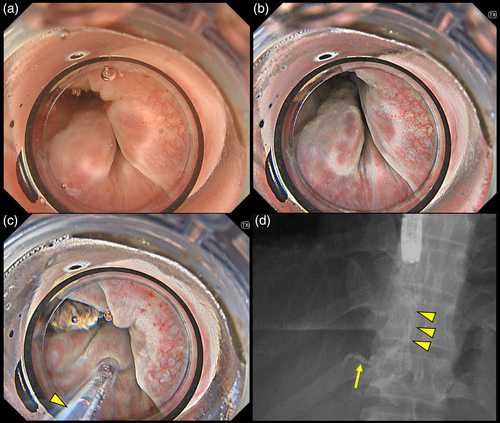
We conducted GI-EIS under TXI in 15 prophylactic cases between September 2023 and October 2024. The high success rate (93.3%) of GI-EIS under TXI can be attributed to the synergistic effects of gel immersion and TXI-enhanced imaging. The gel-immersion maintained the variceal diameter by preventing luminal collapse, while TXI improved visualization of the varices by enhancing texture and contrast. This allowed for precise puncture site selection. Additionally, gel-immersion suppressed peristalsis and minimized esophageal wall tension. These factors collectively contributed to the successful injection (Figure 2).
Details are in the caption following the image
Endoscopic and X-ray fluoroscopic images of gel immersion endoscopic injection sclerotherapy under observation with texture and color enhancement imaging (TXI) for esophageal varices (EVs). (a) EVs are observed using white-light imaging (WLI) with a gel-filled esophageal lumen. A hood with an O-ring for endoscopic variceal ligation is attached to the endoscope. (b) The observation mode is switched to TXI Mode 1, which enhances EV visualization compared with WLI. (c) With the esophageal lumen filled with gel, the EV is punctured, and the sclerosing agent (ethanolamine oleate) is injected under TXI observation. The backflow of blood is visualized (yellow arrowhead). (d) Under X-ray fluoroscopy, the sclerosing agent is injected into the EV (yellow arrowheads) and the left gastric vein (yellow arrow).

Authors declare no conflict of interest for this article.

ACKNOWLEDGMENTS

We thank Dr. Jun Nakamura, Dr. Minami Hashimoto, Dr. Ryoichiro Kobashi, Dr. Mitsuru Otsuka, Dr. Daiki Nemoto, and Professor Hiromasa Ohira for their invaluable contributions to postoperative patient management and insightful advice on article preparation. We also thank the entire medical staff at the Department of Endoscopy, Fukushima Medical University Hospital, for their dedicated assistance with the endoscopic procedures.

      The full text of this article hosted at iucr.org is unavailable due to technical difficulties.