First two cases of Zenker's diverticulum treated with flexible endoscopic septum division in Japan
Abstract
Watch videos of this article.
Brief Explanation
Flexible endoscopic septum division (FESD) is emerging as an effective minimally invasive treatment for Zenker's diverticulum (ZD).1, 2 Overall prevalence of ZD is rare.3 Therefore, ZD was historically treated by surgery,4 and, to date, there are no reports of FESD for ZD in Japan. In this video, we would like to show the first two cases of ZD successfully treated with FESD.
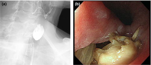
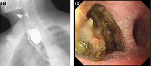
First case is a 59-year-old man referred with symptoms of dysphagia, food sticking in his throat and regurgitation. Barium swallow showed ZD measuring 31 × 18 mm (Fig. 1). Second case is an 83-year-old woman. She also presented with symptoms of dysphagia, drooling and regurgitation for over 20 years, but worsening in the past 3 years. Barium swallow showed ZD measuring 52 × 30 mm (Fig. 2). Both patients were treated by FESD with a scissors-type knife.5


Procedures were carried out under deep sedation with propofol and fentanyl citrate without intubation. After emptying the pouch of food debris, a conventional guidewire was placed through the scope. The endoscope was reinserted with a cap on the tip and saline solutions with indigocarmine injected to create a mucosal bleb over the center of the septum (cricopharyngeus muscle [CP]). With mucosal incision and dissection, the horizontal fibers of the CP were identified and dissected with the knife to achieve a near complete CP myotomy. Clips were deployed at the site of the cut part of the septum to prevent perforation, and no adverse events occurred during the procedure. Procedure time was 40 min for the first and 20 min for the second case. Both patients had complete symptomatic relief at 30 days follow up after FESD (Videos S1, S2).
We hope these two successful cases will raise awareness and pave the way for minimally invasive FESD for ZD in Japan.
Authors declare no conflicts of interest for this article.
Acknowledgments
The authors thank Yuki Sumida and Yuki Miyasako for assistance in data collection and Naoko Matsumoto for administrative support.


