Underwater endoscopic mucosal resection for a superficial polyp located at the anastomosis after surgical colectomy
Underwater endoscopic mucosal resection (U-EMR), as reported by Binmoeller et al., is an effective technique to remove recurrent lesions after piecemeal EMR, as well as large sessile colorectal polyps.1-3 One major advantage of U-EMR is that submucosal injection is not required as: (i) the suctioning of luminal air decreases colonic wall tension; (ii) colonic muscular wall remains circular when it is filled with water; and (iii) water immersion ‘floats’ the mucosa and submucosa.2 This technique is also applied to duodenal ampullary and non-ampullary adenomas.4, 5


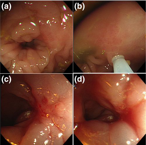
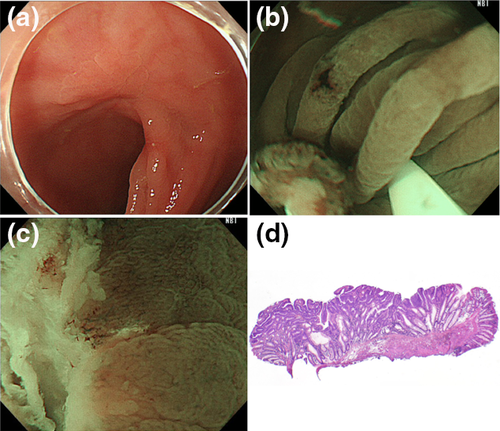
A 77-year-old man presented with a 10-mm-sized superficial colonic polyp located at the anastomosis site after sigmoidectomy for a sigmoid colon cancer (Fig 1a). The lesion was detected during follow-up colonoscopy after surgery in a previous hospital, and the endoscopist attempted to carry out conventional EMR. After several submucosal injections of normal saline, the lesion was not lifted up, whereas the surrounding mucosa was overly lifted (Fig 1b-d). Therefore, the previous doctor opted not to snare the lesion and recommended additional surgery as the biopsy specimen showed high-grade dysplasia. The patient refused surgery and was referred to Osaka Medical Center for Cancer and Cardiovascular Diseases, where we decided to carry out U-EMR. After full immersion in natural saline, we snared and removed the lesion under narrow-band imaging (NBI) observation (Fig 2a,b).3, 5 The procedure was completed in a few minutes without any visible neoplastic tissue on the margin of the mucosal defect with underwater magnifying NBI observation (Fig 2c). The retrieved specimen showed high-grade dysplasia without tumor involvement on the horizontal and vertical margins (Fig 2d). Surveillance colonoscopy will be carried out in the future.
This case clearly demonstrates the drawbacks of submucosal injection for conventional EMR and the efficacy of U-EMR. U-EMR can be easily carried out for lesions with non-lifting sign during a previous attempt, as well as for recurrent lesion after piecemeal EMR or for large sessile colorectal polyps.
Conflicts of Interest
Authors declare no conflicts of interest for this article.


