Precutting endoscopic mucosal resection for colorectal lesions
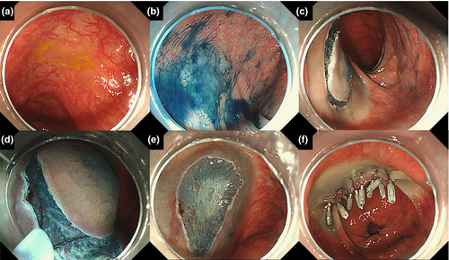
Precutting endoscopic mucosal resection (EMR) is defined by Japan Gastroenterology Endoscopy Society as a technique in which snaring is done without dissecting the submucosal layer after incising the circumference of the lesion by using a knife for endoscopic submucosal dissection (ESD) or the tip of a snare (Fig. 1).1 Conventional EMR is a common technique for resecting colorectal lesions, and the size of the lesion that can be resected safely en bloc using the conventional EMR technique is regarded as a lesion <20 mm in size. However, a recent study reported that even in cases with a lesion <20 mm in size, the incomplete polyp resection rate that was confirmed by obtaining biopsies from the resection margin was relatively high, particularly for lesions 15–20 mm in size (23.3%) and sessile serrated adenomas/polyps (SSA/P) (31.0%).2 Sakamoto et al.3 examined the efficacy of the precutting EMR technique for large colorectal neoplasia and reported an en bloc resection rate of 83.3% for colorectal lesions 20–30 mm in size; in addition, no perforation was observed. As is well known, a higher en bloc resection rate can provide a lower recurrence rate after endoscopic resection.4 Controversy still exists regarding whether the ESD technique should be applied for the treatment of large SSA/P; therefore, the application of precutting EMR for colorectal lesions <30 mm in size, especially large SSA/P, may be feasible for clinical practice. Although the perforation rate of precutting EMR has not been reported for a large number of cases and precutting EMR for colorectal lesions with fibrosis should be applied carefully, precutting EMR does not require an additional device when the circumferential incision is made using the tip of a snare. Thus, precutting EMR should be considered an easy method for resecting large colorectal lesions.

Conflicts of Interest
Authors declare no conflicts of interest for this article.


